
Auscult the chest from back e kon kon space e auscult korte hoi according to macloid
Macleod clinical examination posterior chest auscultation sites intercostal spaces
posterior chest auscultation sites diagram back lung zones
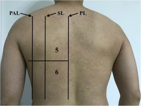
A clinical photograph of a patient's back, serving as an anatomical diagram for lung ultrasound (LUS) zone partitioning. The image illustrates the posterior chest wall with superimposed black lines and numeric labels defining specific diagnostic areas. Vertical anatomical landmarks are indicated by abbreviations: PL (Paravertebral Line), SL (Scapular Line), and PAL (Posterior Axillary Line). These lines delineate the medial and lateral boundaries of the posterior examination zones. A horizontal line bisects the region into two distinct areas: Area 5, representing the upper backside chest area, and Area 6, representing the basal (lower) backside chest area. This standardized 12-zone partition method is utilized in critical care medicine to systematically evaluate the lungs for pathologies such as Acute Respiratory Distress Syndrome (ARDS), interstitial syndrome, and pleural effusions. The visualization provides a reference for medical students and clinicians to ensure consistent probe placement during a longitudinal ultrasound scan of the posterior thoracic segments.
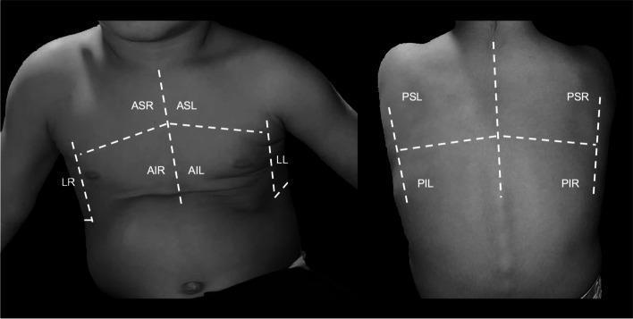
A clinical diagram showcasing a standardized lung ultrasound (LUS) scanning scheme on a pediatric subject. The image is divided into two panels: an anterior-lateral view of the chest and a posterior view of the back. White dashed lines demarcate ten specific scanning regions categorized by anatomical landmarks. Anterior regions include the Anterior Superior Right (ASR), Anterior Inferior Right (AIR), Anterior Superior Left (ASL), and Anterior Inferior Left (AIL), divided by a vertical parasternal line and a horizontal nipple-level line. Lateral areas are marked as Lateral Right (LR) and Lateral Left (LL) along the mid-axillary lines. The posterior panel displays four zones: Posterior Superior Right (PSR), Posterior Inferior Right (PIR), Posterior Superior Left (PSL), and Posterior Inferior Left (PIL), divided by the spinal column and a horizontal mid-scapular line. This protocol is designed for systematic thoracic evaluation, such as monitoring dysphagia-related aspiration or deaeration scores in infants, ensuring comprehensive coverage of all lung lobes during diagnostic imaging.
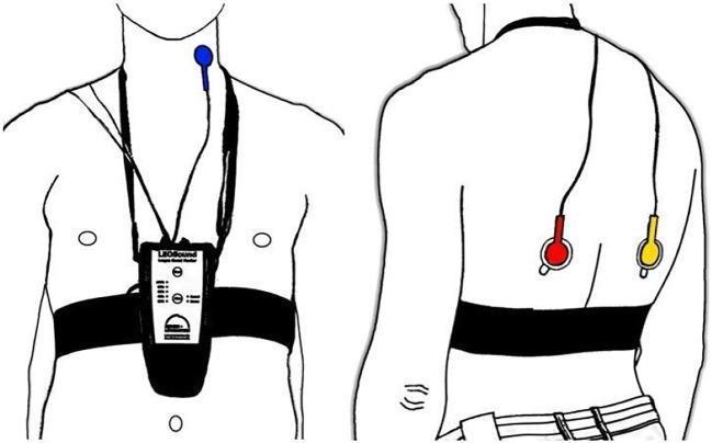
An anatomical diagram illustrating the placement of the LEOSound lung-sound monitor and its bio-acoustical sensors on a patient. The illustration includes two views: an anterior (front) view and a posterior (back) view. In the anterior view, a recording device is secured to the mid-thorax by a black chest belt and a neck strap. A blue tracheal microphone is positioned on the neck near the trachea. In the posterior view, two additional microphones are attached using adhesive pads to the inferior region of the left (red microphone) and right (yellow microphone) scapula. The system is designed for mobile, long-term recording and automated analysis of respiratory sounds such as coughing and wheezing. The diagrams emphasize the standardized anatomical landmarks required for optimal auscultation and data collection in clinical or home settings.
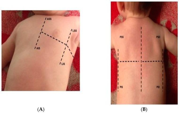
Educational clinical photograph demonstrating the standardized thoracic scanning zones for pediatric Lung Ultrasound (LUS). The image consists of two panels showing an infant's chest and back with superimposed dashed lines defining anatomical quadrants. Panel A illustrates the anterior and lateral hemithorax. The anterior superior (ASS) and anterior inferior (AIS) zones are separated from the lateral superior (LSS) and lateral inferior (LIS) zones. The divisions represent standard LUS protocols delineated by the parasternal, anterior axillary, and posterior axillary lines. Panel B shows the posterior hemithorax divided into four quadrants by vertical and horizontal dashed lines. The regions are labeled: PSS (Posterior Superior Sinister/Left), PSD (Posterior Superior Dexter/Right), PIS (Posterior Inferior Sinister/Left), and PID (Posterior Inferior Dexter/Right). This diagram serves as a clinical guide for systematically evaluating pulmonary areas for conditions such as lung congestion, pleural effusion, or pneumothorax in pediatric patients. It emphasizes a structured approach to ensure comprehensive coverage of the lung fields during ultrasound examination.
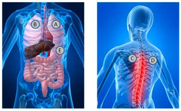
This medical anatomical diagram illustrates the standard stethoscope placement locations for auscultation of heart and lung sounds. The image consists of two panels: an anterior (front) view and a posterior (back) view of a semi-transparent human torso. In the anterior view, labels A and B are positioned over the apical regions of the right and left lungs, respectively, while label E is located at the lower left costal margin, corresponding to the cardiac apex and mitral valve area. Visible internal structures include the lungs, heart, liver, and intestinal tract. In the posterior view, labels C and D are placed in the interscapular region at the level of the upper thoracic spine, typically used for assessing posterior bronchial and vesicular breath sounds. This view highlights the vertebral column, ribs, and scapulae, with a red glow emphasizing the spinal region. This illustration serves as an educational guide for clinical physical examination techniques, demonstrating the relationship between surface landmarks and underlying thoracic and abdominal viscera.
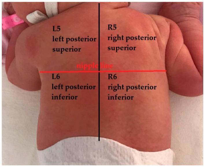
This clinical photograph demonstrates an anatomical mapping of the posterior thorax in a neonate, used specifically for standardized lung ultrasound (LUS) examination. The infant is positioned prone, and the back is divided into four distinct diagnostic zones by two intersecting axes. A vertical black line bisects the back into left and right hemithoraces. A horizontal red line, labeled 'nipple line,' serves as the anatomical landmark to differentiate the superior and inferior segments. The resulting four zones are labeled as follows: L5 (left posterior superior), L6 (left posterior inferior), R5 (right posterior superior), and R6 (right posterior inferior). This visual guide is part of a 12-zone lung ultrasound scoring system designed to semi-quantify pulmonary pathology, such as consolidations or B-lines, in pediatric and neonatal patients. The mapping provides a systematic approach for clinicians to assess and document lung findings consistently across different regions of the posterior chest wall.
Macleod's অনুযায়ী পিছন থেকে Chest Auscultation - কোন কোন Space-এ করতে হয়
পদ্ধতি: "Stepladder" / "W" বা "Zigzag" Pattern
Posterior Chest-এ Auscultation-এর Specific Sites:
| ক্রম | অবস্থান | Intercostal Space | কোন Lobe শোনা যায় |
|---|---|---|---|
| 1 | C7 Vertebra-র নিচে (Supraclavicular area বা Upper trapezius area) | 1st ICS (Posterior) | Upper lobe apex |
| 2 | Scapula-র উপরের বর্ডার বরাবর | 2nd–3rd ICS (Posterior) | Upper lobe |
| 3 | Scapula-র মাঝ বরাবর (Interscapular region) | 4th–5th ICS (Posterior) | Lower lobe (upper part) |
| 4 | Scapula-র নিচের বর্ডার বরাবর | 6th ICS (Posterior) | Lower lobe |
| 5 | Scapula-র নিচে, Paraspinal area | 7th ICS (Posterior) | Lower lobe (base) |
Macleod's-এর মূল নির্দেশনা:
- ১ম ICS (Posterior) থেকে শুরু করো - spine-এর দুই পাশে
- নামতে থাকো - প্রতিটি ICS-এ bilateral comparison করো (আগে ডান পাশ, তারপর বাম পাশ - same level-এ)
- Scapula avoid করো (scapula-র উপর রাখলে ভালো sound পাওয়া যায় না)
- ৭ম ICS (Posterior) পর্যন্ত নামো - এটাই lower limit
- ৫ম–৭ম ICS-এ Posterobasal segment শোনা যায় (effusion, consolidation এখানেই আগে ধরা পড়ে)
Breath Sounds অনুযায়ী কোথায় কী শুনবে:
| Breath Sound | Posterior-এ Location |
|---|---|
| Bronchial | C7-T1 level (behind trachea) |
| Bronchovesicular | Interscapular region (T3-T5), Scapula-র মাঝখানে |
| Vesicular | বাকি সব জায়গায় (lower zones) |
Clinical Tips (Macleod's অনুযায়ী):
- রোগীকে সামনে ঝুঁকে বসান (arms folded across chest) - এতে scapula সরে যায় এবং আরো বেশি area পাওয়া যায়
- Deep breathing করতে বলুন - mouth দিয়ে
- প্রতিটি site-এ কমপক্ষে একটি পূর্ণ respiratory cycle (inspiration + expiration) শুনুন
- Bilateral comparison সবচেয়ে গুরুত্বপূর্ণ - একই level-এ দুই পাশ তুলনা করুন
সহজে মনে রাখার উপায়:
← Spine →
1st ICS  •  •   ← Upper lobe apex
2nd ICS  •  •   ← Upper lobe
3rd ICS  •  •   ← Upper lobe (lower)
    [SCAPULA - avoid]
4th ICS  •  •   ← Lower lobe
5th ICS  •  •   ← Lower lobe
6th ICS  •  •   ← Lower lobe base
7th ICS  •  •   ← Posterobasal (last point)
Na na ami bolci paravertebral level e koto sspace theke auscult suru korb
Paravertebral Line - Auscultation Sites:
2nd ICS  •  •   ← এখান থেকে শুরু
3rd ICS  •  •
4th ICS  •  •
5th ICS  •  •
6th ICS  •  •
7th ICS  •  •   ← এখানে শেষ
কেন 1st ICS-এ না?
- Trapezius muscle ঢেকে রাখে
- Shoulder girdle-এর কারণে stethoscope ঠিকমতো বসানো যায় না
মনে রাখার সহজ নিয়ম (Macleod's):
"2 থেকে 7" - Paravertebral line-এ posterior chest-এ ৬টি site, bilateral comparison করতে করতে নিচে নামো।