
Based on information in this pdf make notes on “mechanism and management of nerve damage and neuritis in leprosy”
cd /tmp/workspace && python3 -c " import pdfplumber path='/tmp/workspace/attachments/dbf1f115-213e-4e7a-b2aa-d86f1b3e739a/Neuritis IAL.pdf' with pdfplumber.open(path) as pdf: print('pages', len(pdf.pages)) text='' for i,page in enumerate(pdf.pages): t = page.extract_text() or '' text += f'\n--- PAGE {i+1} ---\n' + t with open('/tmp/workspace/neuritis_extracted.txt','w') as f: f.write(text) print('done') "
"steroid" in ~/neuritis_extracted.txt
"treatment" in ~/neuritis_extracted.txt
"manage" in ~/neuritis_extracted.txt
Mechanism and Management of Nerve Damage and Neuritis in Leprosy
1. Definitions
- Neuritis = inflammation of nerves. In leprosy usage the term is applied both pathologically and clinically, and is used interchangeably with "neuropathy."
- Leprosy neuritis is typically a subacute, demyelinating, non-remitting process affecting cutaneous nerves and larger peripheral nerve trunks.
- Nerve Function Impairment (NFI) = the sensory, motor and/or autonomic deficit resulting from M. leprae-induced nerve pathology; used interchangeably with "nerve damage."
- Peripheral neuropathy of leprosy is classified as "mononeuritis multiplex" — widespread but patchy/non-homogeneous. A single nerve trunk can be affected without skin lesions, giving rise to "pure neuritic leprosy," a recognized clinical type in India.
2. Mechanism of Nerve Damage
A. Bacterial entry and Schwann cell invasion
- M. leprae invades Schwann cells and axons, causing demyelination and axonal degeneration.
- Entry is receptor-mediated: the bacillus's 21 kDa laminin-binding protein (LBP-21, gene ML1683) binds the G domain of laminin-α2 chain expressed on the Schwann cell-axon unit.
- A 25 kDa phosphorylated glycoprotein on human peripheral nerve also binds M. leprae.
- Phenolic glycolipid-1 (PGL-1), a bacillus-specific surface antigen, binds laminin-2, further promoting Schwann cell invasion.
- M. leprae-induced demyelination also occurs via direct ligation and activation of the ErbB2 receptor of neuregulin-1, a pathway that normally regulates myelination.
- Schwann cells are actively phagocytic and can engulf bacilli (seen as intact AFB within swollen Schwann cells of unmyelinated fibers in lepromatous leprosy, LL); it remains unclear whether they also act as antigen-presenting cells to T cells.
- Experimental (armadillo) data suggest bacilli may also enter and multiply via endoneurial blood vessels, not just direct Schwann cell binding.
B. Immune-mediated injury (three broad mechanisms proposed)
- Interference of M. leprae cell wall proteins with host macrophage metabolism.
- Immune-mediated inflammation from T-cell/Schwann cell interactions (cell-mediated immunity, CMI).
- "Bystander" nerve injury from the bulk influx of inflammatory cells and edema during immune/inflammatory responses — nerve damage as collateral effect of the host response rather than the organism itself.
C. Reaction-related mechanisms (major driver of acute damage)
- Type 1 (reversal) reaction: an upregulated CMI/delayed-type hypersensitivity response to M. leprae antigens — most damaging to nerves, more frequent and severe than Type 2.
- Type 2 reaction (ENL): driven by circulating immune complex deposition, a humoral mechanism; nerve damage tends to be less severe per episode but recurrent/chronic ENL in BL/LL patients causes cumulative damage.
- Damage occurs at three anatomical levels: (1) cutaneous nerve endings, (2) subcutaneous nerves, (3) major nerve trunks. The more proximal the site of damage, the more extensive the resulting deficit (since nerves give off branches distally).
- Damage to a major trunk carrying sensory + motor + autonomic fibers (e.g., posterior tibial, ulnar, median, lateral popliteal, facial) causes the most disabling NFI.
- Facial skin patches in Type 1 reaction carry a higher risk of ocular nerve involvement and need special monitoring.
D. Patterns of neuritis across the disease spectrum
- Neuritis associated with the disease itself — chronic, low-grade, due to granuloma formation and ongoing inflammatory response; subperineurial edema and loss of unmyelinated axons occur very early, often before any clinical symptoms/signs appear.
- Neuritis associated with reactions — acute and severe (Type 1 > Type 2).
- Silent neuritis / "quiet nerve paralysis" — progressive sensory/motor impairment without pain, tenderness, paresthesia, or visible signs of reaction; can develop even while the patient is on multidrug therapy (MDT) — e.g., a Bangladesh cohort showed 7.9% of MB patients developed NFI while still on MDT.
- Neuropathic pain (NP) — seen especially in pure neuritic leprosy and tuberculoid-spectrum patients with Type 1 reactions, though LL patients with recurrent severe Type 2 reactions can also have intense multi-nerve pain. NP can persist for months to years even after treatment is completed (in patients "released from treatment"), and includes paresthesia, dysesthesia, hyperesthesia and allodynia.
E. Order of sensory loss in a developing lesion
F. Clinical course
- Damage can occur both during and after completion of treatment.
- Recurrent episodes of neuritis, though less common than a single episode, can cause severe cumulative nerve damage.
- Deep tendon reflexes remain normal (CNS not involved) except in a small proportion with severe neuritis showing abnormal reflexes/joint position sense.
- Muscle wasting in leprosy is neurogenic atrophy, not primary muscle disease.
3. Consequences of Untreated Nerve Damage
4. Management
A. Early detection is the cornerstone
- Because much of the neuropathology (subperineurial edema, axonal loss) precedes symptoms, regular clinical nerve function assessment is essential — the text recommends monthly testing for the first 6 months of treatment and every 3 months thereafter, using:
- Nerve palpation for thickening/tenderness (comparative, needs clinical expertise).
- Voluntary muscle testing (VMT) for motor NFI.
- Semmes-Weinstein (SW) monofilaments for graded sensory testing (0.05 g to 300 g pressure) at standardized sites on hands, feet and face.
- Nerve conduction studies are sensitive but the combination of palpation + VMT + SW filament testing is comparably effective and more practical clinically.
- This periodic surveillance is what allows silent neuritis to be caught before irreversible damage occurs.
B. Pharmacological management
- Multidrug therapy (MDT) for the underlying leprosy infection is the base treatment; note NFI can still develop while a patient is on MDT, so nerve monitoring must continue throughout.
- Corticosteroids (e.g., prednisolone) are the mainstay for treating neuritis and reactions:
- Indicated whenever there is acute NFI, especially during Type 1 reactions, and in children where a high index of suspicion is needed (in one pediatric cohort, neuritis was the presenting feature of Type 1 reaction in 65% of cases with reaction).
- Trials show nerve function can also recover spontaneously, and that prednisolone is safe but has limits to its usefulness in preventing/reversing disability — i.e., it helps but is not curative for established damage.
- Immediate institution of corticosteroid therapy is indicated once significant sensory threshold changes are detected (specific SW filament threshold criteria given in the text for triggering treatment).
C. Preventing disability (secondary prevention)
- The best strategy overall: early diagnosis and prompt treatment of leprosy and of leprosy reactions — this limits the degree of impairment and eventual disability.
- Patient education and self-care: teaching daily self-care for insensitive hands and feet (protection from injury, soaking/oiling, appropriate footwear) and eye care/protection to prevent secondary complications (ulcers, injuries) in already-anesthetic areas.
- WHO disability grading (proposed 1988) should be applied periodically to each limb and eye separately (highest grade recorded overall) to track progression and direct care/rehabilitation resources appropriately.
- Special attention to facial patches/Type 1 reaction given the added risk of ocular nerve (facial/trigeminal branch) involvement — proactive eye examination and protection.
D. Summary principle from the chapter
"The best way to prevent disabilities occurring as a result of nerve damage is early diagnosis and treatment of leprosy and leprosy reactions" — supported by regular structured nerve function assessment (palpation, VMT, SW monofilament testing), prompt corticosteroid use for reactional neuritis, and patient self-care education to prevent secondary deformity once sensory/motor loss has occurred.
Yes do the management of neuritis and nerve damage from extended research
leprosy neuritis treatment corticosteroid
erythema nodosum leprosum treatment
WHO leprosy reaction neuritis management guidelines prednisolone regimen
PMID: 27210895
https://ijdvl.com/leprosy-reactions-new-knowledge-on-pathoph…
Management of Nerve Damage and Neuritis in Leprosy — Extended Notes
1. Overarching Goals of Management
- Control skin and nerve inflammation
- Relieve pain
- Prevent further reactional episodes and cumulative nerve damage
- Reduce morbidity and prevent permanent deformity/disability
- Continue/complete antimicrobial cure of the underlying infection (Harrison's 22E, p. Ch. "Leprosy")
2. General Principle: Early, Aggressive Treatment
- "Management of leprosy reactions is complex and expert guidance should be sought. Reactions should be treated aggressively to prevent peripheral nerve damage." - Red Book 2021 (AAP Committee on Infectious Diseases), p. 763
- All leprosy patients should be educated to recognize signs of neuritis (nerve pain, tenderness, new numbness/weakness) and told to report immediately so corticosteroids can be started without delay - Red Book 2021, p. 763
- A practical clinical rule (HRSA/NHDP guidance): the initial steroid dose should be large enough to relieve nerve pain/tenderness within 24-48 hours; the maintenance dose should be enough to prevent recurrence of nerve pain.
3. Management of Type 1 (Reversal) Reaction and Associated Neuritis
- Mild T1R (skin involvement only, no neuritis): NSAIDs (aspirin, indomethacin, ibuprofen, diclofenac, acetaminophen) for several weeks, plus reassurance/counseling (Harrison's 22E).
- Moderate-to-severe T1R or any neuritis: Oral corticosteroids are the mainstay.
- Dose: prednisolone 0.5-1 mg/kg/day (≈30-40 mg/day for most adults), continued and then gradually tapered over ~20 weeks (a standard 12- to 20-week course) (Harrison's 22E; Park's PSM; IJDVL review, Lockwood et al.).
- Medscape/US regimens cite an initial dose of prednisolone 40 mg/day with taper.
- The drug of choice is prednisolone - cheap, widely available, and the field-standard for national leprosy programs (Park's PSM).
- Steroid-sparing / second-line agents for non-responders or steroid-limiting adverse effects: methotrexate, cyclosporine, azathioprine, mycophenolate mofetil, topical tacrolimus (Harrison's 22E). Azathioprine + prednisolone was not superior to prednisolone alone, and azathioprine + dapsone increased anemia risk (IJDVL review).
- Local/physical measures for the acutely inflamed nerve: rest/splinting and padding of the affected limb to reduce mechanical stress; surgical nerve decompression is indicated if there is persistent raised intraneural pressure, tenderness, and acute inflammation threatening ischemic damage (Harrison's 22E).
- Long-standing prednisolone/PSM guidance for children: a high index of suspicion for neuritis at diagnosis is emphasized, since neuritis can be the presenting feature of a reaction in a substantial proportion of pediatric cases.
4. Management of Type 2 Reaction (Erythema Nodosum Leprosum, ENL) and Neuritis
- Mild ENL (no/minimal neuritis): rest and NSAIDs are first-line.
- Severe ENL (fever, systemic symptoms, and/or acute neuritis with or without loss of nerve function - a defining criterion of severe ENL, Park's PSM): systemic anti-inflammatory therapy required.
- Corticosteroids: oral prednisolone, generally not exceeding 1 mg/kg/day, total course around 12 weeks for severe reactions (Goldman-Cecil Medicine, Table 301-3). Splitting the daily dose can reduce steroid adverse effects (IJDVL review).
- Thalidomide: highly effective and rapid-acting for severe/recurrent ENL, acting via TNF-α inhibition; typical dosing up to 400 mg/day in tapering doses. Restricted by teratogenicity and requires strict reproductive precautions/registry supervision (in the US, via Celgene/REMS program) (Red Book 2021; Cochrane review PMID 19588412; multiple historical trials PMID 4124808, 4125333).
- Clofazimine: useful, particularly for chronic/recurrent ENL, e.g., 100 mg TDS x12 weeks → 100 mg BD x12 weeks → 100 mg OD x12-24 weeks (IJDVL review).
- Refractory ENL: case reports/series support biologic/targeted therapy - anti-TNF agents (infliximab, etanercept) and apremilast for recurrent or treatment-resistant ENL, though evidence is limited to case reports/letters (PMID 16914716, 28954119, 34890096) - these are not first-line and should be reserved for expert-managed refractory cases.
- Methotrexate has also been studied at lower-than-rheumatologic doses in combination with low-dose steroids for both T1R and T2R (IJDVL review).
5. Key Trial-Level Evidence on Corticosteroids for Nerve Damage
- Cochrane systematic review (Van Veen et al. 2016, PMID 27210895, updated from 2007/2009/2011) — 5 RCTs, 576 participants:
- Two placebo-controlled trials (mild sensory impairment <6 months; NFI of 6-24 months) showed no significant difference in nerve function improvement at 12 months between prednisolone and placebo (moderate-to-low quality evidence, limited by small sample sizes).
- One trial (334 participants) comparing three corticosteroid regimens for severe Type 1 reaction found a longer (5-month) steroid regimen had significantly better response than a 3-month regimen (fewer needed rescue/additional corticosteroids), with no excess serious adverse events.
- A trial of low-dose vs high-dose prednisone for ulnar neuropathy found more adverse effects with the higher dose.
- Conclusion: Steroids clearly have a role in acute reactional neuritis, but evidence for reversing established/longstanding or mild nerve function impairment is weak; optimal regimen (dose/duration) is not firmly established, and further high-quality RCTs are needed.
- TENLEP trial (Wagenaar et al., PLoS NTD 2017) — compared 20 vs 32 weeks of prednisolone in patients with recent NFI: no difference in clinical outcome between the shorter and longer course, supporting that a 20-week taper is adequate for most patients (avoids unnecessary prolonged steroid exposure).
- TRIPOD trials (Richardus et al., Leprosy Review 2003, PMID 14750576/14750577) examined standardized corticosteroid regimens for both prophylaxis and treatment of NFI, and characterized adverse event rates with those regimens — useful for dosing/safety benchmarking though not proving superiority over placebo for long-standing impairment.
- Neurolysis (surgical) systematic review (Gonçalves et al. 2023, PMID 37531518): evaluated surgical nerve decompression for complications of leprosy neuritis; evidence remains limited/low-certainty, so surgical decompression is generally reserved for cases with mechanical/compressive features (e.g., persistent pain, abscess, unresponsive to medical therapy) rather than as routine first-line care.
6. Steroid Safety and Monitoring
7. Rehabilitative and Preventive (Non-Pharmacological) Management
- Patient education and self-care: daily inspection and care of insensitive hands/feet, protective footwear, wound care, and eye protection to prevent secondary ulceration and injury once sensory loss has occurred (Fitzpatrick's Dermatology; Red Book 2021).
- Counseling: addressing the psychological and social impact of nerve damage/deformity and stigma is considered an integral part of management, not an adjunct (Fitzpatrick's Dermatology, "Counseling").
- Physical/occupational therapy and reconstructive surgery: for established deformities (claw hand, foot drop, lagophthalmos) — tendon transfer surgery, physiotherapy, and splinting are used once damage has stabilized (Red Book 2021).
- Topical insulin has shown some benefit in promoting healing of trophic skin ulcers, though other local therapies have been disappointing (Goldman-Cecil Medicine).
- Long-term follow-up: even after MDT completion and "release from treatment," patients need continued monitoring, since nerve function impairment or reactions can cause disfigurement and discomfort after microbiological cure — patients at risk should be counseled about this possibility (Harrison's 22E).
8. Quick-Reference Summary Table
| Scenario | First-line | Escalation / alternative |
|---|---|---|
| Mild Type 1 reaction (no neuritis) | NSAIDs, reassurance | — |
| Severe Type 1 reaction / acute neuritis | Prednisolone 0.5-1 mg/kg/day, tapered over ~12-20 weeks | Methotrexate, cyclosporine, azathioprine, MMF, tacrolimus (topical); splinting/rest; surgical decompression if compressive/ischemic |
| Mild ENL (Type 2) | Rest + NSAIDs | — |
| Severe ENL / neuritis | Prednisolone ≤1 mg/kg/day ~12 weeks, often + clofazimine or thalidomide | Thalidomide (teratogenic - strict controls), clofazimine tapering regimen; refractory cases: anti-TNF (infliximab/etanercept), apremilast (case-level evidence only) |
| Established/longstanding NFI | Steroids show limited benefit (Cochrane) | Rehabilitation, self-care, reconstructive surgery, physiotherapy |
| Underlying infection | MDT continued unchanged through reactions | Adjust only for confirmed drug resistance (second-line: clarithromycin/minocycline/quinolone + clofazimine) |
Evidence Caveats
Make notes on nerve abscess for md derma theory exam
leprosy nerve abscess
leprosy nerve abscess ulnar nerve swelling
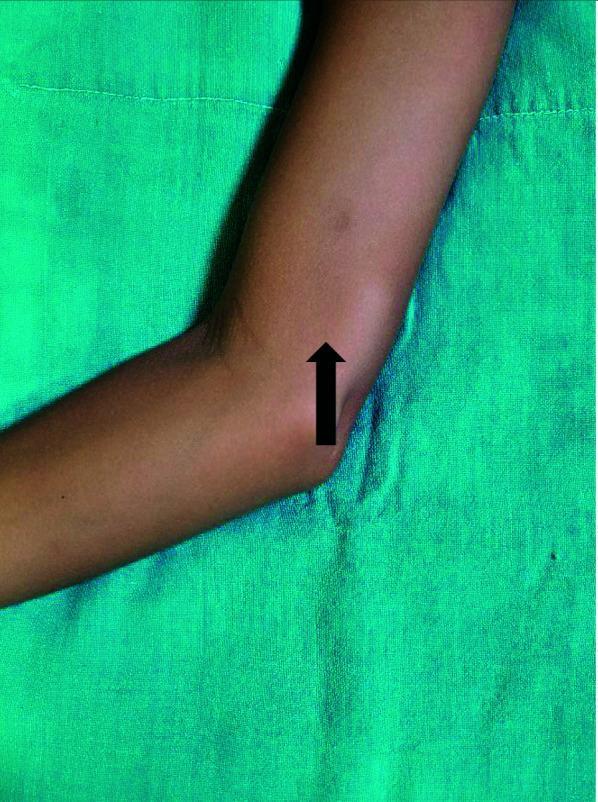
This clinical photograph shows a right arm resting on a green sterile drape, flexed at the elbow. The primary finding is a localized nodular swelling situated on the inner aspect of the distal medial arm, approximately 1 x 2 cm in size. A black arrow indicates the protrusion, which is located in the subcutaneous plane proximal to the elbow joint. The overlying skin appears intact without visible erythema, ulceration, or hypopigmented patches. Clinically, such a nodule in a patient with a history of Borderline Tuberculoid (BT) leprosy and neurologic symptoms (pain/tingling) is highly suggestive of a silent nerve abscess or a localized lepromatous reaction involving the ulnar nerve. The image illustrates a focal neurological manifestation of leprosy relapse or reaction, emphasizing the importance of examining common sites of nerve enlargement and abscess formation in post-treatment follow-ups.
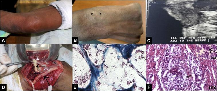
A multi-panel figure illustrating the clinical, diagnostic, and histopathological features of nerve abscesses in leprosy (Hansen's disease). (A-B) Clinical photographs show localized swelling and signs of inflammation at the ulnar aspect of the forearm and enlargement of the superficial radial nerve. (C) An ultrasonogram depicts an ill-defined, heterogeneous, hypoechoic lesion adjacent to the nerve, consistent with a subcutaneous and ulnar nerve abscess. (D) An intraoperative photograph displays a thickened ulnar nerve and the surgical drainage of purulent material (pus). (E) A high-power micrograph of a Fite-Faraco or Ziehl-Neelsen stain reveals numerous acid-fast bacilli (red rod-shaped bacteria) against a blue counterstained background. (F) An H&E stained histopathology slide shows a granulomatous inflammatory response with a characteristic Langhans giant cell (multinucleated cell with peripheral nuclei arrangement) highlighted in the 20x magnification inset. This composite image demonstrates the progression from clinical peripheral nerve enlargement to surgical confirmation and microscopic identification of Mycobacterium leprae.
Nerve Abscess in Leprosy — Exam Notes (MD Derma)
1. Definition
2. Epidemiology / Spectrum Association
- More frequent in the tuberculoid pole of the spectrum — seen in pure neuritic (primary neural) leprosy and tuberculoid/borderline tuberculoid (BT) leprosy.
- Less common toward the lepromatous pole (LL), where the immune response is weaker and granulomas are less well-organized (Fitzpatrick's Dermatology).
- Reflects the underlying immunopathology: high cell-mediated immunity (CMI) in the TT/BT end produces vigorous granulomatous reactions capable of caseating, whereas LL nerves show diffuse bacillary infiltration without organized caseating granulomas.
3. Pathogenesis
- Occurs as an exaggerated granulomatous immune response (akin to a Type 1/reversal reaction confined to a nerve) against M. leprae antigens within the nerve.
- Epithelioid granulomas with Langhans giant cells form around bacillary antigen within the nerve fascicle/perineurium.
- Central caseous necrosis develops within the granuloma (analogous to tuberculous cold abscess pathogenesis, but in leprosy) → liquefaction → abscess formation.
- Intense inflammatory edema within the tight osteofibrous/fibro-osseous tunnels through which many peripheral nerves pass (e.g., ulnar nerve at the elbow, posterior tibial at the tarsal tunnel) raises intraneural/intraneural-tunnel pressure, contributing to ischemic axonal damage and abscess formation.
- Histopathology (as illustrated in reference images): granulomatous inflammation with Langhans giant cells, and numerous acid-fast bacilli on Fite-Faraco/Ziehl-Neelsen stain within the necrotic nerve tissue.
4. Common Sites
- Ulnar nerve (above/at the elbow) — most commonly reported site (case literature repeatedly cites ulnar nerve abscess).
- Lateral popliteal (common peroneal) nerve
- Greater auricular nerve
- Posterior tibial nerve
- Superficial radial nerve and medial cutaneous nerve of forearm (less common, reported in recent case literature)
5. Clinical Features
- A firm-to-fluctuant, tender, localized swelling along the course of a peripheral nerve, often abrupt in onset.
- Overlying skin may be normal or mildly erythematous; usually no ulceration or discharging sinus unless it ruptures spontaneously.
- Associated with signs of nerve trunk involvement: nerve thickening, tenderness, and variable sensory/motor deficit distal to the abscess.
- May present with acute nerve pain (neuritis) or, paradoxically, can be relatively "silent" with painless swelling.
- Can rarely rupture through skin producing sporotrichoid ulcerated nodules along the nerve (an atypical presentation reported recently) or track along fascial planes causing wider soft-tissue swelling (e.g., brachial plexopathy from a ruptured ulnar nerve abscess tracking proximally).
- May be the sole presenting sign of leprosy in an otherwise unremarkable-looking patient (pure neuritic leprosy), making it a diagnostic pitfall for surgeons/orthopedists who may mistake it for a pyogenic or tuberculous abscess.
6. Investigations
- Clinical palpation of the thickened, tender nerve — first step.
- High-resolution ultrasonography (HRUS) of the nerve — shows an ill-defined, heterogeneous, hypoechoic collection adjacent to/within the nerve, with nerve enlargement; useful, accessible, first-line imaging (Zhao et al. 2021, PMID 33680276).
- MR neurography — superior for delineating extent, especially with proximal tracking/plexopathy (e.g., brachial plexus involvement from a ruptured ulnar abscess) (Mittal et al. 2022, PMID 35389025).
- Fine needle aspiration cytology (FNAC) or surgical aspirate of the abscess content: shows caseous/necrotic material; smear for AFB (Fite-Faraco/ZN stain) often strongly positive, especially at the tuberculoid-borderline end where bacilli may be scanty in skin but demonstrable in the nerve lesion.
- Histopathology of excised/drained tissue: granulomatous inflammation with epithelioid cells, Langhans giant cells, caseation necrosis, and AFB within nerve tissue.
- Routine culture (bacterial/fungal) and AFB culture for tuberculosis to rule out mimics (see differentials below) — M. leprae cannot be cultured in vitro, so diagnosis rests on histology/staining/clinical correlation.
- Skin smear/slit-skin smear and skin biopsy from any associated lesion to classify leprosy type.
7. Differential Diagnosis
- Tuberculous (cold) abscess of soft tissue/nerve sheath
- Pyogenic abscess (staphylococcal/streptococcal)
- Peripheral nerve sheath tumor (schwannoma/neurofibroma) — especially if painless and slow-growing
- Ganglion cyst (near joints/tendons)
- Filarial or other granulomatous nerve swellings
- Soft tissue sarcoma (if atypical presentation)
8. Management
A. Medical (backbone therapy)
- Continue/initiate WHO-MDT for the underlying leprosy (rifampicin, clofazimine, dapsone as per PB/MB classification) — treats the causative infection.
- Systemic corticosteroids (oral prednisolone, standard reactional dosing ~0.5–1 mg/kg/day tapered over weeks) to control the surrounding granulomatous/reactional inflammation and protect residual nerve function, similar to management of Type 1 reaction/neuritis.
- Analgesics/NSAIDs for pain control.
B. Surgical
- Incision and drainage (or needle aspiration) of the abscess is mandatory once identified — this is the definitive step, both therapeutically (relieves pressure on the nerve, preventing further ischemic axonal loss) and diagnostically (contents sent for AFB smear, histopathology, and culture) (Fitzpatrick's Dermatology).
- Surgical nerve decompression (neurolysis/epineurotomy) may be used in cases with persistent raised intraneural pressure or where the abscess is causing ongoing compressive damage — although robust clinical trial evidence for decompression improving long-term neuropathy/motor outcome is still lacking (Fitzpatrick's Dermatology; Marques 2021, PMID 34550195; Gonçalves et al. systematic review 2023, PMID 37531518, found evidence for neurolysis in leprosy neuritis complications to be limited/low-certainty).
- Care must be taken during surgery to avoid injury to adjacent neurovascular structures, particularly at classic entrapment sites (e.g., ulnar nerve at the cubital tunnel).
C. Supportive / Rehabilitative
- Splinting/rest of the limb during the acute phase.
- Physiotherapy to maintain range of motion and prevent contractures once the acute process settles.
- Patient education on self-care of the (often already hypoesthetic) limb to prevent secondary trauma/ulceration.
- Long-term follow-up for nerve function (voluntary muscle testing, sensory testing) since abscess formation is itself a marker of severe neuritis with a high risk of residual deficit.
9. Recent Literature / Illustrative Case Reports (for exam citation)
- Zhao H et al., Radiology Case Reports 2021 (PMID 33680276) — sonographic evaluation of leprosy affecting the ulnar nerve, useful for describing HRUS findings.
- Mittal A et al., IJDVL 2022 (PMID 35389025) — brachial plexopathy secondary to ulnar nerve abscess, demonstrating value of MR neurography with clinico-radio-pathological correlation.
- Yadav P et al., Int J Dermatol 2025 (PMID 41063498) — medial cutaneous nerve abscess as an uncommon presentation of borderline tuberculoid leprosy.
- Yadav P et al., Int J Dermatol 2026 (PMID 40833220) — ruptured ulnar nerve abscess presenting with sporotrichoid ulcerated nodules, an atypical mimicker.
- Gonçalves LC et al., systematic review 2023 (PMID 37531518) — evidence base for neurolysis in leprosy neuritis complications remains limited.
10. Key Exam Points to Remember
- Nerve abscess = caseating granulomatous necrosis within a nerve trunk, a complication predominantly of the tuberculoid/BT and pure neuritic forms of leprosy.
- Commonest site: ulnar nerve.
- Can be the sole presenting feature of leprosy — important diagnostic pitfall.
- Diagnosis: clinical + HRUS/MRI + AFB stain/histology of aspirate.
- Management = MDT + steroids (medical) + incision/drainage ± decompression (surgical) — drainage is considered mandatory once diagnosed.
- Differentiate from tuberculous and pyogenic abscess, and nerve sheath tumors.