
Cardiac reflex for 10 marks
cardiac reflexes baroreceptor Bainbridge Bezold-Jarisch diagram
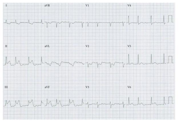
A standard 12-lead diagnostic electrocardiogram (ECG) demonstrating a transient ST-segment elevation in the inferior leads (II, III, and aVF). The morphology in these leads shows marked ST elevation with an upright T wave, consistent with an acute inferior wall injury pattern. The QRS complexes are narrow and preceded by visible P waves, indicating a sinus rhythm. Reciprocal ST-segment depression is visible in leads I and aVL. This specific tracing illustrates a Bezold-Jarisch-like reflex, a physiological phenomenon that can occur due to mechanical irritation of the interatrial vagal network during transseptal puncture, leading to coronary artery spasm. The image serves as a clinical example for recognizing procedure-related cardiac changes and differentiating them from primary atherosclerotic myocardial infarction in the context of electrophysiology interventions.
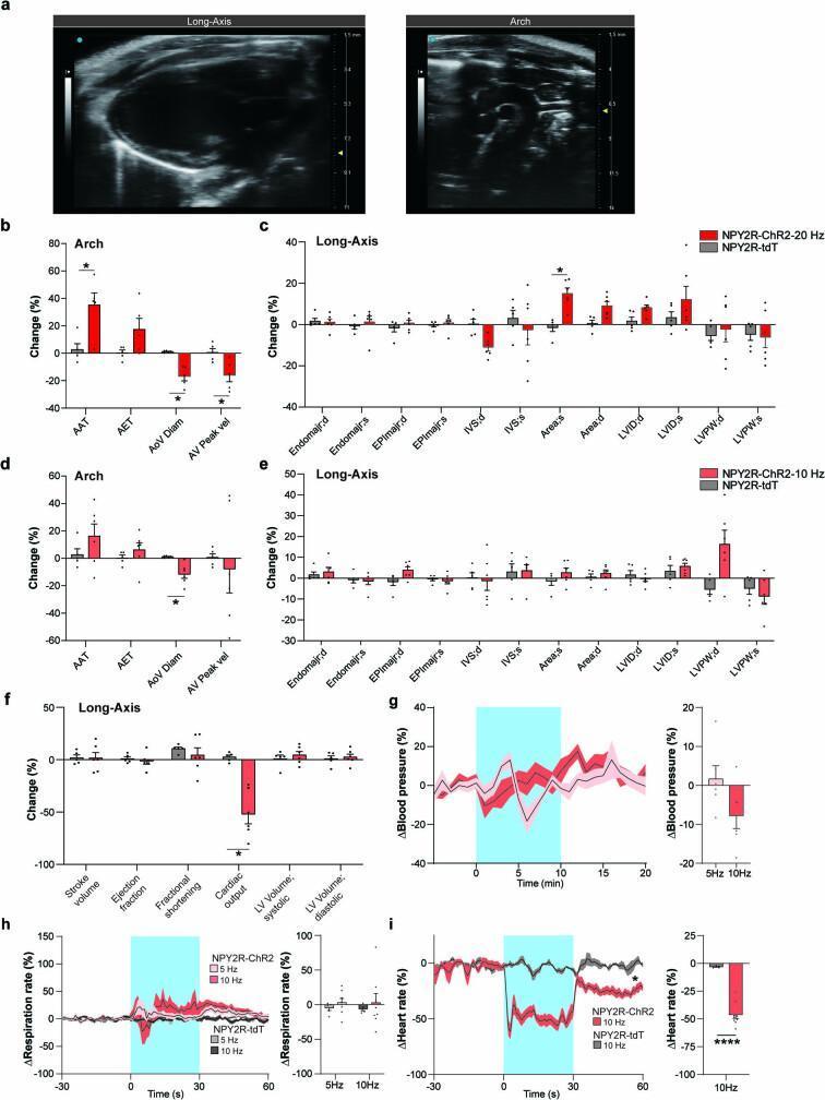
This composite educational infographic illustrates the physiological characterization of vagal sensory neurons (NPY2R VSNs) in response to optogenetic photostimulation, particularly focusing on mechanisms of syncope. Panel a shows representative B-mode ultrasound diagnostic images of a mammalian heart in the parasternal long-axis view and the aortic arch. Panels b through f present comparison bar charts showing the percentage change in various cardiovascular parameters after 20 Hz and 10 Hz photostimulation. Specifically, panel b demonstrates that 20 Hz stimulation significantly decreases aortic valve diameter (AoV Diam) and peak blood velocity (AV Peak vel) while increasing aortic acceleration time (AAT). Panel c reveals an increase in the left ventricular end-systolic area (Area;s). Panel f indicates a significant drop in cardiac output. Panels g, h, and i provide time-course line graphs and summary bar charts demonstrating time-locked reductions in blood pressure, respiration rate, and heart rate during photostimulation. The data collectively illustrate the physiological triad of the Bezold-Jarisch reflex (BJR) and its relevance to syncope research.
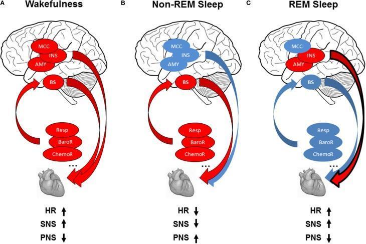
A multi-panel pathophysiology diagram illustrating autonomic cardiac regulation across three vigilance states: (A) Wakefulness, (B) Non-REM Sleep, and (C) REM Sleep. Each panel displays a sagittal brain view, reflex loop components (Respiration, Baroreflex, Chemoreflex), and the heart. Color-coding (red for increased activity/influence, blue for decreased) highlights state-dependent shifts. Wakefulness (A) shows high central autonomic network (CAN) activity in the midcingulate cortex (MCC), insula (INS), and amygdala (AMY), alongside active reflex loops, resulting in increased heart rate (HR) and sympathetic (SNS) tone with decreased parasympathetic (PNS) tone. Non-REM Sleep (B) depicts decreased CAN activity (blue) with maintained reflex loop dominance (red), leading to decreased HR/SNS and increased PNS tone. REM Sleep (C) shows a complex mix of high cortical activity (MCC/INS/AMY) and inhibited reflex loops/brainstem (BS) modulation, returning to a state of increased HR and SNS activity. The diagram demonstrates the dynamic interplay between central command and homeostatic reflexes in cardiovascular control during sleep-wake cycles.
Cardiac Reflexes
- Baroreceptor Reflex
- Chemoreceptor Reflex
- Bainbridge Reflex
- Bezold-Jarisch Reflex
- Cushing Reflex
- Oculocardiac Reflex
1. Baroreceptor Reflex (Carotid Sinus Reflex)
Receptors
- Carotid sinus baroreceptors - located at the bifurcation of the common carotid artery; respond to both increases and decreases in pressure.
- Aortic arch baroreceptors - primarily respond to increases in arterial pressure.
- Both are mechanoreceptors (stretch-sensitive). A rapid change in pressure is the strongest stimulus.
Afferent Pathway
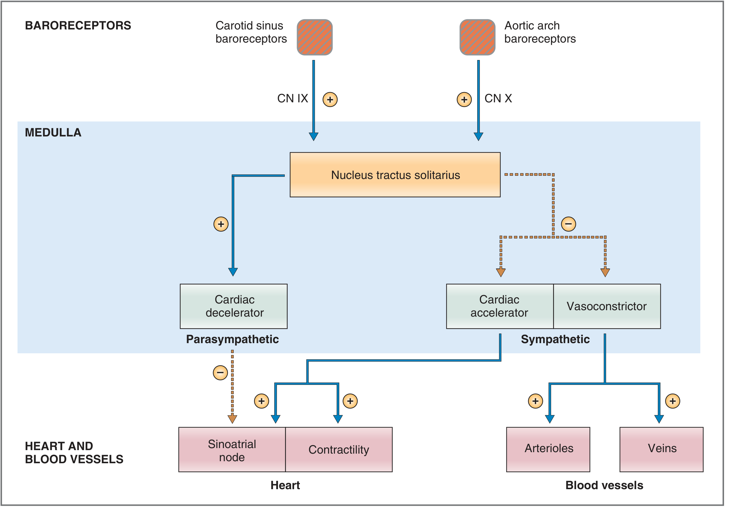
- Carotid sinus - carotid sinus nerve (Hering's nerve) - glossopharyngeal nerve (CN IX)
- Aortic arch - vagus nerve (CN X)
- Both synapse in the nucleus tractus solitarius (NTS) in the medulla.
Central Integration
- Vasopressor/accelerator area (lateral and rostral) - increases BP
- Vasodepressor/decelerator area (central and caudal) - lowers BP; also integrates input from hypothalamus and limbic system
Efferent Pathway and Effectors
- Vasoconstrictor centre (C1) - upper medulla/lower pons - sympathetic fibres to arterioles and venules
- Cardiac accelerator centre - sympathetic fibres to SA node (↑ HR), AV node (↑ conduction), myocardium (↑ contractility)
- Cardiac decelerator centre - parasympathetic fibres (vagus) to SA node (↓ HR)
Response to Increased Arterial Pressure
- ↑ Pa → ↑ stretch on baroreceptors → ↑ firing rate in CN IX and X
- NTS activated → ↑ parasympathetic outflow + ↓ sympathetic outflow
- Results: ↓ heart rate, ↓ contractility, vasodilation → Pa returns to normal
Response to Decreased Arterial Pressure (e.g., hemorrhage)
- ↓ Pa → ↓ baroreceptor firing → ↓ parasympathetic + ↑ sympathetic activity
- Results: ↑ heart rate, ↑ contractility, ↑ TPR (arteriolar vasoconstriction), ↓ venous unstressed volume → ↑ venous return
- Coronary and cerebral beds are spared from vasoconstriction
- In chronic hypertension, the baroreceptors are reset (resensitized to the elevated pressure as "normal"), maintaining rather than correcting the hypertension.
- The reflex loses functional capacity when MAP falls below 50 mmHg.
- Volatile anesthetics (especially halothane) inhibit the heart rate component.
2. Chemoreceptor Reflex
Peripheral Chemoreceptors
- Located in carotid bodies and aortic body
- Sensitive to: ↓ PaO₂ (< 50 mmHg), ↑ PaCO₂, and ↓ pH (acidosis)
Afferent Pathway
- Carotid bodies → sinus nerve of Hering (branch of CN IX)
- Aortic bodies → vagus nerve (CN X)
- Both reach the chemosensitive area of the medulla
Response
- Primary effect: stimulation of respiratory centres → ↑ ventilatory drive
- Cardiovascular effect: ↑ parasympathetic tone → ↓ heart rate and ↓ myocardial contractility
- In persistent hypoxia: direct CNS stimulation → ↑ sympathetic activity (overrides the initial bradycardia)
3. Bainbridge Reflex (Atrial Reflex / Right Heart Reflex)
Stimulus
- ↑ right-sided filling pressure (increased venous return / volume overload)
Receptors
- Stretch receptors in the right atrial wall and cavoatrial junction
Pathway
- Vagal afferent signals → cardiovascular centre in medulla
Response
- Inhibition of parasympathetic activity → ↑ heart rate (tachycardia)
- Also: direct stretch of the atrium affects the SA node locally
4. Bezold-Jarisch Reflex
Stimulus
- Noxious stimuli in the left ventricular wall - mechanical or chemical (ischemia, reperfusion, thrombolysis, serotonin, veratrum alkaloids)
Receptors
- Chemoreceptors and mechanoreceptors within the LV wall
Afferent Pathway
- Unmyelinated vagal afferent type C fibres → medulla → ↑ parasympathetic tone
Classic Triad (Response)
- Hypotension
- Bradycardia
- Coronary artery dilatation
Significance
- Considered cardioprotective - slows the heart and reduces work during myocardial injury
- Implicated in: myocardial infarction, thrombolysis, revascularization, vasovagal syncope
- Natriuretic peptide receptors (ANP/BNP) may modulate this reflex
- Less pronounced in patients with cardiac hypertrophy or atrial fibrillation
5. Cushing Reflex (Vasopressor Response)
Stimulus
- Raised intracranial pressure (ICP) → cerebral ischemia at the medullary vasomotor centre
Response - Biphasic:
- Phase 1 - Cerebral ischemia activates sympathetic nervous system:
- ↑ heart rate, ↑ arterial BP, ↑ myocardial contractility (to perfuse the brain)
- Phase 2 - The resultant high BP activates baroreceptors:
- Reflex bradycardia (via parasympathetic activation)
Clinical Presentation (Cushing's Triad)
- Hypertension (widened pulse pressure)
- Bradycardia
- Irregular respirations
6. Oculocardiac Reflex
Stimulus
- Pressure on the globe of the eye, traction on extraocular muscles, or orbital injury
Afferent Pathway
- Short and long ciliary nerves → ciliary ganglion → ophthalmic division of trigeminal nerve (CN V₁) → Gasserian (trigeminal) ganglion → medulla
Efferent Pathway
- Vagus nerve → SA node
Response
- Bradycardia (sometimes severe enough to cause cardiac arrest)
- Incidence during ophthalmic surgery: 30-90%
Clinical Management
- Administration of atropine or glycopyrrolate (antimuscarinic agents) before surgery reduces the incidence of bradycardia.
Summary Table
| Reflex | Stimulus | Receptor Location | Afferent | Response |
|---|---|---|---|---|
| Baroreceptor | ↑/↓ arterial pressure | Carotid sinus, aortic arch | CN IX, CN X | ↓ HR, BP with ↑ BP; ↑ HR, BP with ↓ BP |
| Chemoreceptor | ↓ PaO₂, ↑ PaCO₂, acidosis | Carotid body, aortic body | CN IX, CN X | ↑ Ventilation; ↓ HR initially |
| Bainbridge | ↑ Venous return / atrial filling | Right atrial wall, cavoatrial junction | Vagal afferents | ↑ Heart rate |
| Bezold-Jarisch | LV noxious stimuli (ischemia, reperfusion) | LV wall (C-fibres) | Unmyelinated vagal C fibres | ↓ HR, ↓ BP, coronary dilation (triad) |
| Cushing | ↑ ICP / cerebral ischemia | Medullary vasomotor centre | Direct CNS | ↑ BP, then reflex bradycardia |
| Oculocardiac | Eye pressure / muscle traction | Extraocular muscles | CN V₁ → medulla | Bradycardia |
- The baroreceptor reflex is the primary short-term blood pressure regulator; it is reset in chronic hypertension.
- The Bainbridge reflex is the "atrial stretch → tachycardia" reflex - opposite to the baroreceptor in direction.
- The Bezold-Jarisch triad (hypotension + bradycardia + coronary dilation) is cardioprotective and relevant to MI and syncope.
- Cushing's triad = hypertension + bradycardia + irregular breathing - signals raised ICP and impending herniation.
- The oculocardiac reflex is afferent via CN V, efferent via CN X - a trigemino-vagal reflex; blocked by atropine.