
elaborate on ascending tracts of spinal cord: 1. anterior and lateral spino thalamic tract, 2. Ventral and dorsal spinocerebellar tract, 3. spinotectal tract, 4. fasiculus dorsolateralis, 5. spinoreticular, 6. spino olivary, 7. spino vestibular, 8. fasciculus gracilis and 9. fasciculus cuneatus. Give the detailed origin, termination, course and its functions respectively.
ascending tracts spinal cord spinothalamic spinocerebellar diagram
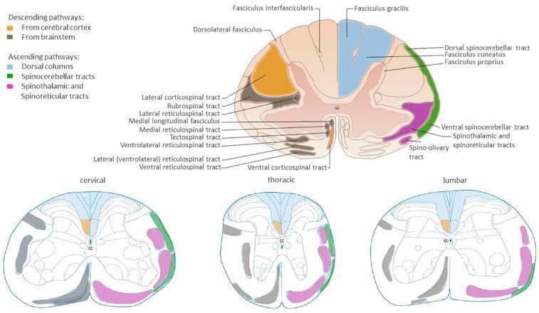
Anatomical diagrams illustrating the organization of major white matter tracts in the human spinal cord across cervical, thoracic, and lumbar cross-sections. A primary large-scale diagram provides detailed labeling of pathways, while three comparative smaller diagrams show regional variations. Descending (motor) pathways are color-coded in orange (corticospinal tracts from the cerebral cortex) and grey/black (brainstem-derived tracts including rubrospinal, reticulospinal, and tectospinal tracts). Ascending (sensory) pathways are highlighted in blue for dorsal columns (fasciculus gracilis and cuneatus), green for dorsal and ventral spinocerebellar tracts, and pink for the spinothalamic and spinoreticular tracts. The diagrams illustrate key neuroanatomical landmarks such as the central canal (CC), dorsal horns, and ventral horns. Educational focus is placed on the somatotopic arrangement and the relative change in white matter volume versus grey matter across different spinal levels, highlighting the clinical relevance for localized spinal cord injury and neurological deficit mapping.
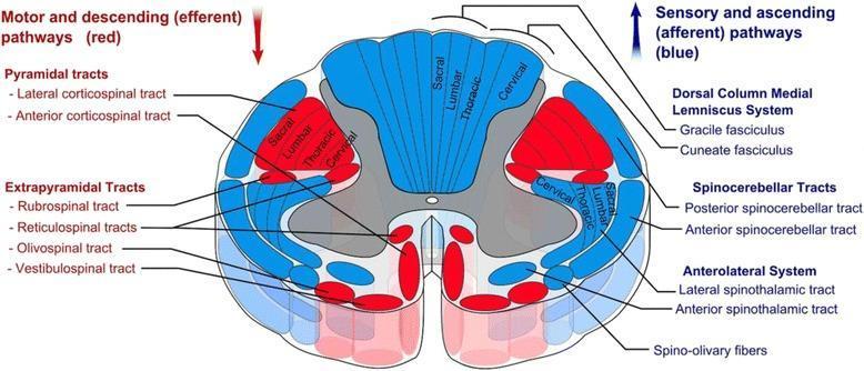
This medical anatomical diagram illustrates a transverse section of the human spinal cord, specifically detailing the organization of white matter tracts. The visual uses a color-coded schema to differentiate between motor and sensory pathways: descending (efferent) motor tracts are highlighted in red, while ascending (afferent) sensory tracts are shown in blue. Key anatomical structures identified include the Pyramidal tracts (lateral and anterior corticospinal tracts), Extrapyramidal tracts (rubrospinal, reticulospinal, olivospinal, and vestibulospinal), the Dorsal Column-Medial Lemniscus system (gracile and cuneate fasciculi), Spinocerebellar tracts, and the Anterolateral system (lateral and anterior spinothalamic tracts). A significant educational feature of the diagram is the depiction of somatotopic organization, showing the concentric arrangement of sacral, lumbar, thoracic, and cervical fibers within the lateral corticospinal and dorsal column tracts. The image provides a comprehensive overview of spinal cord neuroanatomy, useful for understanding clinical localization of spinal cord lesions and the functional topography of the central nervous system.
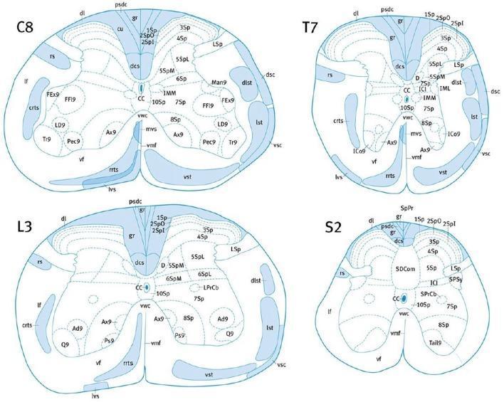
This medical anatomical diagram illustrates the cross-sectional neuroanatomy and white matter tract localization within the mouse spinal cord at four distinct levels: Cervical (C8), Thoracic (T7), Lumbar (L3), and Sacral (S2). Each level depicts the characteristic morphology of the spinal cord, showing the transition from a rounded C8/T7 shape to a more laterally expanded L3 and a flattened, compressed S2. Key ascending (sensory) and descending (motor) pathways are highlighted in blue. Notable structures include the dorsal corticospinal tract (dcs) located centrally along the dorsal midline, flanked by the gracile fasciculus (gr) and cuneate fasciculus (cu, prominent in higher levels). Lateral tracts shown include the rubrospinal (rs), dorsal spinocerebellar (dsc), and lateral spinothalamic (lst) tracts. Ventral regions display the vestibulospinal (lvs, mvs) and reticulospinal (crts, rrts) tracts. The diagrams also delineate Rexed laminae (1Sp-10Sp) within the gray matter and the central canal (CC). This educational resource is designed for neuroanatomical mapping and comparative spinal pathology research.
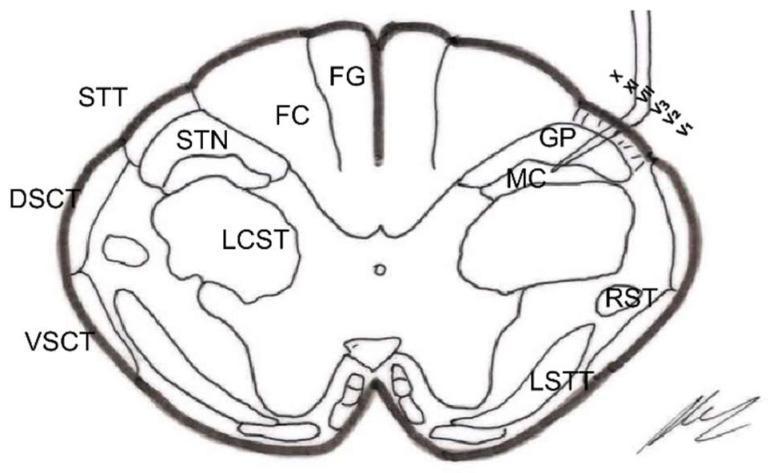
This medical anatomical diagram illustrates a cross-section of the spinal cord (medulla oblongata level, approximately 18 mm below the obex), specifically focusing on the neuroanatomical structures targeted during a trigeminal nucleotomy. The diagram highlights both white matter tracts and gray matter nuclei. In the posterior column, the Fasciculus Gracilis (FG) is medially positioned, while the Fasciculus Cuneatus (FC) is lateral to it. The lateral white matter contains the Lateral Corticospinal Tract (LCST), Dorsal Spinocerebellar Tract (DSCT), and Ventral Spinocerebellar Tract (VSCT). On the right side of the diagram, a surgical probe is shown penetrating the Spinal Trigeminal Tract (STT/STrigT) to target the Spinal Trigeminal Nucleus (STN), specifically indicating the Gelatinosa Portion (GP) and the Magnocellular Portion (MC). Other notable structures include the Rubrospinal Tract (RST) and the Lateral Spinothalamic Tract (LSTT). This illustration is designed for neurosurgical education, demonstrating the spatial relationships necessary for selective destruction of pain-transmitting nuclei in the brainstem and upper cervical spinal cord.
Ascending Tracts of the Spinal Cord
- Anterolateral pathways - pain, temperature, crude touch
- Posterior column pathways - fine touch, vibration, proprioception
- Spinocerebellar pathways - unconscious proprioception
1. Anterior (Ventral) Spinothalamic Tract
Origin
Course
- Second-order axons decussate through the ventral (anterior) white commissure (crossing takes 2-3 spinal segments)
- They then ascend in the anterior funiculus as the ventral spinothalamic tract
- Pass through the brainstem (medulla, pons, midbrain) alongside the lateral spinothalamic tract
- Terminate in the ventral posterolateral (VPL) nucleus of the thalamus
- Third-order neurons project through the posterior limb of the internal capsule to reach the primary somatosensory cortex (areas 3, 1, 2 of postcentral gyrus)
Termination
Function
- Conveys light (crude) touch and pressure sensation from the contralateral side of the body
- Note: light touch is also simultaneously transmitted via the posterior column-medial lemniscal pathway, providing a "backup" system
2. Lateral Spinothalamic Tract
Origin
Course
- Second-order axons cross obliquely in the ventral commissure (over 2-3 segments below entry level)
- Ascend in the lateral funiculus as the lateral spinothalamic (neospinothalamic) tract
- Somatotopic arrangement: sacral fibers are most lateral (ventrolateral), cervical fibers are most medial (dorsomedial)
- Pass laterally through the medulla (between inferior olive and inferior cerebellar peduncle), then enter the pontine and midbrain tegmentum just lateral to the medial lemniscus
- End in the VPL nucleus of the thalamus
- Third-order neurons project to the primary somatosensory cortex
Termination
Function
- Conveys sharp, well-localized pain and temperature sensation from the contralateral body
- Temperature fibers are situated dorsolateral to pain fibers within this tract
- Mediates the discriminative/sensory-discriminative aspect of pain (location, intensity)
- Basis of the surgical procedure cordotomy (cutting this tract to relieve intractable pain)
3. Ventral (Anterior) Spinocerebellar Tract
Origin
Course
- Axons cross immediately in the ventral white commissure to the contralateral side
- Ascend in the anterolateral funiculus, ventral to the dorsal spinocerebellar tract and peripheral to the anterolateral systems
- Travel through medulla and pons
- Enter the cerebellum via the superior cerebellar peduncle
- Most fibers cross a second time within the cerebellum, reaching the ipsilateral cerebellar cortex (vermis and intermediate zone) - this "double crossing" ensures information ultimately reaches the cerebellum ipsilateral to the limb of origin
Termination
Function
- Carries unconscious proprioceptive information from the lower extremities and trunk
- Specifically conveys information about spinal cord interneuron activity, reflecting the level of activity in descending motor pathways
- Provides the cerebellum with a "copy" of spinal cord motor commands for coordination
- Upper-limb equivalent = rostral spinocerebellar tract
4. Dorsal (Posterior) Spinocerebellar Tract
Origin
Course
- Second-order axons do not cross - they ascend ipsilaterally in the dorsolateral funiculus (near the surface, just lateral to the lateral corticospinal tract)
- Travel the entire length of the spinal cord and brainstem
- Enter the cerebellum via the inferior cerebellar peduncle
- Terminate in the vermis and intermediate zone of the ipsilateral cerebellar cortex
Termination
Function
- Carries unconscious proprioceptive information (from muscle spindles, Golgi tendon organs, joint receptors) from the ipsilateral lower limb
- Provides rapid feedback about ongoing movements so the cerebellum can make fine adjustments
- Upper-limb equivalent = cuneocerebellar tract (from the external/accessory cuneate nucleus in the medulla)
5. Spinotectal Tract
Origin
Course
- Axons cross the midline through the ventral white commissure
- Ascend in the anterolateral funiculus, closely associated with (and sometimes considered part of) the spinothalamic tract
- Travel through medulla, pons, and midbrain
- End in the superior colliculus (tectum) and periaqueductal gray (PAG) of the midbrain
Termination
Function
- Mediates spinovisual reflexes - triggers involuntary turning of the head and eyes toward a painful or sudden somatosensory stimulus (orienting reflex)
- The superior colliculus integrates visual, auditory, and somatosensory inputs to coordinate gaze and head orientation
- The PAG connection participates in descending pain modulation (endogenous opioid system, gate control)
- Together with the spinoreticular tract, forms part of the paleospinothalamic pain pathways
6. Fasciculus Dorsolateralis (Lissauer's Tract / Posterolateral Tract)
Origin
- Finely myelinated (Aδ) and unmyelinated (C) fibers from the lateral division of the dorsal root
- Also contains some endogenous intrinsic spinal cord fibers
Course
- Located in the posterolateral white matter between the dorsal horn and the surface of the cord
- Fibers ascend and descend 1-2 spinal cord segments before entering the gray matter to synapse in laminae I, II (substantia gelatinosa), and V of the dorsal horn
Termination
Function
- Acts as a relay/distribution zone for incoming pain and temperature fibers before they synapse
- The substantia gelatinosa (lamina II) neurons here are central to the gate control theory of pain (Melzack & Wall)
- Allows fine-fiber afferents to spread input over multiple spinal levels, contributing to the phenomenon of referred pain and the mild mismatch in dermatomal localization of pain
- Contains enkephalinergic and other inhibitory interneurons important for modulating pain transmission
7. Spinoreticular Tract (Paleospinothalamic)
Origin
Course
- Axons ascend bilaterally (both crossed and uncrossed) in the anterolateral funiculus, intermingled with spinothalamic fibers
- Travel through the spinal cord and brainstem
- Synapse on the reticular formation of the medulla and pons (medullary-pontine reticular formation)
- Reticular neurons then project to the intralaminar thalamic nuclei (centromedian nucleus) and hypothalamus
Termination
Function
- Conveys the emotional, affective, and arousal aspects of pain ("suffering component") - accounts for why pain is unpleasant
- Mediates poorly localized, burning, diffuse pain (paleospinothalamic pain)
- Responsible for the arousal/wakefulness response to noxious stimuli via reticular activating system connections
- Involved in autonomic responses to pain (tachycardia, sweating) via hypothalamic connections
- Anatomically older than the spinothalamic tract; forms the phylogenetically primitive pain pathway
8. Spino-Olivary Tract
Origin
Course
- Axons travel in the anterolateral funiculus and at the anterior border of the anterior funiculus
- Ascend through the spinal cord
- Synapse in the inferior olivary nuclear complex (accessory olivary nuclei) in the medulla
Termination
Function
- Relays cutaneous and proprioceptive information to the inferior olive, which then sends climbing fiber projections to the contralateral cerebellar cortex via the inferior cerebellar peduncle
- Part of the indirect spinocerebellar pathway - a second route by which spinal sensory information reaches the cerebellum
- Involved in the fine motor coordination feedback circuit
- May contribute to nociceptive processing within the cerebellum
9. Spino-Vestibular Tract
Origin
Course
- Axons travel in the lateral and anterior funiculi (also partly in the dorsolateral funiculus, near the dorsal spinocerebellar tract)
- Ascend ipsilaterally
- End directly in the lateral, inferior, and medial vestibular nuclei in the medulla-pons
Termination
Function
- Provides the vestibular nuclei (and indirectly the cerebellum) with proprioceptive information from the spinal cord
- Contributes to postural control and balance regulation in coordination with vestibular input from the inner ear
- Enables reflex adjustments of trunk and limb muscles in response to positional changes
- Some fibers reaching the cerebellum via vestibular relay contribute to the vestibulocerebellum (flocculonodular lobe) function
10. Fasciculus Gracilis
Origin
Course
- Axons enter the posterior funiculus and ascend ipsilaterally in the medial part of the posterior column (the more medially placed of the two posterior column fasciculi)
- Run the entire length of the cord and brainstem (staying ipsilateral throughout the spinal cord)
- Synapse in the nucleus gracilis of the ipsilateral caudal medulla
- Second-order axons from nucleus gracilis cross as internal arcuate fibers and form the medial lemniscus in the contralateral medulla
- The medial lemniscus ascends to the VPL nucleus of the thalamus
- Third-order neurons project via the posterior limb of the internal capsule to the primary somatosensory cortex (areas 3, 1, 2)
Termination
Function
- Vibration sense (pallesthesia)
- Conscious proprioception / position sense (kinesthesia)
- Discriminative (fine) touch and two-point discrimination
- Pressure and weight perception
- Stereognosis (object recognition by feel)
- Graphesthesia (recognition of numbers/letters traced on skin)
- Damage → loss of these modalities ipsilaterally below the lesion level (before decussation in medulla)
11. Fasciculus Cuneatus
Origin
Course
- Axons ascend ipsilaterally in the lateral part of the posterior column, lateral to the fasciculus gracilis (hence "cuneate" = wedge-shaped, occupying the lateral wedge of the posterior funiculus)
- Ascend to the nucleus cuneatus of the dorsal medulla
- Second-order fibers cross as internal arcuate fibers → medial lemniscus (contralateral)
- Medial lemniscus ascends to the VPL thalamus
- Third-order → posterior limb internal capsule → primary somatosensory cortex
Termination
Function
- Vibration, conscious proprioception, fine touch, two-point discrimination, stereognosis, graphesthesia from the upper limb, upper trunk, and neck
- Clinical importance: Subacute combined degeneration (B12 deficiency) affects the posterior columns, causing loss of vibration and proprioception bilaterally; Brown-Séquard syndrome causes ipsilateral loss of these modalities below the lesion
Summary Table
| Tract | Origin (1st neuron) | Decussation | Termination | Modality |
|---|---|---|---|---|
| Anterior spinothalamic | Dorsal root ganglion | Ventral commissure (crosses) | VPL thalamus → cortex | Crude touch, pressure |
| Lateral spinothalamic | Dorsal root ganglion | Ventral commissure (crosses) | VPL thalamus → cortex | Pain, temperature |
| Dorsal spinocerebellar | Clarke's nucleus (C8-L3) | Does NOT cross | Ipsilateral cerebellum via ICP | Unconscious proprioception (lower limb) |
| Ventral spinocerebellar | Spinal border cells | Crosses twice (net ipsilateral) | Ipsilateral cerebellum via SCP | Spinal interneuron activity (lower limb) |
| Spinotectal | Dorsal horn (I, IV, V, VII) | Crosses via ventral commissure | Superior colliculus, PAG | Pain, orienting reflex |
| Fasciculus dorsolateralis | Dorsal root (C & Aδ fibers) | Stays ipsilateral | Dorsal horn (I, II, V) | Distributes pain/temp over 1-2 segments |
| Spinoreticular | Post/ant horn (VII, VIII) | Bilateral (crossed + uncrossed) | Reticular formation → intralaminar thalamus | Arousal, affective pain |
| Spino-olivary | Spinal gray (V-VIII) | Crosses | Inferior olivary nucleus | Indirect cerebellar proprioception |
| Spino-vestibular | Dorsal horn/intermediate zone | Ipsilateral | Vestibular nuclei | Proprioception for postural reflexes |
| Fasciculus gracilis | Dorsal root ganglion (below T6) | At nucleus gracilis (medulla) | VPL thalamus → cortex | Fine touch, vibration, proprioception (lower body) |
| Fasciculus cuneatus | Dorsal root ganglion (above T6) | At nucleus cuneatus (medulla) | VPL thalamus → cortex | Fine touch, vibration, proprioception (upper body) |