
Pyloric stenosis, IHPS, duodenal stenosis
Reading File
Searching sources...
Finding Sources
Finding Sources
Reading File
Reading File
Reading File
Reading File
Reading File
Reading File
Loading Image
Loading Image
Now let me get the HPS ultrasound image and also fetch any additional imaging for the US signs:
Reading File
Finding Sources
Reading File
Loading Image
I now have comprehensive information from multiple authoritative textbooks. Here is the full structured answer:
Infantile Hypertrophic Pyloric Stenosis (IHPS) and Duodenal Stenosis
PART 1: Infantile Hypertrophic Pyloric Stenosis (IHPS)
Epidemiology
- Incidence: ~1 in 300-900 live births; the most common surgical cause of vomiting in infants
- M:F ratio = 4-5:1 (boys far more commonly affected)
- Presents between 3rd and 6th weeks of life (range 2-10 weeks); rare after 12 weeks
- Associated with: first-born children, prematurity, young maternal age, monozygotic twins (high concordance)
- Risk increased in siblings (~6% vs ~0.2% general population)
- Associated syndromes: Turner syndrome, trisomy 18
- Erythromycin or azithromycin exposure (orally or via breast milk) in the first 2 weeks of life increases risk
Pathology
Hypertrophy and hyperplasia of the pyloric muscularis propria (circular > longitudinal muscles) causes progressive gastric outlet obstruction. This is NOT a congenital anomaly - the muscle grows after birth. Mucosal and submucosal edema and inflammation may also contribute. The hypertrophied pylorus forms a firm, palpable "olive."
Clinical Features
| Feature | Details |
|---|---|
| Vomiting | Forceful, projectile, non-bilious (proximal to bile duct entry) |
| Timing | Typically starts ~3-6 weeks; worsens progressively |
| Feeding | Infant remains hungry after vomiting ("hungry vomiter") |
| Peristaltic waves | Visible left-to-right waves across upper abdomen just before vomiting |
| Palpable mass | Firm, olive-shaped, 1-2 cm epigastric mass - nearly pathognomonic; found in up to 90% if stomach decompressed first (now found in <30% in contemporary practice due to early presentation) |
| Dehydration | Sunken fontanelles, dry mucous membranes, decreased urine output |
Metabolic Derangement
Persistent vomiting of gastric acid leads to:
Hypochloremic, hypokalemic metabolic alkalosis
- Loss of HCl → low Cl⁻, elevated HCO₃⁻
- Paradoxical aciduria (kidneys preferentially retain Na⁺ over H⁺ in severe alkalosis)
- Bicarbonate > 30 mEq/L = respiratory depression risk → must be corrected before anesthesia
Diagnosis
Ultrasound is the gold standard (sensitivity and specificity up to 98-100%):
| Ultrasound Criterion | Threshold |
|---|---|
| Pyloric muscle thickness | ≥ 4 mm |
| Pyloric canal length | ≥ 15-17 mm |
| Other signs | Shoulder sign (muscle bulging into antrum), nipple sign (hyperechoic mucosa protrudes into stomach), cervix sign |
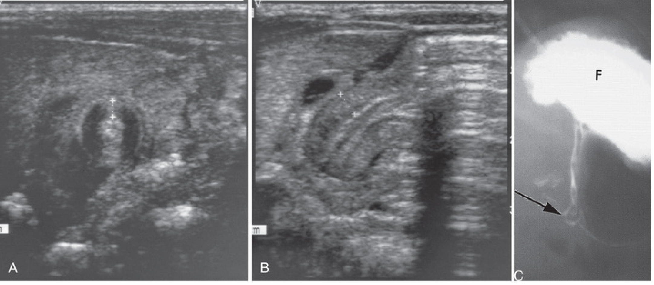
A: Ultrasound - pyloric muscle wall thickness >4mm. B: Pyloric channel length >14mm. C: Contrast study showing narrowed pyloric canal (arrow) with markedly distended fundus (F). From The Developing Human - Clinically Oriented Embryology.
Upper GI contrast series (UGI) is used when ultrasound is equivocal (can show elongated "string sign" of narrowed pyloric channel) and to evaluate for other causes (GERD, malrotation, antroduodenal webs).
Preoperative Resuscitation
Surgery is NEVER a true emergency. Resuscitate first:
- One to two boluses of normal saline 20 mL/kg
- D5/NS at 1.5x maintenance with electrolytes
- Check electrolytes every 6-12 hours
Endpoints for readiness for surgery:
- Wet diapers (adequate urine output)
- Chloride ≥ 90-100 mEq/L
- Bicarbonate ≤ 30 mEq/L
Do NOT proceed to anesthesia with bicarbonate > 30 mEq/L (risk of postoperative apnea/respiratory arrest).
Surgical Treatment: Fredet-Ramstedt Pyloromyotomy
The operation of choice - open (umbilical or RUQ incision) or laparoscopic (now standard at most children's hospitals):
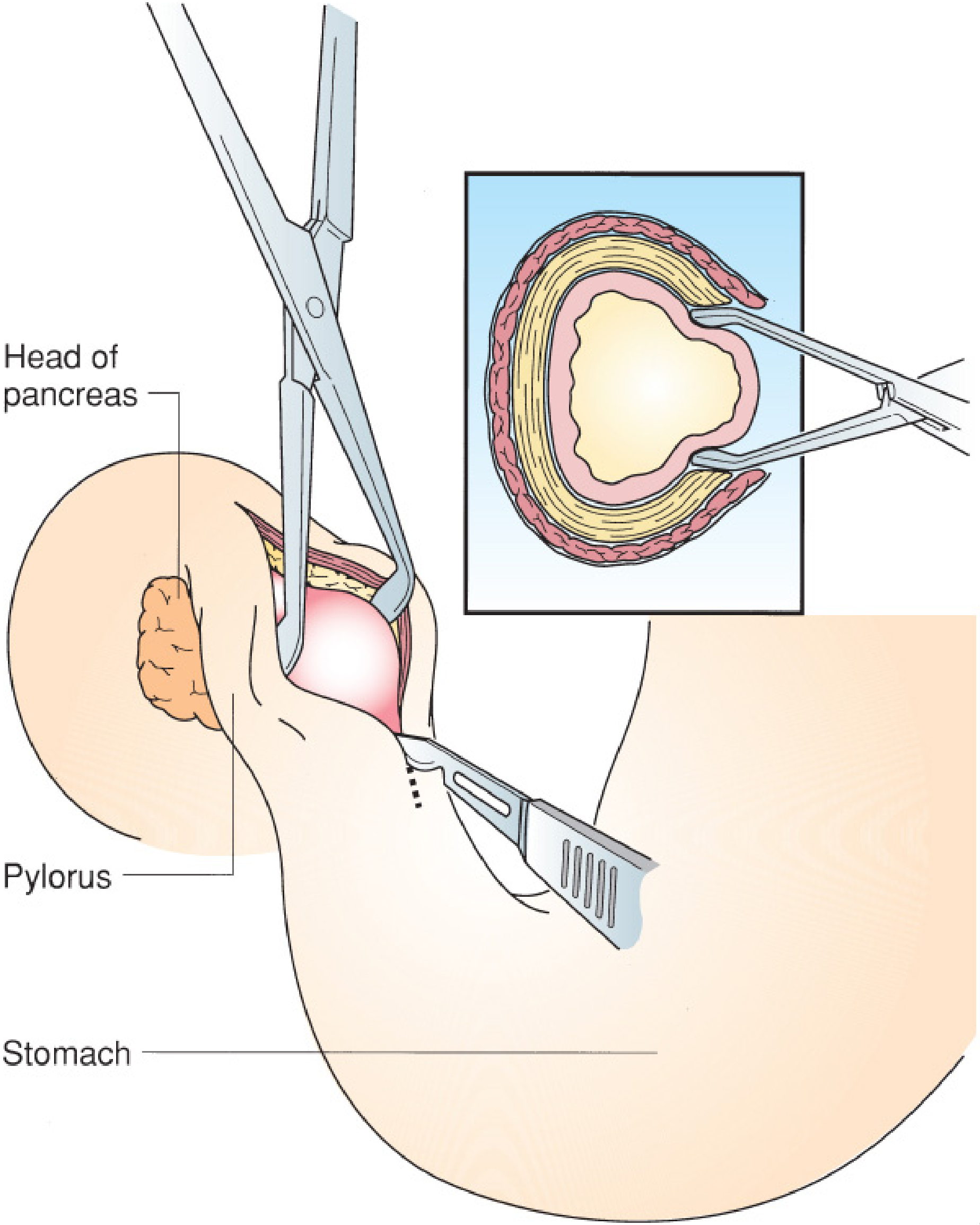
Ramstedt pyloromyotomy. The hypertrophied pyloric muscle is incised longitudinally. The inset shows submucosal herniation into the myotomy site, confirming adequate division. From Mulholland & Greenfield's Surgery.
- A single longitudinal incision through the anterior wall of the pyloric canal, from stomach to duodenal junction
- Muscle is spread until submucosa bulges up freely = complete myotomy
- A leak test (30-60 mL air via orogastric tube) confirms no mucosal perforation
- Laparoscopic approach: 3 ports (umbilical + two 3-mm stab incisions)
Complications:
- Incomplete myotomy - ongoing projectile vomiting; requires return to OR
- Mucosal perforation - repair + buttress with omentum, or rotate pylorus 180° and re-myotomize
- Both are rare; mortality is distinctly unusual
Postoperative feeding: Start at 6-8 hours; ad lib with 60-mL limit; discharge usually within 24-36 hours.
Acquired Pyloric Stenosis (Adults)
Occurs due to antral gastritis, peptic ulcers near the pylorus, or carcinoma of the distal stomach/pancreas causing fibrosis or malignant infiltration.
PART 2: Duodenal Stenosis (and Atresia)
Embryology & Etiology
The duodenum develops in the 4th week. During weeks 5-6, luminal obliteration occurs due to mucosal proliferation, followed by gradual recanalisation over subsequent weeks. Duodenal atresia (DA) and stenosis result from failure of this recanalisation process.
Forms of obstruction:
- Complete membrane obstructing the lumen (most common)
- Blind-ending pouch with fibrous cord to distal duodenum
- Blind-ending pouch with no connection
- Perforate membranes / webs (cause partial stenosis)
- Extrinsic compression: annular pancreas or preduodenal portal vein
Location: In 80% of cases, obstruction is just distal to the ampulla of Vater → bilious vomiting
- Pre-ampullary obstruction (15-20%) → non-bilious vomiting
Incidence & Associated Anomalies
- ~1 per 200,000 live births (combined atresia, stenosis, web); slight female preponderance
- Associated anomalies present in >50% of patients:
| Association | Frequency |
|---|---|
| Down syndrome (Trisomy 21) | 25-30% (some series >50%) |
| Congenital heart disease | ~20% |
| Malrotation | 20-30% |
| VACTERL (incl. esophageal atresia) | OA in ~10% |
| Renal anomalies, imperforate anus, CNS lesions | Variable |
Any infant with Trisomy 21 and bilious vomiting must be evaluated for duodenal stenosis/atresia.
Clinical Features
Prenatal:
- Dilated stomach + duodenal cap on US (double bubble in utero)
- Polyhydramnios in 33-50% (fetus cannot swallow/absorb amniotic fluid)
- IUGR, prematurity
Postnatal:
- Early bilious vomiting (first day of life in atresia; may be delayed months to years in partial stenosis/web)
- Upper abdominal distension
- Duodenal stenosis/partial membrane may present at any age depending on degree - can even go undiagnosed until adulthood
Diagnosis
Plain X-ray - "Double Bubble" sign:
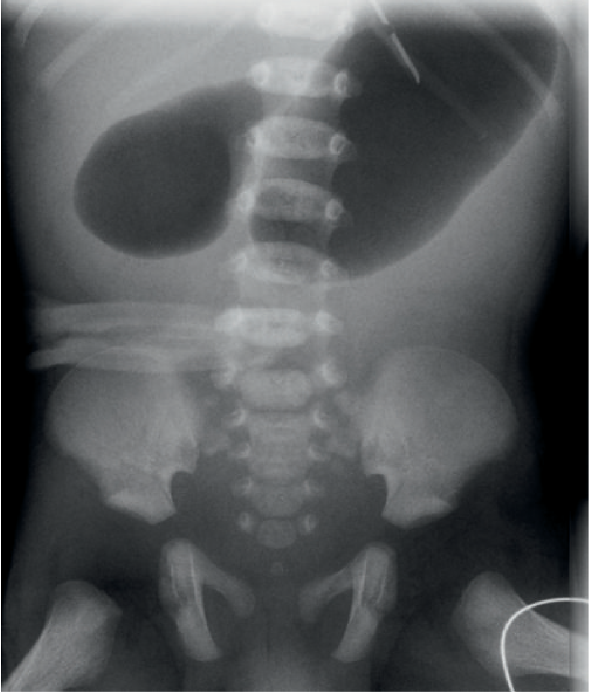
Classic "double-bubble" sign. Two dilated gas-filled structures (stomach + first part of duodenum). No distal gas confirms complete atresia. From Grainger & Allison's Diagnostic Radiology.
- Two air bubbles = dilated stomach + dilated proximal duodenum
- No distal gas = complete atresia; classic double bubble in a stable child does not require further imaging before surgery
- If distal gas is present = partial obstruction (stenosis, web) - warrants upper GI contrast study
Upper GI contrast study (for partial obstruction):
- Duodenal stenosis: narrowing in the second part of duodenum
- Duodenal web: thin filling defect extending across lumen with pre-stenotic dilatation
Ultrasound: Can demonstrate duodenal obstruction, especially with clear fluids given first to distend proximal gut.
Treatment
Surgical correction after stabilization:
- Duodenoduodenostomy (anastomosis of proximal to distal duodenum) - operation of choice for atresia
- Web excision / duodenoplasty for membranous obstruction
- Must evaluate for and address malrotation intraoperatively (present in 20-30%)
Comparison at a Glance
| Feature | IHPS | Duodenal Stenosis/Atresia |
|---|---|---|
| Cause | Post-natal muscle hypertrophy | Failed recanalisation of duodenum (failure of vacuolisation, 8-10 wks) |
| Onset | 3-6 weeks after birth | Day 1 (atresia) or any age (stenosis) |
| Vomiting | Non-bilious, projectile | Bilious (80%) or non-bilious (20% if pre-ampullary) |
| Key X-ray sign | Normal (US is gold standard) | Double bubble on plain film |
| Key US finding | Pyloric muscle ≥4 mm, length ≥15-17 mm | Dilated stomach + duodenum |
| Associated anomalies | Erythromycin exposure; rarely Turner/T18 | Down syndrome (25-50%), VACTERL |
| Metabolic disturbance | Hypochloremic hypokalemic alkalosis | Dehydration ± alkalosis (less specific) |
| Surgery | Fredet-Ramstedt pyloromyotomy | Duodenoduodenostomy |
| Emergency? | No - resuscitate first | No - stabilize first |
Sources: Current Surgical Therapy 14e; Robbins & Cotran Pathologic Basis of Disease; The Developing Human - Clinically Oriented Embryology; Grainger & Allison's Diagnostic Radiology; Mulholland & Greenfield's Surgery 7e; Sleisenger & Fordtran's Gastrointestinal and Liver Disease.
Duodenal stenosis and pyloric stenosis in adults
Searching sources...
Finding Sources
Finding Sources
Finding Sources
Finding Sources
Reading File
Reading File
Reading File
Reading File
Reading File
Reading File
Finding Sources
Reading File
Finding Sources
Reading File
Finding Sources
Finding Sources
Reading File
Reading File
Reading File
I now have comprehensive coverage from all major textbooks. Here is the complete answer:
Pyloric Stenosis and Duodenal Stenosis in Adults
PART 1: Pyloric Stenosis in Adults (Gastric Outlet Obstruction)
Overview
Adult pyloric / gastric outlet obstruction (GOO) is a different entity from infantile HPS. It is broadly classified as benign or malignant, and the underlying cause determines management. Historically, peptic ulcer disease (PUD) accounted for ~90% of all GOO; with effective H. pylori eradication and modern acid suppression, that figure has fallen to only ~5%, and malignancy now accounts for 50-80% of cases.
Bailey & Love: "With the decreasing incidence of peptic ulceration, gastric outlet obstruction should be considered malignant until proven otherwise."
Causes of Adult Gastric Outlet Obstruction
| Category | Specific Cause | Notes |
|---|---|---|
| Benign - Peptic (~5%) | Duodenal ulcers, pyloric channel ulcers, gastric ulcers | Edema (reversible) or fibrotic scar (fixed, mechanical) |
| Malignant (50-80%) | Gastric carcinoma | Most common malignant cause |
| Pancreatic carcinoma (local extension) | Typically palliation | |
| Gastric lymphoma | Often responds to chemotherapy | |
| Ampullary / proximal duodenal neoplasms | ||
| Gallbladder / biliary tree neoplasms | ||
| Inflammatory (~5%) | Crohn's disease | Antral/duodenal involvement |
| Chronic/acute pancreatitis | ||
| Large pancreatic pseudocyst | ||
| Eosinophilic gastroenteritis, amyloid, TB, sarcoid | ||
| Miscellaneous | Adult hypertrophic pyloric stenosis | Rare (~200 cases reported |
| Caustic injury | ||
| Annular/ectopic pancreas | Extrinsic compression | |
| Large pedunculated gastric polyp | ||
| Bouveret syndrome (gallstone impacted in pylorus/duodenum) | ||
| Gastric bezoars | ||
| Postsurgical complications |
Adult Hypertrophic Pyloric Stenosis (AHPS)
A rare entity - approximately 200 cases in the world literature. Anatomically identical to the infantile form (circumferential hypertrophy of the muscularis propria), but aetiology differs:
- Pyloric thickening associated with PUD, hypertrophic gastropathy, or carcinoma
- In a subset, no cause is identified - may represent missed infantile cases or late-onset hypertrophy
- Family history of IHPS in some cases (genetic predisposition)
- 80% occur in men
- Microscopy: variable inflammation, edema, degenerative changes in myenteric plexus ganglion cells
Clinical features: Similar to infantile form but presenting in adults - nausea, mild vomiting, early satiety, epigastric pain after eating. Palpable pyloric mass is not usually found in adults.
Diagnosis:
- Ultrasound (screening): abnormal if pylorus ≥1 cm thick and persistent elongation >2 cm
- Contrast radiography: elongated narrow pyloric channel, delayed gastric emptying, dilated stomach
- EGD is mandatory to exclude carcinoma and chronic peptic ulcer disease (cannot assume benign without tissue)
Treatment:
- Surgical pyloromyotomy or pyloric resection (preferred because of risk of focal carcinoma)
- Endoscopic balloon dilation: effective but 80% recurrence within 6 months
- Palliative endoscopic stent for GOO from gastric cancer
Peptic Ulcer-Related GOO (Benign)
Two mechanisms operate:
- Inflammatory/edematous obstruction - from an active pyloric channel or duodenal ulcer; reversible with ulcer healing
- Fixed, fibrotic/scarred obstruction - from long-standing or recurrent ulceration; mechanical, requires intervention
Higher risk with: Zollinger-Ellison syndrome, long-standing ulcers, NSAID-associated ulcers, DU and pyloric channel ulcers. GOO is the least common ulcer complication (1-2% of PUD patients), while bleeding and perforation are far more common.
Clinical Features (All-Cause GOO)
- Progressive vomiting of undigested food (often containing food eaten hours or days earlier)
- Vomiting is non-bilious (obstruction proximal to duodenum) - characteristically lacks bile
- Early satiety, epigastric fullness, bloating
- Weight loss, malnutrition
- Visible gastric distension
- Succussion splash on examination (retained fluid sloshing in distended stomach)
- In malignancy: less pronounced acid-base disturbance due to hypochlorhydria
Metabolic Consequences
Exactly as in infants, loss of gastric HCl produces:
Hypochloremic, hypokalemic metabolic alkalosis
Sequential progression:
- Vomiting HCl → low Cl⁻, elevated HCO₃⁻, initially normal Na⁺ and K⁺
- Urine: initially low chloride, high bicarbonate (reflecting primary metabolic disturbance)
- With progressive dehydration: Na⁺ retained by kidney → K⁺ and H⁺ excreted → paradoxical aciduria develops
- Progressive hyponatraemia and hypokalaemia
- Alkalosis lowers ionized calcium → tetany can occur
In malignant GOO (hypochlorhydria), the acid-base disturbance is typically less pronounced.
Diagnostic Work-up
- Nasogastric tube (NGT) - large bore; aspirate retained contents (>400 mL after 750 mL saline load at 30 min = mechanical obstruction on saline load test, now declining in use)
- Cross-sectional imaging (CT scan) - first-line; dilated stomach suggests obstruction
- Endoscopy (EGD) - definitive; required in ALL patients to:
- Visualize the obstruction
- Take biopsies to exclude malignancy
- Potentially treat (balloon dilation, stent)
- Contrast upper GI series - shows dilated stomach, narrowed pyloric channel
- Endoscopic ultrasound (EUS) + deeper biopsies if malignancy strongly suspected but not confirmed
Management
Step 1 - Resuscitation (always first):
- NPO
- IV isotonic saline + potassium (replaces Cl⁻ and allows kidney to correct alkalosis)
- IV PPI (reduce gastric secretion)
- Nutritional support (parenteral if needed)
- Large-bore NGT or orogastric tube lavage to decompress stomach
Step 2 - Definitive treatment by cause:
| Cause | Treatment |
|---|---|
| PUD (edema/active ulcer) | PPI + H. pylori eradication + stop NSAIDs; often resolves without surgery |
| PUD (fixed fibrotic stricture) | Endoscopic balloon dilation ± biodegradable stent; surgical stricturoplasty or gastrojejunostomy |
| Malignant GOO (gastric/pancreatic cancer) | Endoscopic self-expanding metal stent (SEMS) (90% allow oral intake); or surgical gastrojejunostomy (open or laparoscopic) |
| Crohn's disease | Anti-inflammatory therapy; endoscopic balloon dilation (40% failure rate) |
| Pancreatitis / pseudocyst | Drainage (percutaneous, endoscopic, or surgical) |
| AHPS | Surgical resection (preferred); pyloromyotomy; balloon dilation (high recurrence) |
For malignant duodenal obstruction with concurrent bile duct obstruction:
- Biliary stenting first, then duodenal SEMS (reversed order is technically difficult)
- Or palliative gastrojejunostomy
PART 2: Duodenal Stenosis in Adults
Adult duodenal stenosis is an obstruction of the duodenal lumen that may be intrinsic or extrinsic. It presents as a post-pyloric, usually bilious obstruction (in contrast to pyloric stenosis, which is non-bilious).
Causes of Adult Duodenal Stenosis
| Category | Causes |
|---|---|
| Malignant | Pancreatic carcinoma (most common), ampullary carcinoma, duodenal carcinoma, cholangiocarcinoma, gallbladder carcinoma, gastric carcinoid |
| Peptic | Duodenal ulcers (scarring and fibrosis, especially D1/D2) |
| Inflammatory | Crohn's disease (duodenal Crohn's), pancreatitis, pancreatic pseudocyst compressing D2 |
| Vascular | Superior mesenteric artery (SMA) syndrome - compression of D3 |
| Congenital (undiagnosed in childhood) | Annular pancreas, duodenal web/membrane, preduodenal portal vein (usually present in childhood but occasionally diagnosed in adults with partial obstruction) |
| Radiation-induced | Post-radiation fibrosis |
| Post-surgical | Anastomotic stricture |
Superior Mesenteric Artery (SMA) Syndrome
A distinct and important cause of adult duodenal stenosis - extrinsic compression of the third part of the duodenum (D3) between the SMA and the aorta.
Mechanism: The SMA branches off the aorta at an acute angle. Under certain conditions this angle narrows, trapping the duodenum between the two vessels.
Precipitating conditions:
- Rapid growth spurts in adolescents
- Profound weight loss (loss of mesenteric/retroperitoneal fat pad that normally holds the SMA away from the aorta)
- Prolonged immobilization in a body cast (increased lordosis)
- Abdominal surgery or inflammatory disease
- Prolonged bed rest
Symptoms:
- Episodic postprandial epigastric pain, nausea, bilious vomiting
- May improve in the prone or knee-chest position (postural relief is characteristic)
- Acute or chronic/relapsing course
Diagnosis:
- Plain film/CT: dilated stomach and duodenum up to D3
- Upper GI contrast series / CT: dilation and abrupt cut-off at D3, at the level of the SMA
- CT angiography / lateral aortography: narrowed aortomesenteric angle
- Antroduodenal manometry: distinguishes mechanical obstruction from motility disorder
- Enteral feeding trial (catheter past obstruction into jejunum): relief of vomiting supports diagnosis
Treatment:
- Correct precipitating factor first (weight gain, remove cast, mobilize)
- Conservative: small frequent meals, prone/left lateral positioning after eating
- Gastric decompression + IV fluids for acute episodes
- Refractory cases: laparoscopic duodenojejunostomy (gastrojejunostomy alone is insufficient as it doesn't decompress the proximal duodenum); occasionally laparoscopic lysis of the ligament of Treitz
Annular Pancreas (Extrinsic Duodenal Stenosis)
Annular pancreas causes extrinsic compression of D2 by a ring of pancreatic tissue surrounding the second part of the duodenum. Classically a paediatric diagnosis, but partial annular pancreas may present in adults with:
- Chronic intermittent postprandial vomiting
- Upper abdominal pain
- Associated peptic ulceration (common in adults with annular pancreas)
- Pancreatitis (obstruction of pancreatic duct)
Treatment: Surgical bypass (duodenojejunostomy or duodenoduodenostomy); the pancreatic tissue itself should not be divided (risk of pancreatic duct injury/fistula).
Clinical Features of Adult Duodenal Stenosis (General)
| Feature | Details |
|---|---|
| Vomiting | Bilious (80-85%, post-ampullary); non-bilious if pre-ampullary |
| Pain | Epigastric or periumbilical, postprandial |
| Distension | Upper abdominal, less prominent than pyloric stenosis |
| Weight loss | Common, especially with malignancy |
| Succussion splash | May be present |
Key Comparisons: Adult Pyloric vs. Duodenal Stenosis
| Feature | Pyloric Stenosis (Adult) | Duodenal Stenosis (Adult) |
|---|---|---|
| Main causes | PUD, gastric cancer, AHPS | Pancreatic/periampullary cancer, PUD, SMA syndrome, annular pancreas, Crohn's |
| Vomiting character | Non-bilious (pre-pyloric) | Bilious (post-ampullary, 80-85%) |
| Metabolic upset | Hypochloremic hypokalemic alkalosis (classical) | Less pronounced / variable |
| Key investigation | EGD + CT + biopsies | CT + EGD ± EUS + contrast series |
| Malignancy concern | Gastric cancer until proven otherwise | Pancreatic/ampullary cancer until proven otherwise |
| Benign treatment | Balloon dilation, H. pylori eradication, gastrojejunostomy | Balloon dilation, duodenojejunostomy (for SMA/annular) |
| Malignant palliation | SEMS or gastrojejunostomy | SEMS or gastrojejunostomy ± biliary stent |
Sources: Sleisenger & Fordtran's Gastrointestinal and Liver Disease; Yamada's Textbook of Gastroenterology 7e; Bailey & Love's Short Practice of Surgery 28e; Harrison's Principles of Internal Medicine 22e; Current Surgical Therapy 14e.
This is a shared conversation. Sign in to Orris to start your own chat.