
ostetomy around hip for osteoarthritis conditions
osteotomy hip osteoarthritis
hip osteotomy intertrochanteric periacetabular osteoarthritis
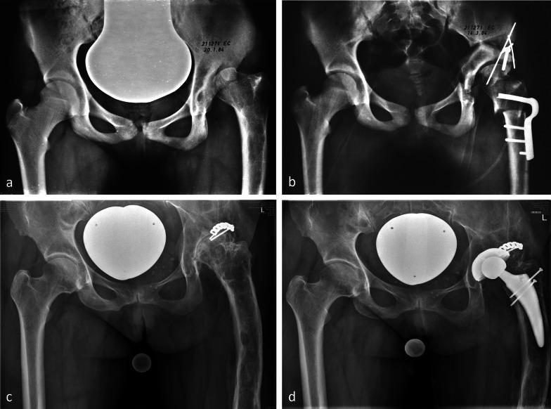
A longitudinal series of four anteroposterior (AP) pelvic radiographs (a–d) demonstrating the 36-year clinical progression and surgical management of a left hip with Proximal Femoral Focal Deficiency (PFFD). Image (a) shows a baseline varus deformity of the proximal femur following a prior intertrochanteric osteotomy. Image (b) illustrates post-surgical results of a periacetabular osteotomy (PAO) combined with intertrochanteric revalgisation, featuring stabilizing hardware including an angled blade plate, screws, and Kirschner wires used to improve femoral head coverage and joint congruity. Image (c) shows the long-term natural history 36 years post-correction, revealing advanced degenerative joint disease (osteoarthritis), joint space loss, and subchondral sclerosis, with hardware from a subsequent posterior shelf procedure still visible. Image (d) displays the final state after Total Hip Replacement (THR), showing a well-positioned prosthetic acetabular cup and femoral stem, restoring anatomical alignment. This series serves as a pedagogical timeline for joint-preserving osteotomies versus eventual arthroplasty in complex pediatric hip deformities.
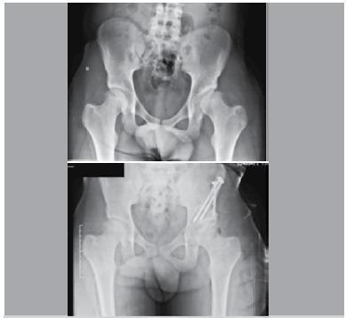
Anteroposterior (AP) pelvic radiographs demonstrating pre-operative and post-operative comparisons for a Periacetabular Osteotomy (PAO). The upper image shows developmental dysplasia of the hip (DDH), specifically on the left side, characterized by a shallow acetabulum and decreased lateral coverage of the femoral head, reflected by a low center-edge (CE) angle of 7 degrees. The lower image presents the post-surgical result where the left acetabulum has been reoriented to improve femoral head coverage, achieving an increased CE angle of 22 degrees. Two large metallic cortical screws are visible in the left ilium, providing surgical fixation of the osteotomized acetabular fragment. The anatomical reorientation is evident by the increased lateral and superior coverage of the 'sourcil' over the femoral head. Key structures visible include the pelvic ring, sacroiliac joints, bilateral hip joints, and the proximal femurs. This visual comparison illustrates the radiological objectives of PAO: correcting acetabular orientation to improve joint mechanics and prevent secondary osteoarthritis.
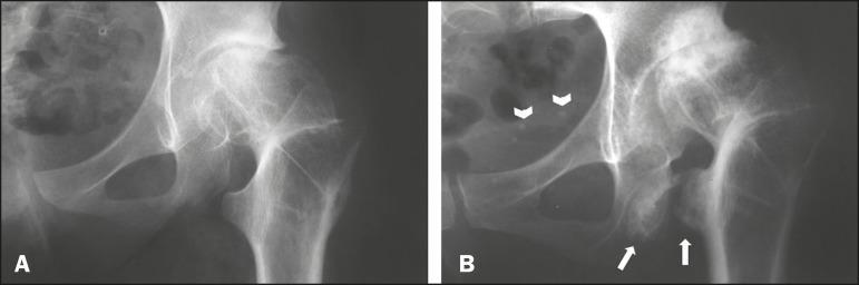
Diagnostic Image: This side-by-side comparison includes two anteroposterior (AP) radiographs of the left hip (A and B) demonstrating long-term progression of orthopedic pathology following a valgus intertrochanteric osteotomy. Panel A, taken one year post-surgery, shows early signs of osteoarthritis with relatively preserved hip joint space and a baseline ischiofemoral distance between the lesser trochanter and the ischium. Panel B, obtained 30 years post-operatively, shows significant advancement of hip osteoarthritis characterized by joint space narrowing and subchondral sclerosis. Notably, Panel B exhibits hallmarks of ischiofemoral impingement, highlighted by white arrows indicating a marked narrowing of the ischiofemoral space. Distinctive visual features include cortical irregularity, reactive bone remodeling, and osteosclerosis on the opposing surfaces of the lesser trochanter and the ischial tuberosity. The image serves as a clinical reference for secondary hip osteoarthritis and the mechanical consequences of altered proximal femoral anatomy leading to extra-articular impingement.
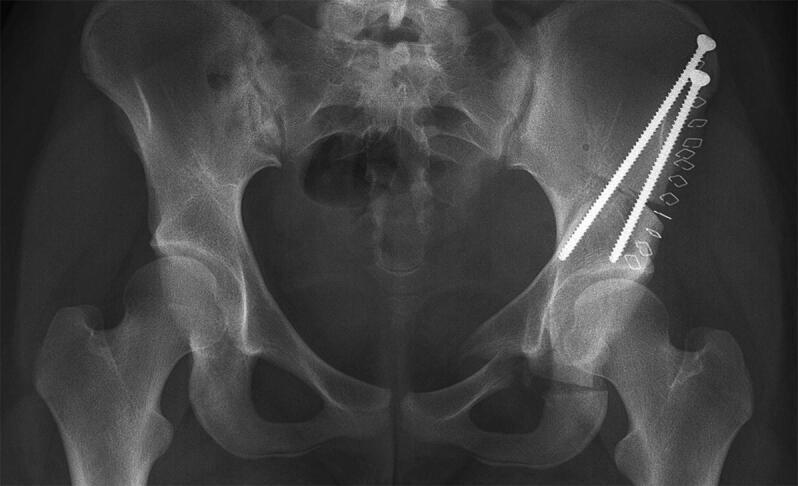
Anterior-posterior (AP) pelvic radiograph demonstrating the postoperative state of a minimally invasive periacetabular osteotomy (PAO) on the patient's right side (anatomical right, image left side shows native anatomy). The diagnostic image reveals metallic surgical hardware used for internal fixation, specifically two long, threaded cortical screws oriented in a superior-lateral to inferior-medial trajectory. These screws bridge the iliac osteotomy site to stabilize the repositioned acetabular fragment. A row of approximately eight metallic surgical staples or skin clips is visible laterally along the soft tissue surgical approach. The bony anatomy shows clear osteotomy lines through the ilium, ischium, and pubis, with the right acetabulum redirected to increase femoral head coverage compared to the untreated contralateral side. The left hip remains undisturbed with normal joint space and acetabular morphology. This image illustrates surgical management for hip dysplasia, focusing on mechanical stabilization and joint realignment to prevent secondary osteoarthritis. Orthopedic surgeons and radiology residents can use this for understanding pelvic fixation and pelvic ring reconstructive geometry.
Osteotomies Around the Hip for Osteoarthritis
Overview and Rationale
Key principle: Osteotomy should be considered in a young patient who maintains a good range of movement of the hip and whose imaging shows a joint devoid of significant degenerative change.
- Bailey and Love's Short Practice of Surgery, 28th Ed
Types of Osteotomy Around the Hip
1. Femoral Osteotomies
A. Intertrochanteric Varus Osteotomy
- Indication: Excessive valgus neck-shaft angle (coxa valga) with lateral uncovering of the femoral head
- The neck-shaft angle is reduced, bringing the femoral head into better contact with the acetabulum
- Redirects load-bearing to a healthier portion of articular cartilage
- Used in early/moderate OA in young adults, Perthes' disease, SCFE
B. Intertrochanteric Valgus Osteotomy
- Indication: Coxa vara, medial joint space narrowing with a steep Pauwels angle
- Redirects forces from the medial pole to the more lateral superior cartilage
- The "valgus-extension osteotomy" (Bombelli technique) was popular in the 1970s-1990s for medial pole OA - long-term results at 25 years have been reported
C. Valgus-Extension Osteotomy (Bombelli)
- Combines valgus angulation with extension
- Aims to move loading to the posterior-lateral articular cartilage
- Reported long-term results (J Bone Joint Surg Br, 2000) showed benefit in selected patients
D. Transtrochanteric Rotational Osteotomy (Sugioka)
- Rotates the femoral head anteriorly or posteriorly
- Used mainly in avascular necrosis (AVN) and in select OA cases
- Moves the necrotic/degenerate zone out of the weight-bearing area
2. Periacetabular Osteotomy (PAO) - Bernese PAO
| Feature | Detail |
|---|---|
| Primary indication | Developmental dysplasia of the hip (DDH) |
| Secondary OA prevention | Corrects under-coverage before cartilage damage becomes irreversible |
| Acetabular retroversion | Reverse PAO to treat anterior over-coverage (pincer FAI) |
| Age group | Typically young adults (<40 years); results worsen with age >40 |
| Key measurement | Lateral centre-edge angle (LCEA) corrected from <20° to 25-35° |
- LCEA < 20° (true dysplasia)
- Tonnis OA grade 0 or 1 (minimal degenerative change)
- Good range of motion preserved
- Age < 35-40 years
3. Chiari Pelvic Osteotomy
- A salvage acetabular osteotomy - cuts the ilium horizontally above the hip joint
- Displaces the iliac wing medially, creating a bony "shelf" over the femoral head
- Used when cartilage is too damaged for redirectional PAO
- Reported for OA secondary to dysplasia (Lack et al.)
- Now less commonly performed due to widespread use of PAO and THR
4. Shelf Procedures (e.g., Spitzy, Staheli shelf)
- Augment lateral acetabular coverage by adding bone graft
- Primarily a pediatric/adolescent procedure but occasionally used in young adults
- Indirect joint preservation by reducing lateral subluxation forces
Indications Summary (Adult Hip OA)
| Procedure | Primary Indication |
|---|---|
| Varus intertrochanteric osteotomy | Coxa valga, lateral OA |
| Valgus intertrochanteric osteotomy | Coxa vara, medial OA |
| Valgus-extension osteotomy | Medial/inferomedial pole OA |
| PAO (Bernese) | DDH with secondary OA risk |
| Chiari osteotomy | Dysplasia with early OA (salvage) |
| Brackett osteotomy | Correction of fixed deformity/ankylosis |
- Bailey and Love's Short Practice of Surgery 28th Ed, Table 39.4
Patient Selection Criteria
- Age: Young patient, ideally < 40 years (PAO results deteriorate beyond 40)
- Joint space: Preserved (no severe OA - Tonnis grade 0-1)
- Range of motion: Good, functional arc of movement
- Cause: Correctable structural deformity (dysplasia, coxa vara/valga, AVN, Perthes', SCFE)
- Imaging: 3D CT for preoperative planning is increasingly standard
- Diagnostic injection: Confirms symptoms are truly intra-articular before proceeding
Preoperative Planning
- 3D CT scanning for deformity characterization
- 3D printing of models for surgical rehearsal
- Computer navigation and robotics - emerging adjuncts for accurate correction
- Diagnostic hip injection - confirms intra-articular source before osteotomy
Outcomes and Conversion to THR
- Intertrochanteric osteotomies do not impair long-term outcomes of subsequent cemented THR, an important reassurance for patients
- 25-year follow-up data exist for valgus osteotomy in arthritic hips (Clin Orthop 2004)
- PAO: intermediate-to-long-term results (Matheney et al., J Bone Joint Surg Am, 2009) show durable outcomes in well-selected patients
- Total Hip Replacement After PAO: Reviewed separately in Campbell's - technically more demanding but achievable (Campbell's Operative Orthopaedics 15th Ed, 2026)
Illustrative Radiographs
Key Takeaway
conservative management that can be done in osteoarthritis hip
hip osteoarthritis conservative management guidelines
hip osteoarthritis conservative management physiotherapy exercise
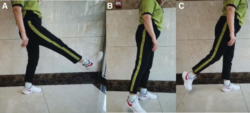
A series of three clinical photographs (labeled A, B, and C) demonstrating a sagittal plane leg swing exercise, a physical therapy technique used in the management of knee osteoarthritis (KOA). The images show a patient standing on an unaffected leg while performing active range-of-motion movements with the affected limb. (A) Depicts hip flexion, with the leg raised forward to approximately 45 degrees from the vertical axis. (B) Shows the leg in a neutral vertical position, slightly abducted to facilitate clearance during the swing. (C) Demonstrates hip extension, with the leg swinging posteriorly behind the coronal plane of the body. The exercise aims to improve joint mobility and periarticular muscle coordination. The standing posture remains relatively upright with a slight forward lean to maintain balance during the dynamic movement. This visual protocol serves as a rehabilitation guide for non-surgical conservative management of degenerative joint disease.
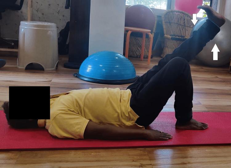
A clinical photograph depicting a male patient in a physiotherapy setting, performing a therapeutic exercise. The patient is positioned supine on a red yoga mat on a wood-laminate floor. The patient is executing a straight leg raise (SLR), with the right lower extremity actively elevated while maintaining knee extension. The contralateral (left) leg is positioned in a crook-lying posture with the knee flexed and the foot flat on the mat to stabilize the lumbar spine and pelvis. The background contains common rehabilitation equipment, including a blue Bosu balance trainer, a gray stability ball, and various stools. This visual demonstrates a post-operative or conservative management exercise focused on strengthening the hip flexors and quadriceps, frequently utilized in rehabilitation protocols for conditions such as avascular necrosis of the femoral head or following total hip replacement.
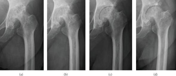
A series of four sequential plain anteroposterior radiographs of a single hip joint, demonstrating the progression of degenerative hip osteoarthritis over a three-year interval. Image (a) represents the baseline, showing terminal-stage osteoarthritis characterized by severe joint space narrowing, subchondral sclerosis, and multiple large bone cysts in the femoral head and acetabulum, resulting in poor joint congruity. Image (b) at one year shows static radiographic findings despite clinical conservative management. Images (c) and (d), corresponding to years two and three, illustrate progressive joint remodeling. Key findings include a visible widening of the radiographic joint space, improved congruity between the femoral head and the acetabular roof, and a reduction in the size and density of the subchondral bone cysts. The final image (d) shows a smoother femoral head contour and more uniform bone density compared to baseline. This series highlights the longitudinal morphological changes possible in advanced osteoarthritis through non-surgical interventions, specifically focusing on joint space recovery and cortical remodeling.
Conservative Management of Hip Osteoarthritis
"There is no specific pharmacological therapy for OA; however, non-operative treatment with non-steroidal anti-inflammatories, regular exercise, physiotherapy and modification of lifestyle with loss of weight does help."
- Bailey and Love's Short Practice of Surgery, 28th Ed
1. Patient Education (Universal - All Guidelines Agree)
- Nature of OA - a degenerative but manageable condition
- Goals of treatment: pain control + function preservation
- Self-management principles: pacing activities, ergonomic training, joint protection
- Discussion of all available treatment options
- Can be delivered individually, in multidisciplinary group programs, or online
2. Non-Pharmacological Interventions
A. Exercise (Strongly Recommended)
| Type | Examples | Benefit |
|---|---|---|
| Aerobic/cardiovascular | Walking, cycling, swimming | Pain reduction, cardiovascular health |
| Strengthening | Hip abductor, quadriceps, core strengthening | Joint stability, offloads articular surfaces |
| Range of motion / flexibility | Gentle hip stretches, yoga | Maintains functional arc, counters capsular contracture |
| Aquatic exercise | Hydrotherapy, pool walking | Reduces joint load while allowing movement - especially useful in obese patients |
| Tai Chi | Balance and proprioceptive training | Reduces fall risk, improves proprioception |
- Straight leg raises (SLR) for quadriceps/hip flexor strengthening
- Hip abductor strengthening (side-lying leg raises, clamshells) - targets the abductor weakness causing Trendelenburg gait
- Sagittal plane leg swings for ROM maintenance
B. Weight Loss
- Obesity is a major modifiable risk factor and significantly accelerates OA progression
- Even modest weight reduction (5-10% of body weight) meaningfully reduces joint load and pain
- Most effective when combined with structured exercise
- Reduces the force transmitted across the hip joint with each step
C. Walking Aids
- A walking stick (cane) held in the opposite hand (contralateral to the affected hip) is recommended
- Mechanism: reduces the workload of the ipsilateral hip abductors and unloads the affected hip joint
- Reduces the resultant force across the hip by approximately 30-40%
- Other aids: rollator frame for severe cases, crutches for acute flares
D. Physiotherapy
- Manual therapy: gentle mobilization of the hip capsule, soft tissue techniques
- Gait retraining - corrects antalgic gait patterns
- Hydrotherapy - warm water reduces muscle spasm and allows pain-free movement
- TENS (transcutaneous electrical nerve stimulation) - short-term analgesia
E. Activity Modification
- Avoid high-impact activities (running, jumping) that load the hip excessively
- Adapt activities of daily living: raised toilet seats, grab rails (aids independence)
- Ergonomic advice: avoid prolonged sitting with hip in extreme flexion/rotation
F. Footwear and Orthotics
- Cushioned footwear to reduce impact transmission
- Lateral wedge insoles have some evidence in lower limb OA
3. Pharmacological Treatment
Step 1 - Topical Agents (First-line, especially elderly/comorbid)
- Deliver anti-inflammatory effect locally with minimal systemic absorption
- Low risk of GI, renal, and cardiovascular side effects
- Effective for peripheral joints; some evidence supports use for hip
- Preferred in older patients and those with GI/cardiovascular risk
- Studies show similarly low adverse event rates even with long-term use (up to 12 months)
- Causes desensitization of skin nerve fibers
- Analgesic effect via modulation of neurotransmitter pathways (substance P depletion)
- Small but significant pain reduction in controlled studies
- Side effect: local burning/stinging - some patients discontinue
Step 2 - Oral Analgesics
Paracetamol (Acetaminophen)
- Historically recommended as first-line oral pharmacotherapy for mild-to-moderate OA pain
- Safe overall, but risk-benefit profile has been debated in recent years
- Current evidence suggests modest efficacy compared to placebo in OA
- Still used widely as baseline analgesia given favorable safety profile at standard doses (max 3-4 g/day)
- Avoid high doses in hepatic impairment or chronic alcohol use
NSAIDs (Oral)
- COX-1/COX-2 inhibitors and selective COX-2 inhibitors (celecoxib)
- Multiple systematic reviews confirm moderate short-term efficacy for hip/knee OA pain
- Key risks: GI bleeding, cardiovascular events (particularly selective COX-2 inhibitors), renal toxicity
- Risk mitigation:
- Use the lowest effective dose for the shortest possible time
- Co-prescribe a proton pump inhibitor (PPI) in high GI-risk patients
- Avoid in heart failure, CKD, prior GI bleeding
- COX-2 selective agents preferred in those with GI risk but no cardiovascular risk
- Educate patients on OTC NSAID availability and interaction with prescribed agents
Duloxetine (SNRI)
- An option for patients with central sensitization / chronic widespread pain component
- Increasingly used in OA where standard analgesics are insufficient or contraindicated
Opioids
- Weak opioids (tramadol, codeine) used when other analgesics fail
- Strong opioids: generally avoided for OA due to dependence risk, adverse effects, and modest functional benefit
- Reserved for severe pain in patients awaiting surgery or when surgery is not possible
Glucosamine / Chondroitin
- Nutraceuticals with anti-inflammatory and chondroprotective claims
- Evidence is mixed; some trials show modest symptom relief, others show no benefit over placebo
- OARSI considers them "inconclusive" for hip; AAOS does not recommend
- Generally safe; some patients report subjective benefit
Step 3 - Intra-Articular Injections
A. Corticosteroid Injection
- Short-to-medium term pain relief (weeks to 2-3 months)
- Most effective for hip OA with an inflammatory component (effusion, warmth)
- Must be performed under image guidance (ultrasound or fluoroscopy) for the hip due to its deep location - confirmed by contrast arthrogram before injection
- Evidence: systematic review confirms efficacy for hip OA under ultrasound guidance
- Repeated injections: caution if >3-4/year due to risk of cartilage damage and soft tissue complications
- Risk of sepsis: rare but serious (estimated ~1 in 77,000 injections)
B. Hyaluronic Acid (Viscosupplementation)
- Restores viscoelastic properties of synovial fluid
- More commonly used in knee OA with moderate evidence
- Evidence for hip OA is weaker but some guidelines support use
- Effect is delayed (2-4 weeks onset) but potentially longer-lasting than corticosteroids (up to 6 months)
- OARSI: conditionally appropriate for hip; AAOS: insufficient evidence to recommend
C. Platelet-Rich Plasma (PRP)
- Emerging treatment; growing evidence base
- Not yet universally recommended by major guidelines
- May provide symptom relief through growth factor-mediated cartilage protection
4. Assistive Devices and Aids
| Device | Role |
|---|---|
| Walking stick (contralateral hand) | Offloads hip abductors, reduces joint reaction force |
| Raised toilet seat | Reduces extreme hip flexion (painful range) |
| Grab rails (bath, toilet) | Safety + reduces hip loading during transfers |
| Shoe horn / stocking aid | Compensates for limited hip flexion |
| Reacher/grabber | Reduces need to bend with a stiff hip |
5. Multidisciplinary and Psychological Approaches
- Cognitive behavioral therapy (CBT): addresses pain catastrophizing and improves coping
- Occupational therapy: home modification, adaptive equipment
- Dietitian referral: structured weight management program
- Pain clinic referral: for chronic, refractory pain with central sensitization
6. Indications to Move to Surgery
- Relentless pain - especially night pain unresponsive to analgesia
- Failure of conservative management after an adequate trial
- Significant limitation of activities of daily living (ADLs) and lifestyle
- Radiological evidence of severe OA with functional decline
Summary Algorithm
Diagnosis confirmed → Patient Education + Exercise Programme + Weight Loss
        ↓
Add: Walking aid + Physiotherapy + Activity modification
        ↓
Add: Topical NSAID → Oral Paracetamol → Oral NSAID (with PPI if needed)
        ↓
Consider: Intra-articular corticosteroid injection (image-guided)
        ↓
Consider: Hyaluronic acid / PRP injection
        ↓
Failure → Surgical referral (osteotomy if young + correctable deformity; THR if advanced OA)