I now have comprehensive content. Let me compile the full answer.
External Carotid Artery
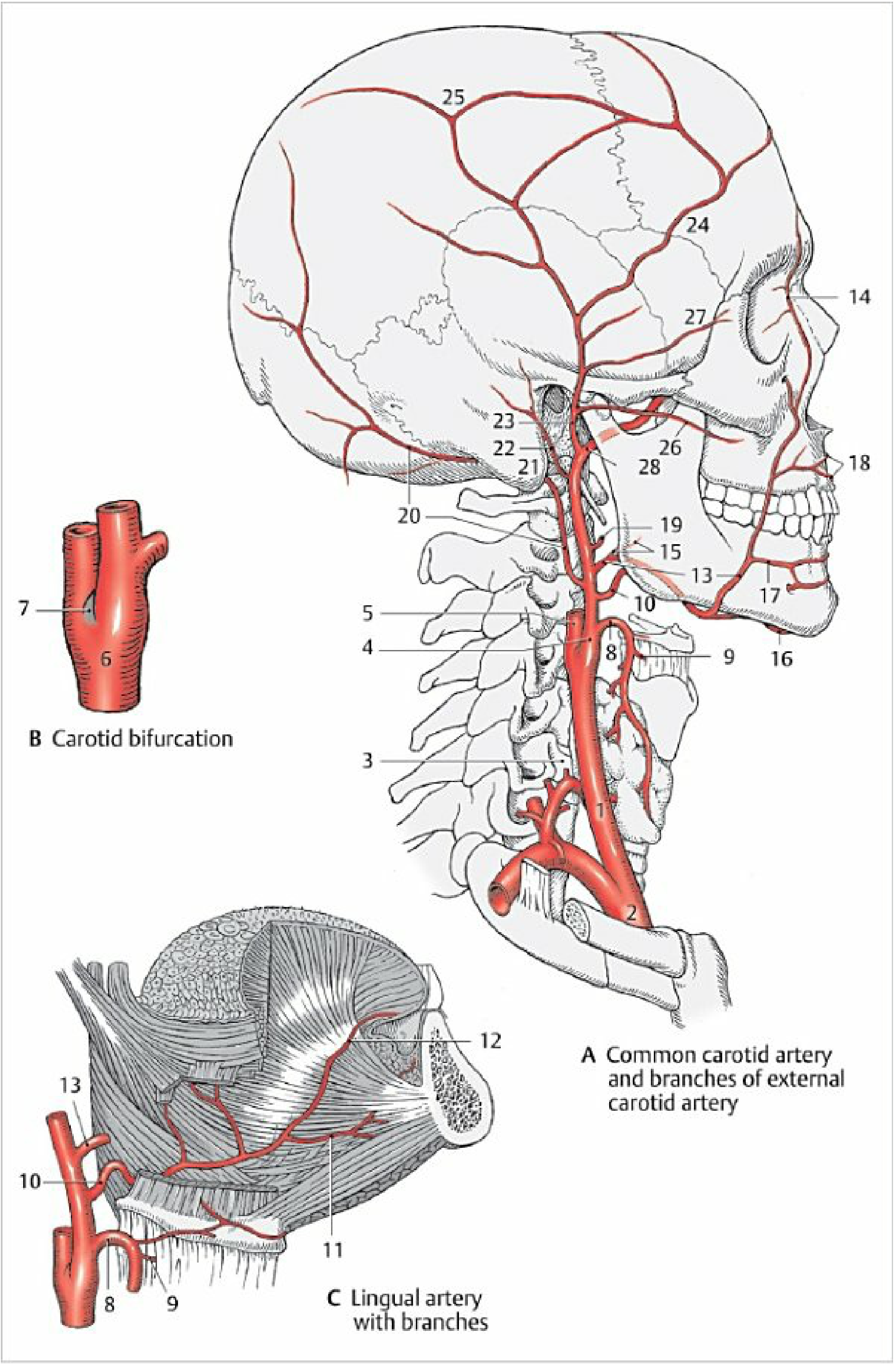
Common carotid artery and branches of the external carotid artery - Color Atlas of Human Anatomy Vol. 2
Origin and Course
The external carotid artery (ECA) is one of the two terminal branches of the common carotid artery, arising at the carotid bifurcation (typically at the level of the superior border of the thyroid cartilage, around C3-C4 vertebral level). At its origin it lies anteromedial to the internal carotid artery, but as it ascends it rotates laterally.
The ECA enters the inferior border of the parotid gland (or passes deep to it), gives off the posterior auricular artery, and then divides into its two terminal branches - the maxillary artery and the superficial temporal artery - near the lower border of the ear. - Gray's Anatomy for Students
Distinguishing ECA from ICA
A key clinical landmark: the hypoglossal nerve (CN XII) lies directly upon the external carotid artery, which helps distinguish the ECA from the internal carotid artery before ligation. The ICA has no branches in the neck, while the ECA gives off multiple branches from the outset. - K.J. Lee's Essential Otolaryngology
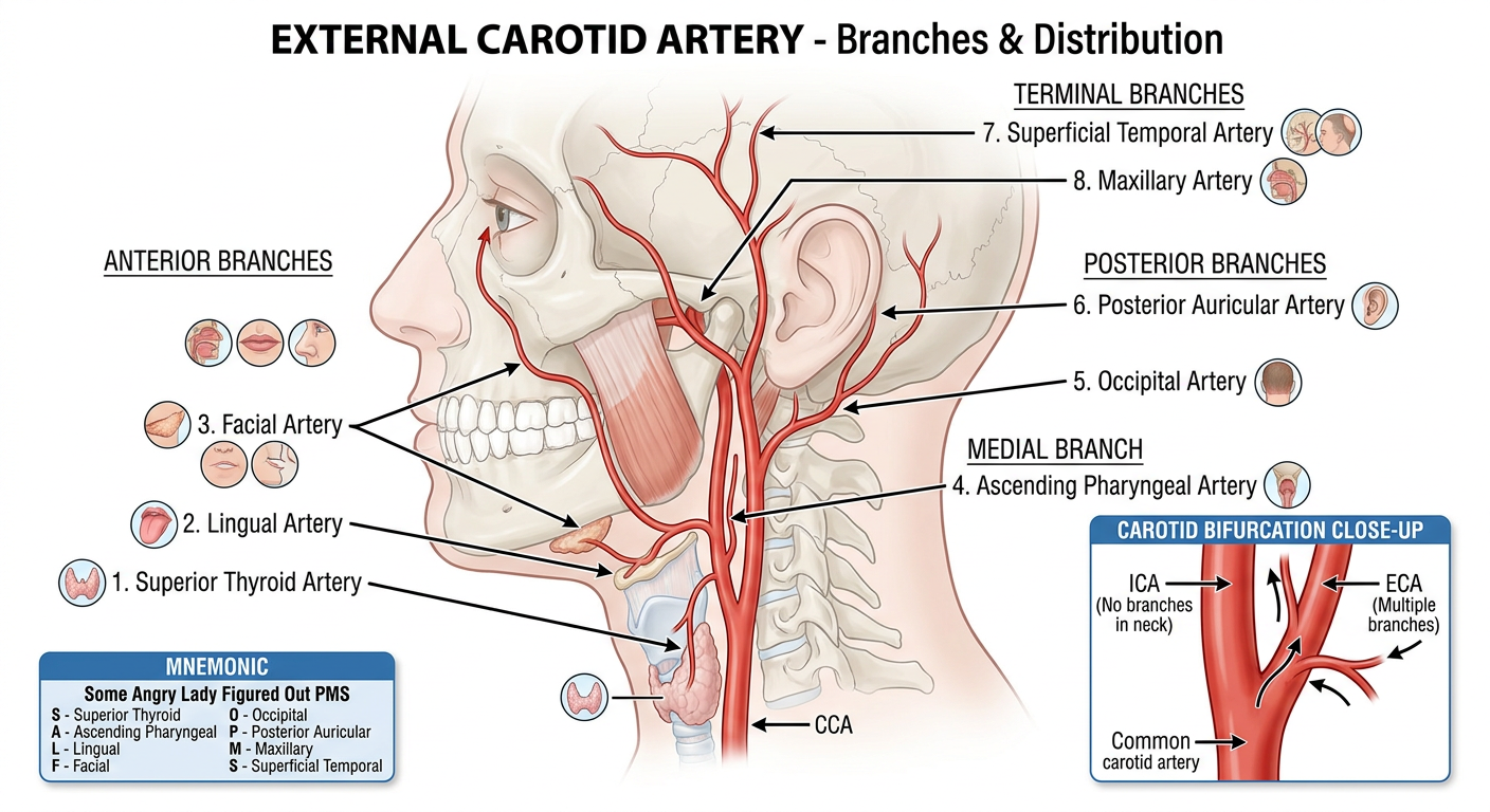
Branches - The "SALFOPMS" Mnemonic
The ECA gives off 8 branches in total (3 anterior, 2 medial/posterior, 2 terminal, 1 posterior):
Anterior Branches
| Branch | Supplies |
|---|
| Superior thyroid artery (1st) | Thyrohyoid muscle, laryngeal structures, sternocleidomastoid, cricothyroid, thyroid gland |
| Lingual artery (2nd anterior) | Tongue muscles, palatine tonsil, soft palate, epiglottis, floor of mouth, sublingual gland |
| Facial artery (3rd anterior) | Face from mandible inferior border to medial canthus, soft palate, palatine tonsil, pharyngotympanic tube, submandibular gland |
- The superior thyroid artery arises near or at the bifurcation, passes downward and forward to the superior pole of the thyroid.
- The lingual artery arises at the level of the hyoid bone, passes deep to CN XII, between the middle constrictor and hyoglossus.
- The facial artery passes deep to stylohyoid and the posterior belly of digastric, loops through/around the submandibular gland, and crosses the mandible just anterior to the masseter muscle to enter the face.
Medial Branch
| Branch | Supplies |
|---|
| Ascending pharyngeal artery | Pharyngeal constrictors, stylopharyngeus, palate, palatine tonsil, pharyngotympanic tube, posterior cranial fossa meninges |
- The smallest branch; arises from the posterior aspect of the ECA and ascends between the ICA and pharynx.
Posterior Branches
| Branch | Supplies |
|---|
| Occipital artery | Sternocleidomastoid, posterior cranial fossa meninges, mastoid cells, deep back muscles, posterior scalp |
| Posterior auricular artery | Parotid gland, nearby muscles, external ear and scalp posterior to the ear, middle and inner ear structures |
- The occipital artery arises near the level of the facial artery origin, passes upward and posteriorly deep to the posterior belly of digastric, and emerges on the posterior scalp.
- The posterior auricular artery is a small branch that ascends posterosuperiorly.
Terminal Branches
| Branch | Supplies |
|---|
| Superficial temporal artery | Parotid gland and duct, masseter, lateral face, anterior ear, temporalis muscle, parietal and temporal scalp regions |
| Maxillary artery | Extensive - external acoustic meatus, tympanic membrane, TMJ, dura mater, meninges, mandibular teeth and skin, infratemporal fossa structures, maxillary sinus, upper teeth and gingivae, infra-orbital skin, palate, pharyngeal roof, nasal cavity |
-
The superficial temporal artery appears as the upward continuation of the ECA, emerges from the upper border of the parotid gland giving off the transverse facial artery.
-
The maxillary artery is the larger terminal branch; it passes deep to the neck of the mandible into the infratemporal fossa and on into the pterygopalatine fossa.
-
Gray's Anatomy for Students, Table 8.13
Clinical Significance
Nasal Blood Supply (Epistaxis)
The ECA supplies the nasal cavity via its facial and maxillary branches:
- The facial artery supplies the anterior septum (nasal septal rami of superior labial artery), vestibule (lateral nasal artery), and a small nasal cavity area.
- The maxillary artery provides the most important supply via the sphenopalatine and greater palatine branches:
- The sphenopalatine artery enters through the sphenopalatine foramen, dividing into posterior septal and posterior lateral rami - it is the most important nasal cavity supply overall.
- The greater palatine artery supplies the anteroinferior nasal floor and septum, with terminal branches anastomosing in Little's area.
- Scott-Brown's Otorhinolaryngology Head & Neck Surgery Vol. 1
Scalp Supply
Three ECA branches supply the majority of the scalp - the superficial temporal, posterior auricular, and occipital arteries supply the lateral and posterior aspects. - Gray's Anatomy for Students
Facial Blood Supply
The blood supply of the face is almost entirely from ECA branches. The facial artery (just posterior and medial to the angle of the mandible) is the primary supply; the ECA system also supplies the largest area of the forehead via the superficial temporal artery. - Fitzpatrick's Dermatology
ECA Ligation
Prior to ligation of the ECA (e.g. for control of hemorrhage), identification of its first few branches is essential to confirm it is not the ICA (the ICA gives no neck branches). The relationship of the hypoglossal nerve is also a key landmark. - K.J. Lee's Essential Otolaryngology
Surgical Relevance (Penetrating Vascular Injury)
In proximal internal carotid artery injuries, the ECA can be transposed to obtain inflow to the native ICA - demonstrating the important collateral and reconstructive role of the ECA in carotid surgery. - Mulholland & Greenfield's Surgery, 7e
Summary Table
| Group | Branch | Mnemonic letter |
|---|
| Anterior | Superior thyroid | S |
| Medial | Ascending pharyngeal | A |
| Anterior | Lingual | L |
| Anterior | Facial | F |
| Posterior | Occipital | O |
| Posterior | Posterior auricular | P |
| Terminal | Maxillary | M |
| Terminal | Superficial temporal | S |
A common mnemonic is "Some Angry Lady Figured Out PMS" - Superior thyroid, Ascending pharyngeal, Lingual, Facial, Occipital, Posterior auricular, Maxillary, Superficial temporal.