
UTI
What aspect of UTI (Urinary Tract Infection) are you interested in?
All of above
"urinary tract infection" AND management
urinary tract infection pathophysiology diagram ascending infection bladder kidney
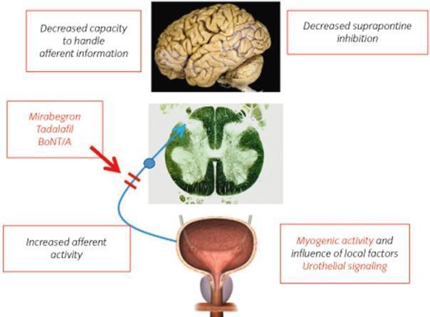
A pathophysiology diagram illustrating the neural pathways and pharmacological targets associated with overactive bladder (OAB). The visual comprises a clinical photograph of a human brain, a histological cross-section of the spinal cord, and an anatomical illustration of the urinary bladder. A blue line indicates the ascending afferent signal pathway originating from the bladder, where text boxes highlight 'increased afferent activity,' 'myogenic activity,' and 'urothelial signaling' as local drivers of dysfunction. The pathway ascends through the spinal cord toward the brain. Central nervous system involvement is annotated with 'decreased suprapontine inhibition' and a 'decreased capacity to handle afferent information,' explaining the loss of cortical control over voiding. Pharmacological intervention is represented by a red arrow pointing to a block in the afferent pathway, listing therapeutic agents including Mirabegron, Tadalafil, and Botulinum neurotoxin A (BoNT/A). This diagram serves as an educational tool for urology and neurology, detailing the communication between the peripheral organ and the central nervous system in lower urinary tract symptoms.
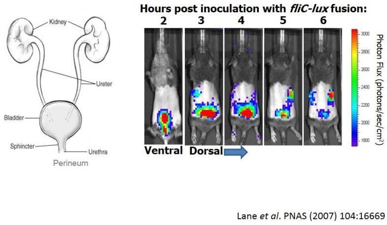
Educational panel illustrating the progression of an ascending urinary tract infection (UTI) using bioluminescence imaging in a murine model. The left panel contains an anatomical diagram of the human urinary tract, labeling the kidneys, ureters, bladder, sphincter, urethra, and perineum. The right panel displays a time-course series of whole-animal bioluminescence imaging at 2, 3, 4, 5, and 6 hours post-inoculation with uropathogenic E. coli (UPEC) strain CFT073 containing a fliC-lux fusion. At 2 hours (ventral view), the light signal (photon flux) is concentrated in the bladder region. From 3 to 4 hours (dorsal view), the signal intensifies, indicating robust flagellin gene expression. By 5 and 6 hours, the signal migrates superiorly, demonstrating the ascent of bacteria through the ureters to the bilateral kidneys. A color-coded scale indicates photon flux intensity, ranging from 1800 (purple/blue) to 3000 (red) photons/sec/cm². The image illustrates bacterial virulence, gene expression during infection, and the transition from cystitis to pyelonephritis.
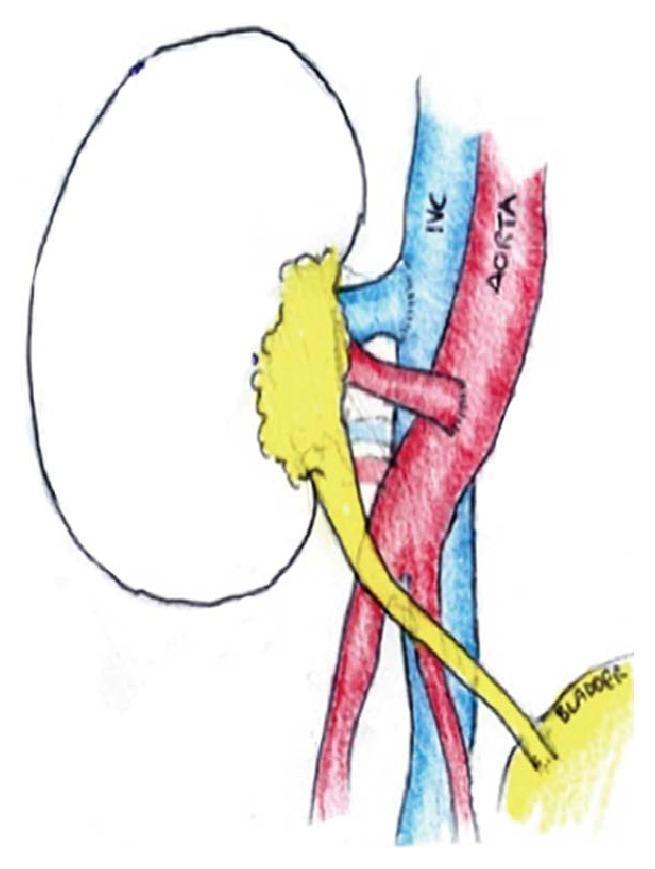
This medical schematic diagram illustrates a kidney transplantation procedure, focusing on the vascular and urinary tract reconstruction. The diagram depicts the donor kidney with its hilum oriented toward the recipient's major vessels. The vascular anastomosis is shown with the donor renal artery (in red) connected to the distal abdominal aorta and the donor renal vein (in blue) connected to the inferior vena cava (IVC). In this specific clinical scenario, the anatomical arrangement reflects a midline laparotomy approach for implantation onto the great vessels rather than the standard iliac fossa placement, often necessitated by complex recipient vascular anatomy. The urinary tract reconstruction is demonstrated by a yellow tubular structure representing the ureter, extending from the renal pelvis and draining into the bladder, labeled at the bottom right. The illustration serves as an educational tool for understanding heterotopic kidney transplant techniques and surgical anatomy in cases of congenital vascular abnormalities.
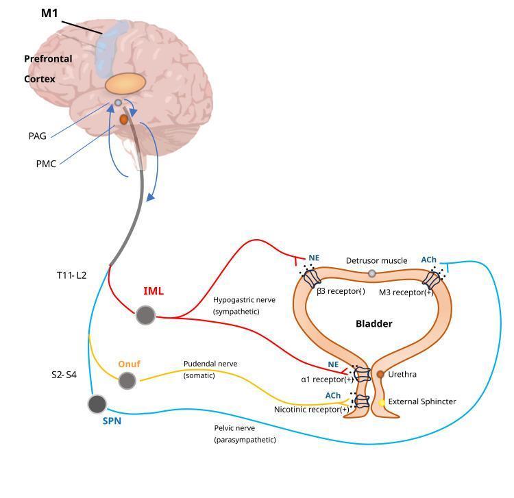
This medical illustration depicts the neural control of the lower urinary tract, specifically the micturition reflex and voluntary bladder control. It shows the hierarchical integration between the brain (primary motor cortex M1, prefrontal cortex, periaqueductal gray PAG, and pontine micturition center PMC) and the spinal cord. The diagram illustrates three distinct pathways: 1) The sympathetic pathway (red), originating from the T11-L2 intermediolateral cell column (IML) via the hypogastric nerve, releasing norepinephrine (NE) to inhibit the detrusor muscle (beta-3 receptors) and contract the bladder neck (alpha-1 receptors). 2) The parasympathetic pathway (blue), originating from the S2-S4 sacral parasympathetic nucleus (SPN) via the pelvic nerve, releasing acetylcholine (ACh) to contract the detrusor muscle (M3 receptors). 3) The somatic motor pathway (yellow), originating from Onuf's nucleus (S2-S4) via the pudendal nerve, releasing ACh to contract the external urethral sphincter (nicotinic receptors). This diagram serves as a pathophysiology and anatomical guide to neurogenic bladder and the physiological mechanisms of urination.
Urinary Tract Infection (UTI) - Comprehensive Overview
1. Definition & Terminology
| Term | Definition |
|---|---|
| Bacteriuria | >10⁵ organisms/mL in urine |
| Asymptomatic bacteriuria (ASB) | Bacteriuria without symptoms - usually does NOT require treatment |
| Cystitis | Infection with lower urinary tract symptoms (frequency, dysuria, strangury) |
| Pyelonephritis | Upper tract infection with parenchymal infiltration - fever, loin pain, systemic upset |
| Urosepsis | Systemic sepsis originating from the urinary tract |
Important: Bacteriuria alone ≠ UTI. Symptoms must be present (exceptions: pregnancy, pre-urologic procedures). - Harrison's Principles of Internal Medicine 22E
2. Classification
Uncomplicated vs Complicated UTI
| Feature | Uncomplicated | Complicated |
|---|---|---|
| Site | Bladder only | Bladder + beyond (prostate, kidney, blood) |
| Setting | Non-catheterized, no structural anomaly | Catheter-associated, structural/functional abnormalities |
| Typical patient | Healthy woman | Any patient with complicating host factors |
| Examples | Acute cystitis in young woman | Pyelonephritis, CAUTI, obstructed system, pregnancy |
- Urologic obstruction (strictures, tumors, diverticula, BPH, urolithiasis)
- Functional abnormalities (neurogenic bladder, VUR, cystocele)
- Urologic instrumentation (catheters, stents, nephrostomy tubes, cystoscopy)
- Metabolic/congenital disease (polycystic kidneys, medullary sponge kidney)
- Immunosuppression (renal transplant)
By Anatomical Location
- Lower UTI: Cystitis, urethritis, prostatitis
- Upper UTI: Pyelonephritis, renal abscess
Recurrent UTI
- Relapse: Same organism within 2 weeks - suggests inadequate treatment or sequestered focus (e.g., prostate)
- Reinfection: Different organism or same organism after >2 weeks
3. Epidemiology
- UTI is the most common bacterial infection overall
- More than 50% of all women will experience at least one UTI in their lifetime; 2-10% have at least one episode per year
- Rare in young men; increases sharply after age 65 due to BPH/prostatitis
- ASB prevalence rises from 1-2% in schoolgirls → 3-5% in premenopausal women → 10-20% in healthy postmenopausal women → 40-50% in elderly institutionalized women
- About 80% of hospital-acquired UTIs are catheter-associated (CAUTI)
- UTI is the most common source of septic shock admissions in the US, with 10-20% mortality
4. Pathophysiology & Microbiology
Route of Infection
- Ascending route (most common): Perineal/rectal bacteria → urethra → bladder → ureter → kidney
- Hematogenous seeding is rare (e.g., S. aureus bacteremia → renal abscess)
Uropathogens
| Organism | Notes |
|---|---|
| Escherichia coli | 80-85% of uncomplicated UTIs; expresses adhesins (type 1 fimbriae, P-fimbriae), toxins, iron-scavenging proteins |
| Staphylococcus saprophyticus | 2nd most common in young sexually active women |
| Klebsiella pneumoniae | More common in complicated/hospital UTIs |
| Proteus mirabilis | Associated with urinary stones (urease-producing) |
| Enterococcus faecalis | Complicated UTIs, catheter-associated |
| Pseudomonas aeruginosa | Hospital-acquired, highly resistant; most P. aeruginosa UTIs are complicated |
Virulence Factors
- Adhesins (fimbriae): Allow attachment to uroepithelium, resisting micturition
- Type 1 fimbriae: Bind uroplakin receptors on bladder umbrella cells → triggers invasion
- P-fimbriae: Bind P-blood group antigen glycolipids on renal tubular epithelium → pyelonephritis
- Toxins (hemolysin, cytotoxic necrotizing factor 1): Cause cell damage
- Iron acquisition (siderophores): Overcome iron-limited urinary environment
- Uromodulin (Tamm-Horsfall protein): The most abundant urinary protein - forms filaments that antagonize uropathogen adhesion and facilitate clearance by micturition (a host defense)
Host Defense Factors
- Regular micturition flushing
- Urine pH, osmolality
- Secretory IgA
- Bladder mucosal glycosaminoglycan layer
- Uromodulin
5. Clinical Features
Lower UTI (Cystitis)
- Dysuria, frequency, urgency
- Suprapubic discomfort/tenderness
- Hematuria (gross or microscopic)
- NO fever, NO systemic signs
- New-onset dysuria + frequency + urgency without vaginal discharge has a positive predictive value of 90% for acute cystitis
Upper UTI (Pyelonephritis)
- Fever (>38°C), rigors
- Flank/loin pain and tenderness (costovertebral angle tenderness - CVAT)
- Nausea, vomiting, malaise
- May have preceding lower UTI symptoms
- In pregnancy: right-sided predominance (due to ureteric compression by gravid uterus)
- Can progress to urosepsis, bacteremia, septic shock
Red Flags / Indicators for Complicated UTI
- Symptoms >4-6 days
- Elderly (risk for bacteremia)
- Ill-appearing / signs of pyelonephritis
- Pregnant women
- Known chronic/recurrent renal infection
- Anatomic urologic abnormalities
- Suspected obstruction (stones, BPH)
- Diabetes, sickle cell, cancer, immunosuppression
- Recent hospitalization or urinary instrumentation
6. Diagnosis & Investigations
Urinalysis
- Pyuria (>10 WBC/hpf): Most sensitive marker of UTI
- Nitrite: Indicates gram-negative bacteriuria (not reliable for Enterococcus, Staph, Candida)
- Leukocyte esterase: Proxy for pyuria
- Microscopy: WBC casts in pyelonephritis; bacteria on Gram stain
- Combined leukocyte esterase + nitrite has good sensitivity for uncomplicated UTI
Urine Culture - Quantitative Interpretation
| Clinical Setting | Significant Colony Count |
|---|---|
| Asymptomatic bacteriuria | ≥10⁵ CFU/mL (×2 consecutive for women) |
| Acute uncomplicated cystitis | ≥10² CFU/mL of E. coli or S. saprophyticus |
| Acute uncomplicated pyelonephritis | ≥10⁴ CFU/mL (95% have ≥10⁵) |
| Complicated UTI | ≥10⁵ CFU/mL |
| In-and-out catheter specimen | ≥10² CFU/mL |
| Suprapubic/percutaneous aspiration | Any organisms isolated |
Culture is NOT required for uncomplicated cystitis in young women; it IS required for complicated UTI, pyelonephritis, pregnancy, treatment failure, and recurrent UTI.
Blood Tests (for pyelonephritis/urosepsis)
- FBC: Leukocytosis
- CRP: Elevated in pyelonephritis (NOT in simple cystitis)
- Blood cultures: In all suspected urosepsis cases
- Creatinine/electrolytes: Assess renal function
Imaging
| Indication | Modality |
|---|---|
| Urosepsis (emergent) | CT abdomen/pelvis with IV contrast (first line) |
| Suspected obstruction | CT KUB or ultrasound |
| Rapid bedside assessment | Ultrasound (can detect hydronephrosis) |
| Concern for gas-forming infection | CT preferred (MRI misses gas/small stones) |
| VUR assessment | Voiding cystourethrogram (VCUG) |
- Imaging is indicated in: urosepsis, suspected obstruction, treatment failure, early relapse after pyelonephritis, functional/anatomic abnormalities
7. Treatment
Uncomplicated Cystitis (Women)
| Agent | Regimen | Notes |
|---|---|---|
| Fosfomycin | 3 g single dose (oral) | Excellent for uncomplicated cystitis; minimal resistance |
| Nitrofurantoin | 100 mg (MR) BD × 5 days | First-line; NOT for pyelonephritis (poor tissue penetration) |
| Trimethoprim-sulfamethoxazole (TMP-SMX) | 160/800 mg BD × 3 days | Use only if local resistance <20% |
| Trimethoprim | 200 mg once daily × 7 days | Alternative |
| Pivmecillinam | 400 mg BD × 3-7 days | Used in Europe; effective |
Fluoroquinolones (ciprofloxacin, levofloxacin) are NOT recommended as first-line for uncomplicated UTI. They achieve excellent renal/prostate tissue levels and should be reserved for complicated UTI and pyelonephritis (or when first-line agents fail/are contraindicated).
Pyelonephritis (Outpatient - uncomplicated)
- Ciprofloxacin 500 mg BD × 7 days OR
- Levofloxacin 750 mg OD × 5 days OR
- TMP-SMX 160/800 mg BD × 14 days (if organism sensitive)
- Oral fluoroquinolones are preferred for outpatient pyelonephritis due to excellent tissue penetration
Pyelonephritis (Inpatient/Severe)
- IV ceftriaxone, IV ciprofloxacin, IV levofloxacin, or aminoglycoside
- Duration: 7-14 days (up to 2 weeks for severe cases)
- For abscesses ≥3 cm: drain + 4-6 weeks antibiotics
Complicated UTI / CAUTI
- 7-14 days treatment
- Remove/replace catheter if possible
- Culture-guided therapy essential
- Agents: fluoroquinolones, cephalosporins, piperacillin-tazobactam, carbapenems (based on organism/sensitivity)
Treatment of Resistant Organisms (P. aeruginosa)
- MDR Pseudomonas UTI: Ceftazidime-avibactam, ceftolozane-tazobactam, imipenem-cilastatin-relebactam, or cefiderocol
- Single-dose aminoglycoside (e.g., amikacin 15 mg/kg) acceptable for cystitis only
8. Special Populations
Pregnancy
- Screen ALL pregnant women for ASB at 12-16 weeks gestation (USPSTF Grade A)
- Up to 30% of women with ASB will develop pyelonephritis if untreated; treatment reduces this by >80%
- ASB/lower UTI in pregnancy: treat for 7-10 days (not just 3-day courses)
- Safe antibiotics in pregnancy: Cephalosporins (e.g., cephalexin 500 mg BD), nitrofurantoin (avoid at term - risk of neonatal hemolysis), amoxicillin; TMP-SMX (avoid in 1st trimester - folate antagonism; avoid in 3rd trimester - neonatal kernicterus)
- Avoid: Fluoroquinolones (fetal cartilage toxicity), tetracyclines
- Acute pyelonephritis in pregnancy: Hospitalize + IV antibiotics (ampicillin + gentamicin); risks include maternal sepsis, preterm labor, permanent renal injury
Elderly Patients
- ASB is common (10-20% community-dwelling women; 35-40% men in nursing homes)
- Do not treat ASB in elderly - associated with antibiotic resistance and adverse drug effects
- Atypical presentations: confusion/delirium (but note - UTI causing isolated delirium without urinary symptoms is overdiagnosed)
- Adjust antibiotic doses for renal function (avoid nitrofurantoin if GFR <30)
Catheter-Associated UTI (CAUTI)
- Most common hospital-acquired infection; 80% of nosocomial UTIs
- Organism spectrum is broader: Candida spp., Pseudomonas, Enterococcus, Klebsiella
- Remove/replace catheter promptly
- Treat only if symptomatic (fever, flank pain, rigors, suprapubic tenderness) - not just bacteriuria
Men
- UTI in a man should be considered complicated until an underlying genitourinary abnormality is excluded
- Prostatitis (ABP or CBP) must be excluded - requires prolonged therapy penetrating prostate tissue
- Acute bacterial prostatitis: peaks in men 20-40 years and again >60 years; very tender swollen prostate; treat with fluoroquinolone × 4-6 weeks
Pediatric UTI
- Female gender, high-grade kidney dilation, intact foreskin (in boys): key risk factors
- Evaluate for vesicoureteral reflux (VUR) after first febrile UTI in young children
- Risk increases linearly with degree of hydronephrosis (grade 1: 4%, grade 4: 40% UTI incidence)
9. Recurrent UTI - Prevention
| Strategy | Details |
|---|---|
| Behavioural | Adequate hydration, void after intercourse, wipe front-to-back |
| Cranberry products | Modest evidence for prevention; shown in 2024 network meta-analysis (PMID 39030132) |
| Postcoital prophylaxis | Single-dose TMP-SMX or nitrofurantoin after intercourse |
| Continuous low-dose prophylaxis | TMP-SMX SS once daily × 6-12 months |
| Patient-initiated (self-start) therapy | Short course at symptom onset - for selected motivated patients |
| Topical vaginal estrogen | Postmenopausal women - restores lactobacillus flora |
10. Differential Diagnosis
- Dysuria DDx: Sexually transmitted infections (gonorrhoea, chlamydia, Mycoplasma), vulvovaginal candidiasis, urethral syndrome, interstitial cystitis, herpes genitalis
- Flank pain DDx: Pyelonephritis, renal colic/nephrolithiasis, appendicitis (right), cholecystitis (right), pelvic inflammatory disease, premature labor (in pregnancy)
- Pyuria without bacteriuria ("sterile pyuria"): TB, chlamydia urethritis, interstitial nephritis, bladder tumor
Summary Table
| Feature | Uncomplicated Cystitis | Pyelonephritis |
|---|---|---|
| Symptoms | Dysuria, frequency, urgency | Fever, CVAT, systemic upset |
| CRP | Normal | Elevated |
| Blood culture | Not needed | Obtain |
| Imaging | Not needed | CT if severe/urosepsis |
| First-line Rx | Fosfomycin / Nitrofurantoin / TMP-SMX | Ciprofloxacin / Levofloxacin |
| Duration | 1-7 days | 7-14 days |