Explain in detail about all types of edema with its example with detailed explaination.

Reading File
Finding Sources
Finding Sources
Reading File
Reading File
Reading File
Reading File
Reading File
Reading File
Finding Sources
Finding Sources
Reading File
Finding Sources
Searching Images

pitting edema lower extremity legs

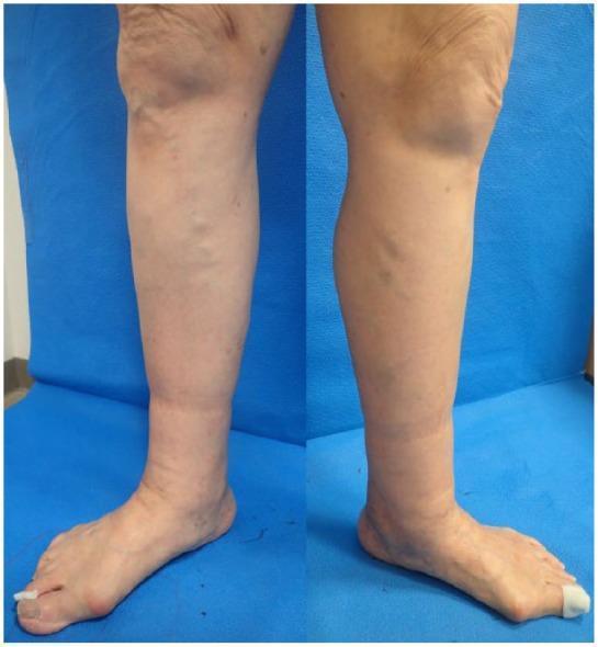
Clinical photograph of the lower extremities of a 79-year-old female presenting with bilateral lower leg edema. The image displays a side-by-side view of the left and right legs from the knee to the foot. Key findings include diffuse non-pitting edema primarily localized below the knees, most prominent in the calf and ankle regions, resulting in an increased limb circumference and a taut, shiny skin appearance. Prominent varicose veins are visible along the medial aspect of both legs, with greater severity noted on the left. The clinical presentation is consistent with CEAP classification grade C3 (venous edema) and secondary lymphedema, indicated by the loss of normal anatomical contours at the ankles. These visual features are characteristic of chronic venous insufficiency and comorbid lymphatic drainage impairment (phlebolymphedema). This material is intended for instruction on vascular assessment, clinical grading of chronic venous disease, and the differential diagnosis of lower extremity swelling.
Clinical photograph of the lower extremities of a 79-year-old female presenting with bilateral lower leg edema. The image displays a side-by-side view of the left and right legs from the knee to the foot. Key findings include diffuse non-pitting edema primarily localized below the knees, most prominent in the calf and ankle regions, resulting in an increased limb circumference and a taut, shiny skin appearance. Prominent varicose veins are visible along the medial aspect of both legs, with greater severity noted on the left. The clinical presentation is consistent with CEAP classification grade C3 (venous edema) and secondary lymphedema, indicated by the loss of normal anatomical contours at the ankles. These visual features are characteristic of chronic venous insufficiency and comorbid lymphatic drainage impairment (phlebolymphedema). This material is intended for instruction on vascular assessment, clinical grading of chronic venous disease, and the differential diagnosis of lower extremity swelling.

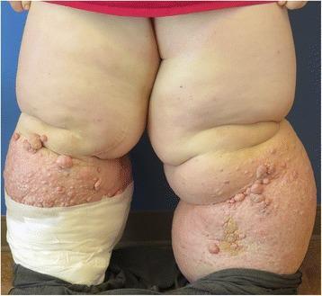
This clinical photograph displays a posterior view of Stage III bilateral lower extremity lymphedema in a young adult. Both legs exhibit massive, non-pitting edema resulting in profound distortion of normal anatomical contour and significant limb enlargement. The skin of the right lower extremity shows advanced integumentary changes, including hyperkeratosis, irregular nodular formations, and verrucous papules consistent with lymphostatic verrucosis. Patches of brownish-red discoloration and textural thickening indicate chronic dermal fibrosis and inflammation. The left lower extremity is partially covered with white compression bandaging or dressings, used for decongestive therapy. Deep skin folds are visible at the knees and thighs, representing areas prone to intertrigo. This case illustrates primary lymphedema praecox progressing to elephantiasis, a severe stage of lymphatic failure characterized by irreversible tissue changes and significant functional morbidity.
This clinical photograph displays a posterior view of Stage III bilateral lower extremity lymphedema in a young adult. Both legs exhibit massive, non-pitting edema resulting in profound distortion of normal anatomical contour and significant limb enlargement. The skin of the right lower extremity shows advanced integumentary changes, including hyperkeratosis, irregular nodular formations, and verrucous papules consistent with lymphostatic verrucosis. Patches of brownish-red discoloration and textural thickening indicate chronic dermal fibrosis and inflammation. The left lower extremity is partially covered with white compression bandaging or dressings, used for decongestive therapy. Deep skin folds are visible at the knees and thighs, representing areas prone to intertrigo. This case illustrates primary lymphedema praecox progressing to elephantiasis, a severe stage of lymphatic failure characterized by irreversible tissue changes and significant functional morbidity.

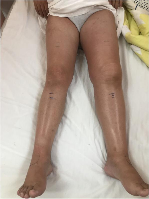
This clinical photograph shows a front-on view of a patient’s lower extremities while lying supine. The image demonstrates relatively symmetrical anatomy with no gross muscular atrophy or significant pitting edema visible. Both legs exhibit minor skin irregularities, including subtle variations in pigmentation and prominent superficial veins in the lower legs. Notably, surgical or diagnostic skin markings (short horizontal lines) are present on the bilateral mid-thighs and shins, and a curved marking is visible on the dorsal aspect of the left ankle. Clinically, these findings are consistent with a pre-diagnostic physical examination for suspected vascular pathology. The left lower extremity appears to have slightly more pronounced skin tone variation compared to the right. In the context of the patient's history, these physical findings precede diagnostic confirmation of a left external iliac vein occlusion, likely related to extrinsic compression or pregnancy-related venous changes such as May-Thurner syndrome.

This clinical photograph shows a front-on view of a patient’s lower extremities while lying supine. The image demonstrates relatively symmetrical anatomy with no gross muscular atrophy or significant pitting edema visible. Both legs exhibit minor skin irregularities, including subtle variations in pigmentation and prominent superficial veins in the lower legs. Notably, surgical or diagnostic skin markings (short horizontal lines) are present on the bilateral mid-thighs and shins, and a curved marking is visible on the dorsal aspect of the left ankle. Clinically, these findings are consistent with a pre-diagnostic physical examination for suspected vascular pathology. The left lower extremity appears to have slightly more pronounced skin tone variation compared to the right. In the context of the patient's history, these physical findings precede diagnostic confirmation of a left external iliac vein occlusion, likely related to extrinsic compression or pregnancy-related venous changes such as May-Thurner syndrome.

Searching Images

pulmonary edema chest X-ray

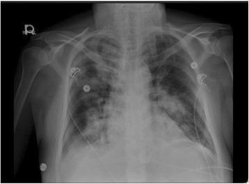
A portable anteroposterior (AP) chest X-ray demonstrating a clinical state of pulmonary edema. The image shows bilateral, patchy reticular opacities with a predominant distribution in the hilar and perihilar regions of both lungs. There is no evidence of cardiomegaly, as the cardiothoracic ratio appears within normal limits. The bony thorax, including the clavicles and ribs, and the great vessel silhouettes appear unremarkable. External medical artifacts, including ECG leads and wires, are visible across the chest wall. The visual findings are consistent with pulmonary congestion, often associated with a depressed ejection fraction or acute heart failure. This diagnostic image serves as a teaching tool for recognizing interstitial and alveolar patterns in the context of cardiovascular and respiratory medicine.
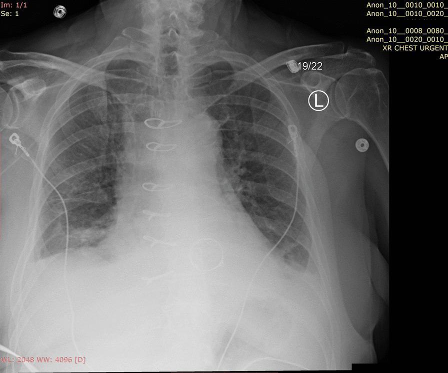
A portable anteroposterior (AP) chest X-ray demonstrating a clinical state of pulmonary edema. The image shows bilateral, patchy reticular opacities with a predominant distribution in the hilar and perihilar regions of both lungs. There is no evidence of cardiomegaly, as the cardiothoracic ratio appears within normal limits. The bony thorax, including the clavicles and ribs, and the great vessel silhouettes appear unremarkable. External medical artifacts, including ECG leads and wires, are visible across the chest wall. The visual findings are consistent with pulmonary congestion, often associated with a depressed ejection fraction or acute heart failure. This diagnostic image serves as a teaching tool for recognizing interstitial and alveolar patterns in the context of cardiovascular and respiratory medicine.

Anteroposterior (AP) chest X-ray demonstrating features of acute pulmonary edema in a clinical setting. The lung fields show bilateral, symmetric, diffuse hazy opacities with a predominant distribution in the lower zones. Increased interstitial markings and patchy airspace opacities suggest alveolar fluid accumulation. The cardiomediastinal silhouette appears slightly enlarged. Several medical monitoring and support devices are present, including multiple external ECG leads and wires superimposed on the thorax, a right-sided central venous catheter with the tip projecting toward the superior vena cava, and a left-sided thin tubular structure consistent with a drainage tube or monitoring line. The bony structures, including the clavicles, ribs, and scapulae, are intact. This imaging is characteristic of pulmonary vascular congestion and fluid overload, often seen in the context of acute heart failure or severe valvular dysfunction such as mitral regurgitation.
Anteroposterior (AP) chest X-ray demonstrating features of acute pulmonary edema in a clinical setting. The lung fields show bilateral, symmetric, diffuse hazy opacities with a predominant distribution in the lower zones. Increased interstitial markings and patchy airspace opacities suggest alveolar fluid accumulation. The cardiomediastinal silhouette appears slightly enlarged. Several medical monitoring and support devices are present, including multiple external ECG leads and wires superimposed on the thorax, a right-sided central venous catheter with the tip projecting toward the superior vena cava, and a left-sided thin tubular structure consistent with a drainage tube or monitoring line. The bony structures, including the clavicles, ribs, and scapulae, are intact. This imaging is characteristic of pulmonary vascular congestion and fluid overload, often seen in the context of acute heart failure or severe valvular dysfunction such as mitral regurgitation.

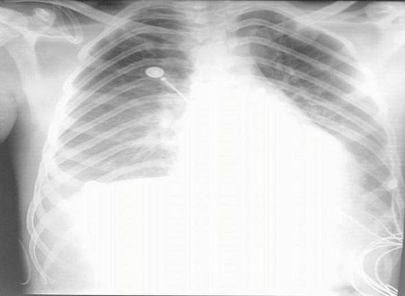
Frontal chest X-ray (radiography) demonstrating classic findings of cardiogenic pulmonary edema and cardiomegaly. The image shows a markedly enlarged cardiac silhouette with indistinct borders, occupying a significant portion of the thoracic cavity. Diffuse, bilateral hazy opacities are present throughout the lung fields, appearing most dense in the perihilar ('bat-wing' distribution) and basal regions, which obscure the normal pulmonary vascular markings. Blunting of the costophrenic angles is visible, particularly on the right, suggesting associated pleural effusions. Several medical devices are present, including a radiopaque central venous catheter entering from the right and terminating near the mediastinum, as well as external electrocardiogram (ECG) leads and wires projected over the chest wall. The clinical significance of these findings relates to acute heart failure or myocarditis, causing fluid extravasation into the lung parenchyma and pleural space. This image serves as a teaching tool for identifying radiological signs of congestive heart failure and cardiomegaly in an acute clinical setting.

Frontal chest X-ray (radiography) demonstrating classic findings of cardiogenic pulmonary edema and cardiomegaly. The image shows a markedly enlarged cardiac silhouette with indistinct borders, occupying a significant portion of the thoracic cavity. Diffuse, bilateral hazy opacities are present throughout the lung fields, appearing most dense in the perihilar ('bat-wing' distribution) and basal regions, which obscure the normal pulmonary vascular markings. Blunting of the costophrenic angles is visible, particularly on the right, suggesting associated pleural effusions. Several medical devices are present, including a radiopaque central venous catheter entering from the right and terminating near the mediastinum, as well as external electrocardiogram (ECG) leads and wires projected over the chest wall. The clinical significance of these findings relates to acute heart failure or myocarditis, causing fluid extravasation into the lung parenchyma and pleural space. This image serves as a teaching tool for identifying radiological signs of congestive heart failure and cardiomegaly in an acute clinical setting.

Searching Images

cerebral brain edema CT scan

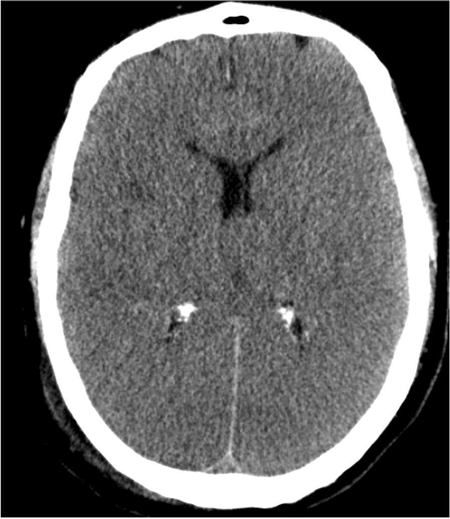
This diagnostic axial non-contrast computed tomography (CT) scan of the brain demonstrates significant signs of global cerebral edema. Key findings include diffuse cortical hypodensity across both cerebral hemispheres and a marked loss of grey-white matter differentiation, a hallmark of severe hypoxic-ischemic injury. The image shows prominent gyral effacement (flattening of the brain's convolutions) and the obliteration of the subarachnoid spaces and basal cisterns, indicating increased intracranial pressure. The ventricular system, particularly the lateral ventricles, appears slit-like and compressed. Hyperdense calcifications are visible in the choroid plexus of the posterior horns of the lateral ventricles, which is a common physiological finding. This CT appearance is clinically significant for neuroprognostication in cases of post-cardiac arrest or global anoxia, where the 'pseudo-delta sign' or generalized brain swelling reflects poor clinical outcomes.
This diagnostic axial non-contrast computed tomography (CT) scan of the brain demonstrates significant signs of global cerebral edema. Key findings include diffuse cortical hypodensity across both cerebral hemispheres and a marked loss of grey-white matter differentiation, a hallmark of severe hypoxic-ischemic injury. The image shows prominent gyral effacement (flattening of the brain's convolutions) and the obliteration of the subarachnoid spaces and basal cisterns, indicating increased intracranial pressure. The ventricular system, particularly the lateral ventricles, appears slit-like and compressed. Hyperdense calcifications are visible in the choroid plexus of the posterior horns of the lateral ventricles, which is a common physiological finding. This CT appearance is clinically significant for neuroprognostication in cases of post-cardiac arrest or global anoxia, where the 'pseudo-delta sign' or generalized brain swelling reflects poor clinical outcomes.

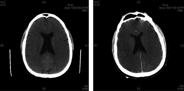
This Comparison Chart features two side-by-side axial non-contrast brain CT scans demonstrating the progression of acute cerebral edema over a short time interval. The initial scan (left) shows preserved sulcal spaces and identifiable gray-white matter differentiation. The follow-up scan (right) reveals significant interval worsening characterized by generalized loss of gray-white matter differentiation and effacement of the cortical sulci, resulting in a 'featureless' appearance of the brain parenchyma. Furthermore, there is a visible decrease in overall tissue density (hypodensity) and compression of the lateral ventricles, consistent with global mass effect from diffuse cerebral edema. These radiological findings are hallmark indicators of severe hypoxic-ischemic brain injury or global metabolic insult. The image serves as a critical diagnostic example for neurology and radiology students to identify radiographic signs of elevated intracranial pressure and evolving brain death patterns.

This Comparison Chart features two side-by-side axial non-contrast brain CT scans demonstrating the progression of acute cerebral edema over a short time interval. The initial scan (left) shows preserved sulcal spaces and identifiable gray-white matter differentiation. The follow-up scan (right) reveals significant interval worsening characterized by generalized loss of gray-white matter differentiation and effacement of the cortical sulci, resulting in a 'featureless' appearance of the brain parenchyma. Furthermore, there is a visible decrease in overall tissue density (hypodensity) and compression of the lateral ventricles, consistent with global mass effect from diffuse cerebral edema. These radiological findings are hallmark indicators of severe hypoxic-ischemic brain injury or global metabolic insult. The image serves as a critical diagnostic example for neurology and radiology students to identify radiographic signs of elevated intracranial pressure and evolving brain death patterns.

Searching Images

angioedema face lips swelling

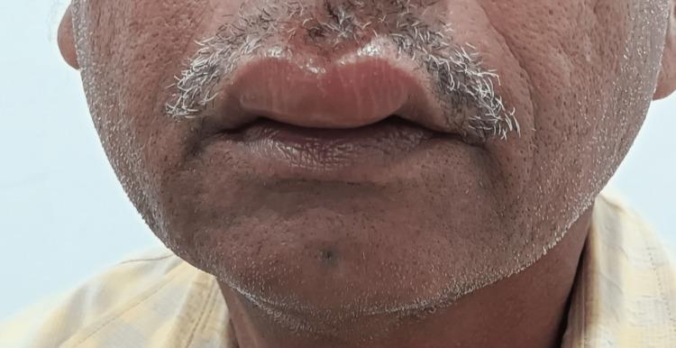
This clinical photograph displays a front-facing view of a patient's lower face, highlighting marked swelling of the lips (cheilitis). The upper lip exhibits significant edematous distension with a smooth, shiny surface and erythematous discoloration, which is characteristic of angioedema. The lower lip also shows moderate swelling. The cutaneous area immediately below the lower lip and extending toward the chin appears mildly inflamed with subtle soft tissue edema. This presentation is consistent with a Cutaneous Adverse Drug Reaction (CADR), specifically drug-induced angioedema secondary to the cephalosporin antibiotic cefixime. The image illustrates the typical localized, non-pitting edema of the deep dermis and subcutaneous tissues frequently seen in Type I hypersensitivity reactions or other drug-mediated inflammatory responses.
This clinical photograph displays a front-facing view of a patient's lower face, highlighting marked swelling of the lips (cheilitis). The upper lip exhibits significant edematous distension with a smooth, shiny surface and erythematous discoloration, which is characteristic of angioedema. The lower lip also shows moderate swelling. The cutaneous area immediately below the lower lip and extending toward the chin appears mildly inflamed with subtle soft tissue edema. This presentation is consistent with a Cutaneous Adverse Drug Reaction (CADR), specifically drug-induced angioedema secondary to the cephalosporin antibiotic cefixime. The image illustrates the typical localized, non-pitting edema of the deep dermis and subcutaneous tissues frequently seen in Type I hypersensitivity reactions or other drug-mediated inflammatory responses.

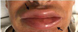
This clinical photograph displays a close-up view of the lower face of an adult patient exhibiting severe, isolated angioedema. The primary finding is significant bilateral edema of both the upper and lower lips, which appear markedly enlarged and erythematous. The arrows in the image highlight the boundaries of the swelling at the vermilion border. The lip surface appears taut and smooth, consistent with acute interstitial fluid accumulation. Notably, the surrounding perioral skin, chin, and nose show no signs of similar swelling or inflammation, indicating the localized nature of the reaction. This visual serves as a classic representation of acute angioedema, a condition often associated with allergic reactions, ACE-inhibitor use, or environmental exposures such as Karenia brevis (red tide) as indicated in the clinical context. The image is intended for medical education in dermatology and emergency medicine to teach the identification of localized soft tissue edema without systemic cutaneous involvement.

This clinical photograph displays a close-up view of the lower face of an adult patient exhibiting severe, isolated angioedema. The primary finding is significant bilateral edema of both the upper and lower lips, which appear markedly enlarged and erythematous. The arrows in the image highlight the boundaries of the swelling at the vermilion border. The lip surface appears taut and smooth, consistent with acute interstitial fluid accumulation. Notably, the surrounding perioral skin, chin, and nose show no signs of similar swelling or inflammation, indicating the localized nature of the reaction. This visual serves as a classic representation of acute angioedema, a condition often associated with allergic reactions, ACE-inhibitor use, or environmental exposures such as Karenia brevis (red tide) as indicated in the clinical context. The image is intended for medical education in dermatology and emergency medicine to teach the identification of localized soft tissue edema without systemic cutaneous involvement.

Finding Sources
Finding Sources
Loading Image
Loading Image
I now have all the material needed for a comprehensive, well-illustrated answer. Let me compose the full detailed response.

Edema: A Detailed Guide to All Types

Edema is the abnormal accumulation of interstitial fluid within tissues. Approximately 60% of lean body weight is water; two-thirds is intracellular and most of the remaining is interstitial. Edema occurs when normal Starling forces - which govern fluid movement between the vascular and interstitial spaces - are disrupted.
Key concept: Fluid leaving capillaries (driven by hydrostatic pressure) is normally balanced by fluid re-entering (driven by colloid osmotic/oncotic pressure). The small net outflow is drained by lymphatics back to the bloodstream. When any of these forces go out of balance, edema results.
  • Transudate - protein-poor fluid; results from hydrostatic pressure increase or osmotic pressure decrease
  • Exudate - protein-rich fluid; results from increased vascular permeability (inflammation)
Severe generalized edema with profound subcutaneous swelling and fluid in body cavities is called anasarca. Fluid in specific cavities has specific names: hydrothorax (pleural), hydropericardium (pericardial), hydroperitoneum/ascites (peritoneal).

Pathophysiology: The Starling Forces

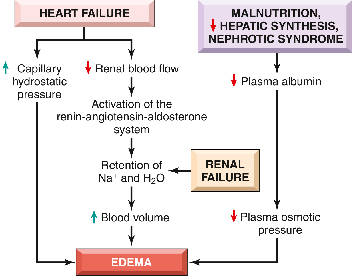
The diagram below (from Robbins & Kumar Basic Pathology) shows how heart failure, renal failure, and hypoproteinemia converge on edema through different but interconnected pathways:
Pathways leading to systemic edema - Robbins

Classification of Edema

Edema is classified in two broad ways:
By DistributionBy Mechanism
LocalizedIncreased hydrostatic pressure
GeneralizedReduced plasma osmotic pressure
Increased vascular permeability
Lymphatic obstruction
Na+ and water retention

TYPE 1: Edema due to Increased Hydrostatic Pressure

Mechanism

When venous pressure rises, the outward filtration force at capillaries exceeds the inward oncotic pull. Fluid accumulates in the interstitium.

Subtypes and Examples

A. Congestive Heart Failure (CHF) Edema

The classic and most common cause of generalized edema.
Pathophysiology:
  • Reduced cardiac output → blood pools in the venous system → increased capillary hydrostatic pressure
  • Reduced cardiac output also hypoperfuses the kidneys → activates the renin-angiotensin-aldosterone axis → Na+ and water retention (secondary hyperaldosteronism)
  • Increased blood volume further raises venous pressure → worsening edema - a vicious cycle
Clinical features:
  • Right heart failure → systemic (peripheral) edema: bilateral pitting edema of the ankles/legs when standing; sacral edema when bedridden (dependent edema); ascites; hepatomegaly
  • Left heart failure → pulmonary edema (see Type 6 below)
  • Finger pressure over edematous tissue leaves a finger-shaped depression - pitting edema
Example: A 65-year-old man with ischemic cardiomyopathy develops bilateral ankle swelling that is worse at the end of the day and better in the morning, along with shortness of breath and orthopnea. Chest X-ray shows cardiomegaly. This is classic dependent pitting edema from right heart failure.
Bilateral lower extremity edema with chronic venous insufficiency

B. Deep Venous Thrombosis (DVT) Edema - Localized

Thrombosis in the deep veins of the leg obstructs venous return from that limb only → unilateral leg edema.
Example: A 45-year-old woman on long-haul flight develops unilateral calf swelling, redness, and tenderness. Duplex ultrasound confirms DVT in the popliteal vein. The edema is localized to the affected leg only.

C. Cirrhosis-Related (Portal Hypertension) Edema

In liver cirrhosis, portal hypertension raises hydrostatic pressure in the portal venous bed, causing fluid to weep into the peritoneal cavity (ascites). Simultaneously, reduced hepatic albumin synthesis lowers oncotic pressure, compounding the problem.
Example: A 52-year-old alcoholic male presents with massive abdominal distension (ascites), bilateral ankle edema, and jaundice. Serum albumin is 2.1 g/dL.

TYPE 2: Edema due to Reduced Plasma Osmotic Pressure (Hypoproteinemia)

Mechanism

Albumin is responsible for nearly half of total plasma protein and is the dominant contributor to colloid osmotic (oncotic) pressure. When albumin falls, the inward osmotic force that retains fluid in capillaries weakens, and fluid leaks into the interstitium.
Low albumin → edema + reduced intravascular volume → renal hypoperfusion → secondary hyperaldosteronism → Na+ and water retention. Unfortunately, this Na+ retention does NOT fix the primary protein deficiency and instead worsens the edema.

Examples

A. Nephrotic Syndrome Edema

Glomerular damage allows albumin to pass into the urine (heavy proteinuria >3.5 g/day). Serum albumin falls. The result is classic hypoalbuminemic edema.
Clinical features:
  • Periorbital edema (especially in children) - because loose connective tissue around the eyes fills first
  • Generalized pitting edema
  • Ascites, pleural effusions
  • Proteinuria, hypoalbuminemia, hyperlipidemia, lipiduria (the nephrotic pentad)
Example: A 4-year-old boy develops puffy eyelids every morning, progressing to abdominal swelling and scrotal edema. Urinalysis shows 4+ protein. Serum albumin is 1.8 g/dL. Diagnosis: Minimal change disease / nephrotic syndrome.

B. Liver Disease / Cirrhosis Edema (Hypoproteinemic Component)

Severe liver disease reduces hepatic albumin synthesis → low oncotic pressure → edema and ascites.

C. Protein Malnutrition (Kwashiorkor)

Children with severe protein deficiency (kwashiorkor) develop marked hypoalbuminemia → generalized edema, classically with a distended abdomen.
Example: A malnourished 2-year-old child in a resource-limited setting presents with bilateral pitting leg edema, abdominal distension, thin hair, and skin changes. Serum albumin is very low. This is kwashiorkor.

TYPE 3: Lymphedema (Lymphatic Obstruction)

Mechanism

Lymphatics normally drain the small amount of fluid that is not reabsorbed into capillaries at the venular end. When lymphatics are obstructed or destroyed, protein-rich interstitial fluid accumulates. Chronically, this stimulates fibroblast proliferation and connective tissue deposition → hard, non-pitting edema.

Subtypes

A. Primary Lymphedema

Congenital or developmental failure of lymphatic vessels.
  • Milroy disease - congenital lymphedema present at birth (autosomal dominant)
  • Lymphedema praecox - develops at puberty, most common in young women

B. Secondary Lymphedema

Obstruction of previously normal lymphatics by:
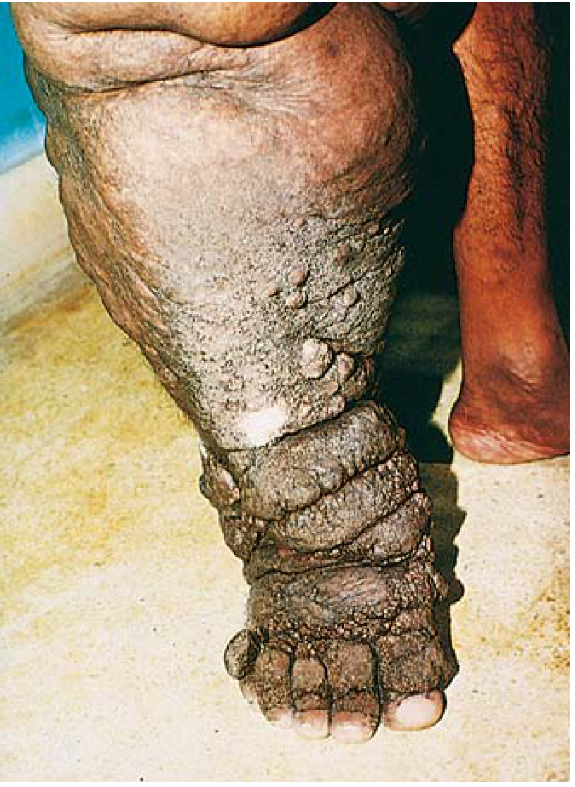
  • Filariasis (Elephantiasis) - the parasitic nematodes Wuchereria bancrofti and Brugia malayi lodge in lymphatics, causing chronic obstruction → massive lymphedema of the lower extremities or scrotum (elephantiasis). A major global health problem in tropical regions.
  • Post-mastectomy - surgical removal of axillary lymph nodes → arm lymphedema (most common cause in developed world)
  • Malignant infiltration - tumor invading lymphatics
  • Post-radiation - fibrosis of lymph channels after radiotherapy
Clinical features of lymphedema:
  • Initially pitting, progresses to non-pitting (brawny) edema
  • Skin becomes thickened, hyperkeratotic, warty (lymphostatic verrucosis)
  • Recurrent cellulitis
Example: A 55-year-old woman treated for breast cancer 3 years ago with axillary node dissection and radiation develops progressive swelling of her left arm that is non-pitting. This is classic secondary lymphedema.
Elephantiasis - the extreme end of lymphedema from filarial infection:
Massive edema and elephantiasis caused by filariasis - Robbins Pathology
Massive edema and elephantiasis of the leg caused by filariasis (Robbins & Kumar Basic Pathology)

TYPE 4: Inflammatory Edema (Increased Vascular Permeability)

Mechanism

In acute and chronic inflammation, cytokines (especially histamine, bradykinin, serotonin, leukotrienes, prostaglandins) cause endothelial cell contraction, widening intercellular junctions. Plasma proteins leak into the interstitium, drawing water with them. The resulting fluid is an exudate - protein-rich (>3 g/dL) and may contain leukocytes.
This is fundamentally different from the other types of edema, which produce a protein-poor transudate.

Examples

A. Acute Inflammation

Any site of infection, trauma, or burn produces local inflammatory edema. The classic cardinal signs of inflammation are: rubor (redness), calor (heat), tumor (swelling = edema), dolor (pain), and functio laesa (loss of function).
Example: A bee sting on the forearm causes local redness, warmth, and swelling within minutes due to histamine-mediated increased vascular permeability. This resolves over 24-48 hours.

B. Angioedema

A specific, potentially life-threatening form of inflammatory/allergic edema affecting the deep dermis and subcutaneous/submucosal tissues.
Subtypes:
  • Allergic angioedema (IgE-mediated) - triggered by allergens (food, drugs, insect venom) → mast cell degranulation → histamine release → permeability increase
  • Drug-induced angioedema - ACE inhibitors cause bradykinin accumulation → angioedema of the face, lips, tongue, larynx. Can be fatal if airway is involved
  • Hereditary angioedema (HAE) - autosomal dominant deficiency of C1-inhibitor → uncontrolled complement activation → bradykinin excess → recurrent episodes of edema in hands, abdomen, face, larynx
Example: A 46-year-old man started on lisinopril for hypertension 2 weeks ago develops sudden swelling of the lips, tongue, and throat - ACE inhibitor-induced angioedema. This is a medical emergency.
Angioedema - acute lip and facial swelling

C. Burn Edema

Severe thermal injury causes massive increase in vascular permeability over the burned area → protein-rich exudate → can cause significant hypovolemia due to plasma loss.

D. Sepsis / SIRS Edema

Systemic inflammatory response causes widespread increased capillary permeability → generalized edema, hypoalbuminemia, hypotension.

TYPE 5: Edema due to Sodium and Water Retention

Mechanism

Excessive retention of Na+ (and water following osmotically) expands intravascular volume → increases hydrostatic pressure AND dilutes plasma proteins → decreases osmotic pressure. Both effects promote edema.
Example: Acute poststreptococcal glomerulonephritis (AGN) - in children, streptococcal throat infection triggers immune complex deposition in glomeruli → acute reduction in GFR → Na+ and water retention → sudden generalized edema, hypertension, hematuria.
Other examples: acute renal failure, certain medications (NSAIDs, corticosteroids, calcium channel blockers which cause ankle edema by arteriolar dilation and secondary Na+ retention).

TYPE 6: Pulmonary Edema

Definition

Accumulation of excess fluid within the lung interstitium and alveoli.

Types

A. Cardiogenic (Hydrostatic) Pulmonary Edema

Left ventricular failure → raised pulmonary venous pressure → fluid transudes from pulmonary capillaries into alveoli.
Pathophysiology:
  • Left ventricular dysfunction → back-pressure in pulmonary veins → pulmonary capillary wedge pressure rises
  • Fluid first accumulates in interstitium (interstitial edema) → then floods alveoli (alveolar edema)
  • The fluid-filled alveoli impair gas exchange → hypoxemia → respiratory failure
Morphology: Lungs are 2-3 times their normal weight; frothy, blood-tinged fluid on section. Microscopically: engorged alveolar capillaries, alveolar septal edema, intra-alveolar hemorrhage. In chronic pulmonary edema: septa are thickened and fibrotic; alveoli contain hemosiderin-laden macrophages ("heart failure cells").
Example: A 70-year-old man with known ischemic heart disease wakes up at 3 AM with sudden severe breathlessness, frothy pink sputum, and cyanosis. He is sat upright. CXR shows bilateral "bat-wing" opacities, cardiomegaly, and pleural effusions. This is acute cardiogenic pulmonary edema (flash pulmonary edema).
Chest X-ray showing cardiogenic pulmonary edema with bat-wing distribution

B. Non-Cardiogenic Pulmonary Edema (ARDS / Increased Permeability)

Acute Respiratory Distress Syndrome (ARDS) - inflammatory injury to the alveolar-capillary barrier causes massive permeability increase → protein-rich exudate floods alveoli.
Causes: Sepsis, aspiration, pneumonia, trauma, pancreatitis, inhaled toxins.
This type differs from cardiogenic pulmonary edema in two key respects:
  1. Pulmonary capillary wedge pressure is normal (heart is not at fault)
  2. The edema fluid is protein-rich (exudate), not a protein-poor transudate
Example: A 35-year-old with gram-negative sepsis from urinary tract infection develops worsening hypoxia, bilateral infiltrates on CXR, and PCWP of 14 mmHg (normal). Diagnosis: sepsis-induced ARDS with non-cardiogenic pulmonary edema.
Portable AP chest X-ray showing bilateral pulmonary edema

TYPE 7: Cerebral Edema

Definition

Accumulation of excess fluid within brain parenchyma. Dangerous because the skull is rigid - even small increases in brain volume raise intracranial pressure (ICP) dramatically.

The Two Principal Types (from Harrison's & Robbins)

A. Vasogenic Edema

Disruption of the blood-brain barrier (BBB) allows protein-rich fluid to shift from the vascular compartment into the brain extracellular spaces.
Mechanism: Tight junctions between cerebral endothelial cells break down → plasma proteins and water leak into white matter.
Causes: Brain tumors, brain abscess, meningitis, hypertensive encephalopathy, trauma.
Features:
  • Predominantly affects white matter (spreads along fiber tracts)
  • Fluid is protein-rich
  • Responds to corticosteroids (reduce BBB disruption)
Example: A 55-year-old woman with glioblastoma multiforme presents with headache and focal neurological deficits. MRI shows a ring-enhancing mass with surrounding "finger-like" edema spreading along white matter tracts. This is vasogenic edema. IV dexamethasone reduces it.

B. Cytotoxic Edema

Failure of the Na+/K+-ATPase pump in neurons and glia (due to energy failure) → cells cannot pump Na+ out → cells swell with water (intracellular swelling).
Mechanism: Cellular energy failure → pump failure → Na+ and water accumulate inside cells → cell swelling → loss of gray-white matter differentiation.
Causes: Global ischemia (cardiac arrest), hypoxia, metabolic toxins, water intoxication.
Features:
  • Predominantly affects gray matter
  • Fluid is intracellular (not truly "interstitial")
  • Does NOT respond to corticosteroids
  • Represents early irreversible cell injury
Example: A patient resuscitated after 10 minutes of cardiac arrest. Head CT 24 hours later shows diffuse cerebral hypodensity with loss of gray-white matter differentiation and effacement of sulci. This is global cytotoxic edema.
Morphology of cerebral edema: The edematous brain is softer than normal. Gyri are flattened, sulci narrowed, ventricular cavities compressed. If severe, herniation can occur through the foramen magnum → compression of medullary respiratory centers → death.
Axial CT brain showing diffuse cerebral edema with loss of gray-white differentiation

TYPE 8: Dependent Edema (Gravitational/Positional Edema)

Mechanism

Hydrostatic pressure is highest in the most dependent (lowest) body parts. Prolonged standing or immobility causes pooling of blood in the lower extremities → increased capillary hydrostatic pressure → edema.
Examples:
  • Ankle edema in people who stand all day (nurses, teachers, surgeons)
  • Sacral edema in bedridden patients
  • Idiopathic edema of women (cyclic edema related to orthostatism)
Features: Pitting edema; resolves with elevation and walking; no underlying systemic disease.

TYPE 9: Idiopathic (Cyclic) Edema

Occurs almost exclusively in women of reproductive age. Characterized by recurrent episodes of edema, often with abdominal bloating, weight gain, and ankle/hand swelling. Worse in hot weather, in the upright position, and premenstrually.
Mechanism: Abnormal fluid redistribution from standing; possible neurohumoral dysregulation; sometimes diuretic abuse perpetuates the cycle.
Example: A 28-year-old woman with unexplained episodes of hand, leg, and facial edema despite negative workup. Edema is worse in the afternoon and better in the morning. No cardiac, renal, or hepatic disease found.

TYPE 10: Myxedema (Hypothyroid Edema)

Mechanism

In severe hypothyroidism, glycosaminoglycans (hyaluronic acid, chondroitin sulfate) accumulate in the dermis and interstitial tissue. They are highly hygroscopic (water-attracting), causing a distinctive type of edema.
Features:
  • Non-pitting edema (does not pit because it is caused by actual tissue deposition, not free fluid)
  • Characteristic locations: face (periorbital), hands, shins (pretibial myxedema in Graves' disease)
  • Doughy, waxy texture
  • Skin is dry, cool, pale or yellowish
Example: A 50-year-old woman presents with fatigue, weight gain, cold intolerance, constipation, and a puffy face. On examination, non-pitting edema is found around the eyes and on the dorsal hands. TSH is markedly elevated. Diagnosis: primary hypothyroidism with myxedema.
Pretibial myxedema is a distinct entity seen in Graves' disease (hyperthyroidism), where TSH receptor antibodies stimulate glycosaminoglycan deposition in the pretibial skin. Clinically, it appears as erythematous, non-pitting plaques on the shins with a classic "peau d'orange" (orange peel) appearance.

TYPE 11: Macular Edema (Retinal)

Definition

Accumulation of fluid in the macula of the retina (the central area responsible for sharp vision). It is a form of localized edema at a specific anatomical site.

Mechanism

Breakdown of the inner blood-retinal barrier (analogous to the BBB) in the retinal capillaries allows fluid and proteins to leak into the outer plexiform layer of the retina.

Main Causes:

  • Diabetic macular edema (DME) - the most common cause of vision loss in working-age adults; hyperglycemia damages retinal capillary endothelium → permeability increase → fluid accumulates in the macula
  • Cystoid macular edema (CME) - post-surgical or inflammatory
  • Central retinal vein occlusion - venous back-pressure
  • Age-related macular degeneration (AMD) - wet form with choroidal neovascularization
Example: A 60-year-old diabetic with poor glycemic control presents with gradual blurring of central vision. Fundoscopy shows retinal thickening at the macula, hard exudates, and microaneurysms. OCT confirms macular edema. Diagnosis: diabetic macular edema.

Summary Table of All Edema Types

TypeMechanismFluid TypePitting?Key Examples
Increased hydrostatic pressureVenous backpressureTransudateYesCHF, DVT, portal hypertension
Reduced osmotic pressureLow albuminTransudateYesNephrotic syndrome, cirrhosis, malnutrition
LymphedemaLymphatic obstructionProtein-richInitially yes, then No (brawny)Filariasis, post-mastectomy
Inflammatory/AllergicIncreased vascular permeabilityExudateVariableAngioedema, burns, infection
Na+/water retentionRenal Na+ retentionTransudateYesPost-streptococcal GN, renal failure
Pulmonary (cardiogenic)Raised pulmonary venous pressureTransudateN/A (lungs)Left heart failure
Pulmonary (ARDS)Increased permeabilityExudateN/A (lungs)Sepsis, aspiration
Cerebral - VasogenicBBB breakdownProtein-richN/ATumor, abscess, meningitis
Cerebral - CytotoxicCell energy failureIntracellularN/ACardiac arrest, ischemia
Dependent/GravitationalOrthostatismTransudateYesProlonged standing, immobility
Idiopathic (cyclic)Dysregulated fluid shiftTransudateVariableWomen of reproductive age
MyxedemaGAG accumulationNon-fluid (tissue)NoHypothyroidism, Graves' dermopathy
Macular edemaRetinal permeabilityExudateN/ADiabetic, CME, AMD

Clinical Approach to a Patient with Edema

  1. Is it localized or generalized?
    • Localized: think DVT, lymphedema, local inflammation, angioedema
    • Generalized: think systemic disease (cardiac, renal, hepatic)
  2. Is there serious hypoalbuminemia (serum albumin <3.0 g/dL)?
    • Yes: think nephrotic syndrome, cirrhosis, severe malnutrition
  3. Is there heart failure?
    • Clinical assessment + BNP/NT-proBNP + Echo
  4. Is there oliguria or renal impairment?
    • Renal failure, glomerulonephritis
  5. Is the edema pitting or non-pitting?
    • Non-pitting: think lymphedema, myxedema
Key sources used: Robbins & Kumar Basic Pathology (2022), Robbins Cotran Pathologic Basis of Disease (2025), Harrison's Principles of Internal Medicine 22E (2025), Frameworks for Internal Medicine, Robbins & Kumar Basic Pathology
This is a shared conversation. Sign in to Orris to start your own chat.