
AKI treatment
"acute kidney injury" AND management
acute kidney injury management algorithm flowchart
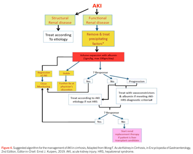
Summary : This flowchart presents a suggested algorithm for the management of acute kidney injury (AKI) in patients with cirrhosis, distinguishing between structural and functional renal disease, and outlining stepwise treatment decisions based on response to interventions. flowchart: # Nodes : • AKI (start, red text) • Structural Renal disease (rectangle, yellow-green) • Functional Renal disease (rectangle, yellow-green) • Treat according to etiology (rectangle, white) • Remove & treat precipitating factors* (rectangle, yellow) • Volume expansion with albumin (rectangle, red) — "1gm/kg, max 100gm/day" • ? Response (diamond, blue) • Regression of AKI (rectangle, yellow) • Stable (rectangle, yellow) • Close Monitoring (rectangle, yellow) • Further Rx at physician’s discretion (rectangle, yellow) • Yes (diamond, blue) • No (diamond, blue) • Progression (rectangle, blue) • Treat according to AKI etiology if not HRS (rectangle, white) • Treat with vasoconstrictors & albumin if meeting AKI-HRS diagnostic criteria# (rectangle, blue) • ? Response (diamond, blue) • Yes (diamond, blue) • No (diamond, blue) • Start renal replacement therapy if patient is liver transplant candidate (rectangle, pink) # Connectors : • AKI splits into two branches: Structural Renal disease and Functional Renal disease. • Structural Renal disease → Treat according to etiology. • Functional Renal disease → Remove & treat precipitating factors* → Volume expansion with albumin. • Volume expansion with albumin → ? Response. • ? Response branches: – Yes → Regression of AKI → Close Monitoring. – Yes → Stable → Further Rx at physician’s discretion. – No → Treat according to AKI etiology if not HRS. – Progression → Treat with vasoconstrictors & albumin if meeting AKI-HRS diagnostic criteria# → ? Response. • Second ? Response: – Yes → Treat according to AKI etiology if not HRS. – No → Start renal replacement therapy if patient is liver transplant candidate. # Layout : • Top-down hierarchical flow. • Initial split into two main branches (structural vs. functional). • Functional branch has a more complex, multi-step decision tree with feedback loops. • Decision diamonds indicate response checkpoints. • Colour coding: yellow for initial steps, red for albumin intervention, blue for response/treatment, pink for renal replacement therapy. # Analysis : • The algorithm prioritises distinguishing structural from functional renal disease in AKI management for cirrhosis. • Functional renal disease is managed by removing precipitating factors and volume expansion with albumin, followed by assessment of response. • If AKI does not regress or progresses, further treatment with vasoconstrictors and albumin is considered if AKI-HRS criteria are met. • Non-responders may be considered for renal replacement therapy if they are liver transplant candidates. • The flowchart emphasises stepwise escalation of therapy and close monitoring, with decision points guiding further management.
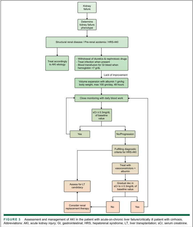
Summary : This flowchart outlines the assessment and management of acute kidney injury (AKI) in patients with acute-on-chronic liver failure or critically ill patients with cirrhosis, including diagnostic steps, treatment options, and decision points for liver transplantation (LT) and renal replacement therapy. flowchart: # Nodes : • Start (rectangle): "Kidney failure" • Decision (rectangle): "Determine kidney failure phenotype" • Process (rectangle): "Structural renal disease / Pre-renal azotemia / HRS-AKI" • Process (rectangle): "Treat accordingly to AKI etiology" • Process (rectangle): - "Withdrawal of diuretics & nephrotoxic drugs" - "Treat infection when present" - "Blood transfusion for GI bleed when hemoglobin <7 g/dL" • Decision (rectangle): "Lack of improvement" • Process (rectangle): "Volume expansion with albumin 1 gm/kg body weight, max 100 gm/day, 48 hours" • Process (rectangle): "Close monitoring with daily blood work" • Decision (diamond): "sCr ≤ 0.3mg/dL of baseline value" • Terminal (rectangle): "Yes" • Terminal (rectangle): "No/Progression" • Process (rectangle): "Fulfilling diagnostic criteria for HRS-AKI" • Process (rectangle): "Treat with vasoconstrictors + albumin" • Decision (diamond): "Gradual dec in sCr to ≤ 0.3mg/dL of baseline value" • Terminal (rectangle): "Yes" • Terminal (rectangle): "No" • Process (rectangle): "Assess for LT candidacy" • Process (rectangle): "Consider renal replacement therapy" # Connectors : • Arrows flow top-down, with decision diamonds splitting into "Yes" and "No/Progression" branches. • After "Kidney failure" → "Determine kidney failure phenotype" → "Structural renal disease / Pre-renal azotemia / HRS-AKI". • "Treat accordingly to AKI etiology" and the box with withdrawal/treatment/transfusion are parallel options. • If "Lack of improvement", proceed to "Volume expansion with albumin". • Then to "Close monitoring with daily blood work". • At "sCr ≤ 0.3mg/dL of baseline value", "Yes" leads to "Assess for LT candidacy", then "Consider renal replacement therapy". • "No/Progression" leads to "Fulfilling diagnostic criteria for HRS-AKI", then "Treat with vasoconstrictors + albumin". • Next decision: "Gradual dec in sCr to ≤ 0.3mg/dL of baseline value" splits to "Yes" (to "Assess for LT candidacy") and "No" (to "Consider renal replacement therapy"). # Layout : • Vertical, top-down arrangement. • Parallel process boxes for initial treatment options. • Decision diamonds create branches for improvement vs. progression. • Merges at "Assess for LT candidacy" and "Consider renal replacement therapy". • Colour coding: green for process/decision nodes, tan for terminal/therapy nodes. # Analysis : • The flowchart provides a stepwise approach for AKI management in cirrhotic patients, emphasizing initial withdrawal of nephrotoxic agents, volume expansion, and close monitoring. • Decision points are based on serum creatinine (sCr) changes relative to baseline. • If AKI does not improve, further diagnostic criteria for hepatorenal syndrome (HRS-AKI) are applied, with escalation to vasoconstrictor therapy and albumin. • Final management options include assessment for liver transplantation and consideration of renal replacement therapy, depending on response to treatment. • The workflow is logical, with clear escalation steps and criteria for moving to advanced therapies.
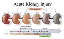
Summary : This figure illustrates the progression of acute kidney injury (AKI) from normal kidney function through increased risk, damage, decreased glomerular filtration rate (↓GFR), kidney failure, and ultimately death. It also highlights the window for early intervention and the stages of clinical management over time. flowchart: # Stages of Acute Kidney Injury : • Normal • Increased risk • Damage • ↓GFR (decreased glomerular filtration rate) • Kidney failure • Death # Complications : • Arrow labeled "Complications" points from the "Damage" stage to the "↓GFR" stage. # Intervention and Management Timeline : • Time axis runs horizontally below the kidney stages. • Phases labeled: Risk assessment, Recognition, Response, Renal support, Rehabilitation. • "Window for early targeted intervention" indicated between "Increased risk" and "Damage" stages. • "Staging" indicated between "↓GFR" and "Kidney failure" stages. # Design Encodings : • Kidneys are depicted in a gradient from healthy red (Normal) to dark gray (Death). • Arrows indicate progression from left (Normal) to right (Death). • Coloured bars below the timeline represent different phases of clinical management. # Analysis : • The figure visually communicates that AKI is a progressive condition with distinct stages, and that early intervention is possible before significant damage occurs. • The timeline emphasizes the importance of risk assessment and recognition before irreversible kidney failure. • The colour gradient and arrows reinforce the concept of worsening kidney function over time, culminating in death if not managed.
Acute Kidney Injury (AKI) - Treatment
Staging (KDIGO Criteria)
| Stage | Serum Creatinine | Urine Output |
|---|---|---|
| 1 | Rise ≥0.3 mg/dL within 48h OR 1.5-1.9× baseline | <0.5 mL/kg/h for 6-12h |
| 2 | 2.0-2.9× baseline | <0.5 mL/kg/h for ≥12h |
| 3 | ≥3× baseline OR ≥4 mg/dL OR initiation of RRT | <0.3 mL/kg/h for ≥24h OR anuria ≥12h |
1. General Management Principles
- Hemodynamic optimization: Autoregulation of renal blood flow is lost in AKI, making the kidney highly susceptible to hypotensive episodes. Maintain adequate MAP (individualized per patient age, hypertension status, and renovascular disease).
- Avoid nephrotoxins: Stop or dose-adjust aminoglycosides, amphotericin B, acyclovir, pentamidine, NSAIDs, iodinated contrast, and gadolinium-based contrast agents (group II cyclic agents preferred if unavoidable; risk of nephrogenic systemic fibrosis with eGFR <30).
- Medication dose adjustment: All renally cleared drugs must be adjusted; critical care pharmacist consultation is invaluable.
- Monitor fluid balance daily.
2. Fluid and Electrolyte Management
Fluids
- In AKI attributed to dehydration: 1-3 L isotonic (balanced crystalloid) solution over 24-48 hours is expected to reduce serum creatinine.
- Balanced crystalloids (e.g., Lactated Ringer's, Plasmalyte) are preferred over normal saline, especially with large volumes, to avoid hyperchloremic acidosis.
- Avoid hydroxyethyl starch (HES) - associated with increased AKI risk.
- After initial resuscitation, adopt a conservative fluid strategy - fluid overload (≥10% body weight gain) is independently associated with increased mortality. Focus shifts to preventing further fluid accumulation and early removal of excess volume.
- Dynamic measures (stroke volume, cardiac output) should guide ongoing fluid management rather than static preload markers.
Hyperkalemia (most immediately life-threatening complication)
| Intervention | Mechanism | Onset |
|---|---|---|
| IV Calcium gluconate/chloride | Cardiac membrane stabilization | Minutes |
| Insulin + Dextrose | Intracellular K⁺ shift | 15-30 min |
| Sodium bicarbonate | Intracellular K⁺ shift (esp. with acidosis) | 15-30 min |
| Inhaled beta-agonist (salbutamol) | Intracellular K⁺ shift | 15-30 min |
| Sodium polystyrene sulfonate (Kayexalate) | GI K⁺ binding/elimination | Hours |
| Dialysis (RRT) | Definitive removal | Immediate on initiation |
Metabolic Acidosis
- Sodium bicarbonate may be used for severe acidosis (pH <7.2) and to promote K⁺ shift.
- Severe refractory acidosis (pH <7.1) is an urgent indication for RRT.
Hypervolemia
- Loop diuretics (furosemide) are useful for managing fluid overload but do NOT improve mortality, dialysis requirement, or kidney recovery - do not use to accelerate renal recovery.
- Avoid concurrent aminoglycosides + loop diuretics (ototoxicity risk).
3. Drugs Used in AKI - Evidence Summary
| Drug | Verdict |
|---|---|
| Loop diuretics | No benefit on mortality/recovery; use only for fluid management |
| Dopamine ("renal dose") | No proven benefit - not recommended |
| Fenoldopam | Inconclusive evidence |
| Atrial natriuretic peptide (ANP) | Initial promising results not confirmed in large RCTs |
| Nesiritide | No mortality benefit in cardiac patients; not established in AKI |
| N-acetylcysteine (contrast prophylaxis) | Meta-analyses inconclusive; may have benefit in CI-AKI prevention |
| Statins | Benefit in AKI prevention controversial; not proven for treatment |
| Insulin-like growth factor (IGF-1) | No benefit in RCTs |
4. Renal Replacement Therapy (RRT)
Urgent (Emergent) Indications - "AEIOU"
- Acidosis - severe refractory metabolic acidosis (pH <7.1)
- Electrolytes - refractory hyperkalemia
- Intoxication - dialyzable drugs/toxins (e.g., lithium, methanol, ethylene glycol, salicylates)
- Overload - refractory fluid overload
- Uremia - uremic pericarditis, uremic encephalopathy, platelet dysfunction (BUN >100 mg/dL)
Timing of RRT
- Remains controversial. The STARRT-AKI RCT (largest trial) compared accelerated vs. standard strategy - 90-day mortality was similar between groups.
- Current guidance: wait for a clear indication before initiating rather than starting empirically (the standard/watchful approach is safe in most patients).
- Early initiation may be reasonable in selected high-risk patients.
Modality: IHD vs. CRRT
| Feature | IHD (Intermittent Hemodialysis) | CRRT (Continuous RRT) |
|---|---|---|
| Preferred in | Hemodynamically stable patients | Hemodynamically unstable patients (ICU) |
| Solute clearance | Rapid, intermittent | Slow, continuous |
| Fluid removal | Rapid | Gradual, well-tolerated |
| Cytokine removal | Limited | High-flux/specialized filters studied but no proven outcome benefit |
Note: Studies using high-flux CRRT or specialized adsorbents (cytokine/endotoxin removal) have NOT demonstrated a mortality benefit.
Platelet Dysfunction in Uremia
- DDAVP (desmopressin) can temporarily improve platelet function when BUN is markedly elevated.
5. Nutritional Support
- AKI patients - especially those receiving RRT - are highly catabolic.
- Target adequate protein intake (dialysis patients lose amino acids across the filter).
- Avoid excessive protein restriction (outdated practice - no benefit in delaying RRT).
- Enteral nutrition preferred over parenteral when GI tract is functional.
6. Complication Management
| Complication | Management |
|---|---|
| Hyperkalemia | As above |
| Metabolic acidosis | Bicarbonate; RRT if refractory |
| Hyperphosphatemia | Dietary restriction; phosphate binders |
| Hyponatremia/hypernatremia | Careful fluid tonicity management |
| Anemia | Transfuse if symptomatic; avoid erythropoietin acutely |
| Infections | Early recognition and appropriate antibiotics (dose-adjusted) |
| Uremic bleeding | DDAVP, conjugated estrogens; dialysis |
7. Specific Causes - Targeted Treatment
| Cause | Specific Treatment |
|---|---|
| Pre-renal (dehydration) | IV crystalloid resuscitation |
| Sepsis-associated AKI | Early antibiotics, source control, vasopressors to maintain MAP ≥65 mmHg |
| Contrast-induced AKI | IV crystalloid hydration pre/post procedure; use lowest contrast volume |
| Rhabdomyolysis | Aggressive normal saline hydration; target high urine output |
| Tumor lysis syndrome | Aggressive crystalloid hydration; allopurinol or rasburicase |
| Obstructive AKI | Relieve obstruction (catheter, nephrostomy, ureteric stent) |
| Glomerulonephritis/vasculitis | Immunosuppression (steroids, cyclophosphamide per etiology) |
| HRS-AKI (cirrhosis) | Vasoconstrictors (terlipressin/norepinephrine) + albumin 1 g/kg/day (max 100 g/day) |
AKI Progression - Stages of Intervention
Key Takeaways
- Remove the cause - identify and correct pre-renal, intrinsic, or post-renal etiology first.
- Protect the kidney - avoid nephrotoxins, maintain perfusion pressure, optimize fluid balance.
- Manage complications - hyperkalemia, acidosis, and fluid overload are life-threatening.
- RRT when indicated - use the AEIOU criteria; CRRT preferred in hemodynamic instability.
- No "magic drug" - dopamine, loop diuretics, ANP, and nesiritide have not proven to accelerate recovery.
- Conservative fluid strategy after initial resuscitation is associated with better outcomes.