
oral mucocele lower lip clinical photo
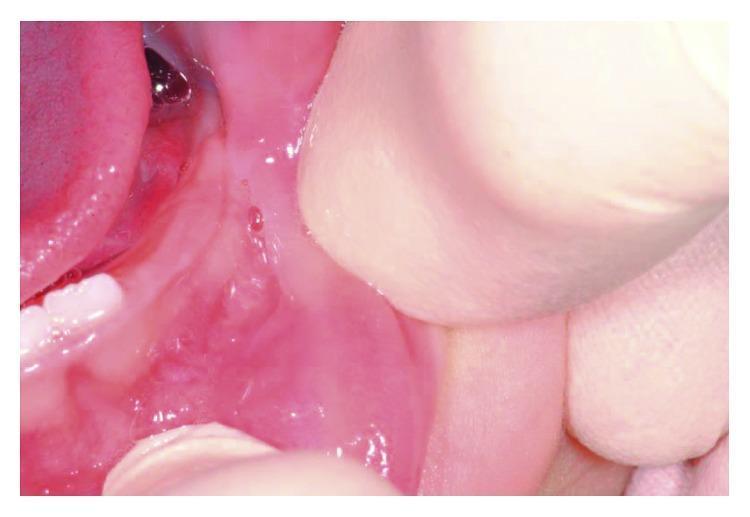
This clinical intraoral photograph shows the lower lip and labial mucosa of a pediatric patient (aged 10 months) during a follow-up examination. The image demonstrates a completely healed surgical site following the laser excision of a mucocele. The mucosa exhibits a healthy, uniform pink color and smooth texture, indicating successful epithelialization without scarring. There are no signs of inflammation, erythema, edema, or mass recurrence in the primary surgical area. The surrounding oral structures, including the deciduous teeth and gingival tissue, appear developmentally normal. The photo illustrates the typical long-term outcome of diode laser surgery for minor salivary gland lesions in infants, highlighting functional and aesthetic recovery of the oral mucosa.
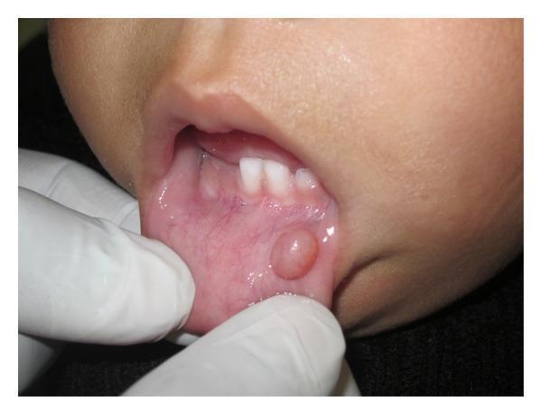
A close-up clinical photograph of an infant's oral cavity showing a mucocele on the lower lip mucosa. The image demonstrates a well-circumscribed, sessile, and roughly spherical nodule approximately 5 mm in diameter. The lesion is located on the labial mucosa of the lower lip and exhibits a smooth, translucent surface with a color similar to or slightly more pale than the surrounding pink mucosa. A gloved hand is visible, gently everting the lower lip to provide clear visualization of the pathology. The upper primary incisors are visible in the background. This clinical presentation is classic for a mucous extravasation cyst (mucocele), typically caused by local trauma or blockage of minor salivary gland ducts. The image serves as an educational example of common pediatric oral soft tissue lesions and their clinical morphology.
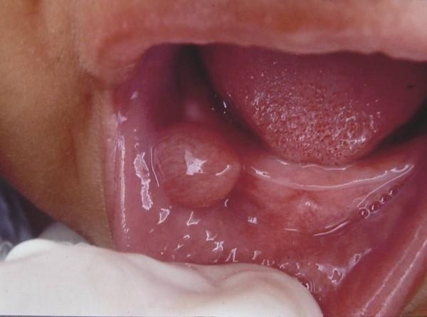
This clinical photograph shows an intraoral view of an infant's lower lip, highlighting a well-circumscribed, turgid nodular lesion characteristic of an oral mucocele (mucous extravasation cyst). The lesion is a raised, dome-shaped mass with a smooth, translucent surface that appears slightly shiny or bluish-pink, suggesting it contains fluid or semi-fluid mucous material. The surrounding labial mucosa is healthy and erythematous with visible fine vascularization. The tongue is partially visible in the background, displaying normal pinkish-red color and anatomical papillae. This image illustrates a common minor salivary gland disorder typically caused by local trauma or ductal obstruction, frequently encountered in pediatric dentistry and oral pathology for the clinical diagnosis of soft tissue swellings.
Oral Mucocele
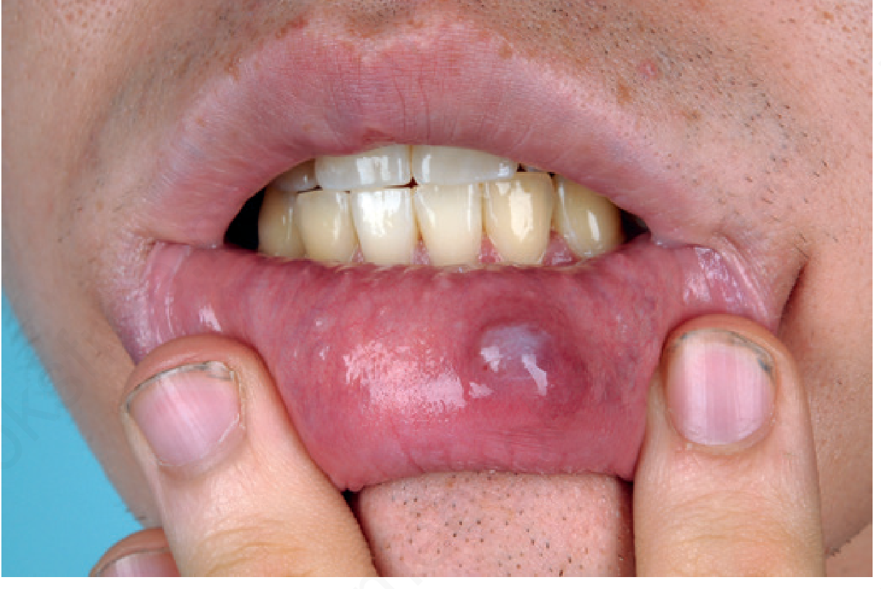
What is a Mucocele?
- Mucous extravasation phenomenon (most common) - trauma or biting causes rupture of a minor salivary duct, and mucin spills into the surrounding connective tissue stroma
- Mucous retention cyst - true ductal obstruction leads to an epithelial-lined cystic cavity (less common; seen more in posterior oral mucosa)
Clinical Features
| Feature | Description |
|---|---|
| Location | Inner lower lip (most common); rarely upper lip or buccal mucosa |
| Appearance | Soft, rounded, smooth, translucent - often with a bluish or pinkish tint |
| Size | 2-10 mm in diameter |
| Consistency | Fluctuant and tense |
| Symptoms | Painless; patient may report the lesion fluctuates in size (especially around meals) |
| Contents | Sticky straw-colored fluid (bluish if hemorrhage has occurred) |
Pathophysiology
Differential Diagnosis
- Ranula - a mucocele of the floor of the mouth arising from the sublingual gland (a "plunging ranula" dissects through the mylohyoid muscle)
- Fibroma - firmer, not fluctuant, not translucent
- Lipoma - soft but not translucent
- Hemangioma - blanches on pressure
- Lymphangioma - similar appearance but often multilobulated
Management
- Local anesthesia with lidocaine + epinephrine (also elevates the lesion for easier access)
- Stab incision (no. 11 blade) to drain mucin
- Cryotherapy (2-3 mm freeze rim) or electrocautery/electrodesiccation of the base
- Shave the roof off with a no. 15 blade, then cryotherapy or electrodesiccation
- Radiofrequency loop excision (cutting 20 W, coagulate 30 W)
- Excisional biopsy with removal of the associated minor salivary gland lobule - this is definitive and also confirms the diagnosis histologically
- Laser ablation (diode or CO2 laser) - increasingly used, especially in children
- Topical corticosteroids (for recurrent mucoceles in graft-versus-host disease or lichenoid inflammation)
Key point: Incomplete excision causes recurrence. The accompanying minor salivary gland lobule must be removed along with the cyst. (Andrews' Diseases of the Skin; Robbins Pathologic Basis of Disease)
Prognosis
Med after surgery
Post-Op Care After Mucocele Removal
1. Medications
| Category | What to Use | Notes |
|---|---|---|
| Pain relief | OTC analgesics - ibuprofen (e.g. Advil 400-600 mg every 6-8 hrs) or paracetamol (500-1000 mg every 6 hrs) | Pain is generally mild and well controlled with OTC drugs; prescription painkillers are rarely needed |
| Topical antibiotic ointment | Apply frequently to the healing site (e.g. mupirocin or bacitracin ointment) | Speeds healing and prevents irritation from adjacent teeth |
| Oral antibiotics | Only if infection develops - cover typical oral pathogens (e.g. amoxicillin 500 mg 3x/day, or amoxicillin-clavulanate if more coverage needed) | Not routinely required for clean excisions; prescribed only if signs of infection appear |
Note: If sutures (absorbable) were placed, they dissolve on their own - no removal needed. If a micromarsupialization silk suture was used, it is removed at 7 days.
2. Wound Care
- Expect some swelling in the lip after surgery - this is normal and resolves in a few days
- Bleeding should be minimal; control with direct pressure if it occurs, and it should stop within hours
- Do not bite or chew on the surgical area - this is tempting but can disturb healing and cause recurrence
- Healing by secondary intent (if no sutures were placed) takes approximately 5-7 days
- Full mucosal healing is expected within 2 weeks
- If the site is not healing by 2 weeks, see your doctor - incomplete healing may signal an underlying issue requiring biopsy
3. Diet
- Eat soft foods for the first few days (yogurt, mashed foods, soups, smoothies)
- Avoid hard, crunchy, or sharp foods that could scratch or irritate the healing mucosa
- Avoid very hot foods/drinks which can increase swelling and discomfort
- Stay well hydrated - adequate saliva flow supports healing
4. Oral Hygiene
- Continue brushing teeth normally but be gentle around the wound site
- Warm saline rinses (half a teaspoon of salt in a glass of warm water) after meals help keep the area clean and reduce bacterial load
- Avoid alcohol-based mouthwashes on the wound - they can irritate healing tissue
5. Warning Signs - See a Doctor If:
- Bleeding that does not stop with pressure
- Increasing pain, redness, or swelling after day 2-3 (signs of infection)
- Pus or foul taste from the wound
- The lesion returns (recurrence) - the most common complication, and can be re-treated more aggressively or referred