
Uh, since I'm supposed to take a workshop as a pediatric physiotherapist on neonatal reflexes and, uh, how are-- why are they important in a neonatal physiotherapy assessment. Also, what is their importance, um, in a, um, you know, in therapeutic, uh, strategy? So give me an outline.
neonatal primitive reflexes pediatric physiotherapy assessment therapeutic importance 2024
primitive reflexes neonatal neurological assessment physiotherapy
primitive reflexes neonatal assessment cerebral palsy early detection
https://pmc.ncbi.nlm.nih.gov/articles/PMC12128709
Workshop Outline: Neonatal Reflexes in Pediatric Physiotherapy Assessment & Therapeutic Strategy
MODULE 1: Introduction - What Are Neonatal Primitive Reflexes?
- Enable feeding, movement, and environmental engagement before cortical maturation
- Generate motor and sensory feedback that drives bottom-up brain development through neuroplasticity
- Each reflex, when expressed at the correct developmental stage, activates sensory receptors that feed back to the brainstem, promoting growth from medulla → pons → mesencephalon → neocortex in a hierarchical sequence
- Regulation originates from multiple brainstem areas (medulla → pons → mesencephalon)
- The frontal lobe ultimately takes over regulation and inhibits primitive reflexes as cortical maturity is achieved
- Reflexes are integrated, not eradicated - they remain in the CNS but are suppressed by higher cortical control
MODULE 2: The Core Neonatal Reflexes - Inventory, Onset, and Normal Extinction
| Reflex | How to Elicit | Appears (Gestational Age) | Normal Extinction |
|---|---|---|---|
| Palmar Grasp | Touch palm with finger | 28 weeks GA | 2-3 months |
| Moro (Startle) | Sudden head drop or loud noise; arms abduct then adduct | 28 weeks GA | 5-6 months |
| Rooting | Stroke cheek/corner of mouth; infant turns to that side | 32 weeks GA | 1 month (awake); 4 months |
| Asymmetric Tonic Neck Reflex (ATNR) | Turn head to one side; ipsilateral arm/leg extend, contralateral flex ("fencer posture") | 35 weeks GA | 6-7 months |
| Symmetric Tonic Neck Reflex (STNR) | Flex/extend neck; upper/lower limbs change tone reciprocally | 4-6 months | ~8-12 months |
| Plantar/Babinski | Stroke plantar surface; toes fan out + big toe dorsiflexes | Birth | 1-2 years |
| Stepping/Walking | Hold upright, sole touches surface; reciprocal stepping | Birth | 2 months (replaced by voluntary walking ~12 months) |
| Galant (Trunk Incurvation) | Stroke paravertebral skin; trunk curves to stimulated side | Birth | 4-6 months |
| Sucking/Rooting | Perioral stimulation or finger in mouth | 28-32 weeks GA | Gradually with voluntary feeding |
| Parachute | Tilt infant forward; arms extend for protection | 7-8 months | Remains for life |
| Tonic Labyrinthine (TLR) | Head position in space (prone → increased flexion tone; supine → increased extension tone) | Birth | ~4-6 months |
- Harriet Lane Handbook, 23rd ed. (Table 20.1) & Textbook of Family Medicine, 9th ed. (Table 21-5)
MODULE 3: Importance in Neonatal Physiotherapy Assessment
3.1 Neurological Integrity Screening
| Finding | Clinical Significance |
|---|---|
| Absent reflex | Lower motor neuron lesion, peripheral nerve injury, muscle disease, or severe CNS depression |
| Asymmetric Moro | Brachial plexus injury (Erb's palsy C5-C6 = arm adducted/internally rotated), hemiplegia, clavicle fracture |
| Obligatory or persistently dominant ATNR | Corticospinal tract dysfunction, early cerebral palsy |
| Absent suck reflex | Indirect sign of neonatal neurological compromise |
| Persistence beyond expected age | Failure of cortical inhibition - marker for CP, developmental delay, ASD-associated motor deficits |
| Upgoing Babinski beyond 1-2 years | Upper motor neuron pathology (note: normal in infants until 1-2 years) - Harriet Lane Handbook, Table 20.1 |
3.2 Assessment of Gestational Age / Developmental Stage
3.3 Tone Assessment Integration
- Resting posture: Full-term infant has strong limb flexion and symmetric movement
- Passive tone: Resistance to passive movement
- Active tone: Head control in pull-to-sit, ventral suspension
- Hypotonia: Frog-legged posture, limb arms near ears = hypotonic infant
- Hypertonia: Spastic or rigid movements
3.4 Standardized Assessment Tools That Use Primitive Reflex Data
- Hammersmith Infant Neurological Examination (HINE): For ages 2-24 months; endorsed by the American Academy of Pediatrics for early detection of cerebral palsy; the Brief-HINE shows high sensitivity and specificity for CP prediction
- Hammersmith Neonatal Neurological Examination (HNNE): For the neonatal period
- Prechtl's General Movements Assessment (GMA): Complements reflex assessment by evaluating spontaneous movement quality
- Dubowitz Neurological Assessment: Includes full primitive reflex inventory for preterm and term neonates
3.5 Retained Primitive Reflexes as Developmental Biomarkers
- Delayed psychomotor development
- Communication and language deficits (67-73% of autistic infants with early motor deficits later had communicative problems)
- Impediments in educational and social development
- The transition from brainstem-reflexive responses to cortically-based voluntary responses is a key neurodevelopmental milestone
MODULE 4: Therapeutic Strategies - Using Reflex Assessment to Guide Intervention
4.1 The Bottom-Up Developmental Model
4.2 Facilitation of Normal Reflex Expression (Preterm / NICU Neonates)
- Positioning therapy: Use of flexion positioning in preterm neonates supports the tonic labyrinthine reflex balance and mimics in-utero posture; reduces hypertonic extensor patterns
- Sensory stimulation protocols: Tactile, proprioceptive, and vestibular input designed to activate appropriate reflexes (e.g., perioral stimulation to facilitate rooting → support oral feeding readiness)
- Non-nutritive sucking facilitation: Utilizes the sucking reflex to improve feeding outcomes and neurodevelopmental state regulation
- Kangaroo care: Provides vestibular and proprioceptive input that supports normal reflex integration
4.3 Primitive Reflex Integration Therapy (Retained Reflexes)
| Retained Reflex | Clinical Problem | Therapeutic Approach |
|---|---|---|
| Moro | Hyper-reactivity, poor sitting balance (children learning to sit independently commonly show retained Moro) | Vestibular modulation, slow rocking, deep pressure input, trunk stability work |
| ATNR | Difficulty with bilateral coordination, rolling, midline crossing | Midline orientation activities; rotational patterns; Bobath handling to break ATNR pattern |
| TLR | Postural instability, gravitational insecurity | Prone weight-bearing, equilibrium/righting reactions facilitation |
| Palmar Grasp | Immature voluntary release; fine motor delay | Graded grasping tasks, proprioceptive hand input, weight-bearing on open hands |
| STNR | Difficulty crawling on hands and knees (arms buckle when head raised) | Quadruped weight-bearing, crawling facilitation, head-trunk dissociation exercises |
4.4 Facilitation of Postural Reactions (The "Higher Level" Successors)
- Righting reactions (head, body)
- Equilibrium reactions (tilting, propping)
- Parachute reaction (appears at 7-8 months; remains for life)
4.5 Handling and Facilitation Techniques (NDT/Bobath Framework)
- Key points of control: Proximal handling to inhibit abnormal reflex-driven postures (e.g., reducing ATNR influence by maintaining midline head position during activities)
- Inhibition of abnormal patterns + simultaneous facilitation of normal patterns
- Developmental sequencing: Intervention follows the normal neurodevelopmental reflex-to-voluntary-movement hierarchy
4.6 Parent/Caregiver Education
- Teach parents to recognize normal vs. abnormal reflex expression at home
- Safe handling techniques that do not reinforce pathological reflex patterns
- Tummy time (prone positioning) to facilitate integration of TLR and development of head/neck control
- Feeding position guidance (avoiding full neck extension that obligates the ATNR)
MODULE 5: Red Flags Summary for the Physiotherapist
| Red Flag | Act On |
|---|---|
| Any primitive reflex absent in newborn | Urgent neurological review |
| Asymmetric Moro or grasp | Rule out brachial plexus injury, hemiplegia |
| Any reflex persisting well beyond expected extinction age | Refer for developmental pediatrics evaluation; begin integration therapy |
| Obligatory ATNR (infant cannot break the pattern) | High suspicion for cerebral palsy |
| No parachute reaction by 10-12 months | Significant developmental concern |
| Loss of previously present reflex | Acquired neurological problem - urgent review |
MODULE 6: Recommended Assessment Tools & Resources
- HINE / Brief-HINE - endorsed by AAP for CP early detection (ages 2-24 months)
- Hammersmith Neonatal Neurological Examination - neonatal period
- Prechtl's General Movements Assessment - complements reflex testing
- Dubowitz Neonatal Neurological Assessment
- Leisman G et al. (2025). "Evaluating Primitive Reflexes in Early Childhood as a Potential Biomarker for Developmental Disabilities." PubMed PMC12128709 - current evidence summary
- StatPearls: Primitive Reflexes - Comprehensive Neurological Assessment - excellent quick reference
Create a visual reflex testing guide as a PDF reference card for clinic
ls -la /tmp/workspace/ 2>/dev/null || mkdir -p /tmp/workspace && ls -la /tmp/workspace/
mkdir -p /tmp/workspace/neonatal-reflex-card && python3 -c "import reportlab; print(reportlab.Version)"
~/neonatal-reflex-card/build_card.py
"""
Neonatal Primitive Reflexes – Clinical Reference Card (A4 Landscape, 2 pages)
Pediatric Physiotherapy Edition
"""
from reportlab.lib.pagesizes import A4, landscape
from reportlab.lib import colors
from reportlab.lib.units import mm, cm
from reportlab.lib.styles import getSampleStyleSheet, ParagraphStyle
from reportlab.lib.enums import TA_CENTER, TA_LEFT, TA_RIGHT
from reportlab.platypus import (
SimpleDocTemplate, Table, TableStyle, Paragraph, Spacer,
HRFlowable, KeepTogether
)
from reportlab.graphics.shapes import Drawing, Rect, String, Line, Circle, Polygon
from reportlab.graphics import renderPDF
from reportlab.platypus import Flowable
import reportlab.lib.colors as lcolors
# ─── Colour palette ───────────────────────────────────────────────────────────
TEAL = colors.HexColor("#1A7A8A")
TEAL_LIGHT = colors.HexColor("#D4EEF2")
TEAL_MID = colors.HexColor("#4AAEBB")
AMBER = colors.HexColor("#E8A020")
AMBER_LIGHT= colors.HexColor("#FDF3DC")
RED = colors.HexColor("#C0392B")
RED_LIGHT = colors.HexColor("#FADBD8")
GREEN = colors.HexColor("#1E8449")
GREEN_LIGHT= colors.HexColor("#D5F5E3")
PURPLE = colors.HexColor("#6C3483")
PURPLE_LIGHT=colors.HexColor("#E8DAEF")
GREY_DARK = colors.HexColor("#2C3E50")
GREY_MID = colors.HexColor("#5D6D7E")
GREY_LIGHT = colors.HexColor("#ECF0F1")
WHITE = colors.white
ORANGE = colors.HexColor("#D35400")
ORANGE_LIGHT=colors.HexColor("#FDEBD0")
PW, PH = landscape(A4) # 297 x 210 mm
MARGIN = 10*mm
# ─── Styles ───────────────────────────────────────────────────────────────────
styles = getSampleStyleSheet()
def S(name, **kw):
return ParagraphStyle(name, **kw)
H_TITLE = S("htitle", fontSize=18, fontName="Helvetica-Bold",
textColor=WHITE, alignment=TA_CENTER, spaceAfter=0, leading=22)
H1 = S("h1", fontSize=9, fontName="Helvetica-Bold",
textColor=WHITE, alignment=TA_CENTER, spaceAfter=0, leading=11)
H1L = S("h1l", fontSize=9, fontName="Helvetica-Bold",
textColor=WHITE, alignment=TA_LEFT, spaceAfter=0, leading=11)
H2 = S("h2", fontSize=8, fontName="Helvetica-Bold",
textColor=GREY_DARK, alignment=TA_LEFT, spaceAfter=0, leading=10)
H2C = S("h2c", fontSize=8, fontName="Helvetica-Bold",
textColor=GREY_DARK, alignment=TA_CENTER, spaceAfter=0, leading=10)
BODY = S("body", fontSize=7.2, fontName="Helvetica",
textColor=GREY_DARK, alignment=TA_LEFT, spaceAfter=0, leading=9.5)
BODYC = S("bodyc", fontSize=7.2, fontName="Helvetica",
textColor=GREY_DARK, alignment=TA_CENTER, spaceAfter=0, leading=9.5)
SMALL = S("small", fontSize=6.5, fontName="Helvetica",
textColor=GREY_MID, alignment=TA_LEFT, spaceAfter=0, leading=8)
SMALLC = S("smallc", fontSize=6.5, fontName="Helvetica",
textColor=GREY_MID, alignment=TA_CENTER, spaceAfter=0, leading=8)
BOLD_TEAL = S("bteal", fontSize=7.5, fontName="Helvetica-Bold",
textColor=TEAL, alignment=TA_LEFT, leading=9.5)
BOLD_RED = S("bred", fontSize=7.5, fontName="Helvetica-Bold",
textColor=RED, alignment=TA_LEFT, leading=9.5)
BOLD_GREEN = S("bgreen", fontSize=7.5, fontName="Helvetica-Bold",
textColor=GREEN, alignment=TA_LEFT, leading=9.5)
ITALIC = S("italic", fontSize=7, fontName="Helvetica-Oblique",
textColor=GREY_MID, alignment=TA_LEFT, leading=9)
# ─── Helper: coloured header cell ─────────────────────────────────────────────
def hdr(text, style=H1, bg=TEAL):
return Paragraph(text, style)
def P(text, style=BODY):
return Paragraph(text, style)
def spacer(h=2):
return Spacer(1, h*mm)
# ─── Inline badge helper ──────────────────────────────────────────────────────
def badge(text, fg=WHITE, bg=TEAL):
"""Small inline text tag"""
return f'<font color="#{bg.hexval()[2:].upper()}">{text}</font>'
# ─── Drawing helpers: small anatomical-style icons ────────────────────────────
class BabyIcon(Flowable):
"""Tiny schematic baby figure"""
def __init__(self, w=18, h=28, color=TEAL):
super().__init__()
self.w, self.h, self.color = w, h, color
self.width, self.height = w, h
def draw(self):
c = self.canv
co = self.color
# head
c.setFillColor(co)
c.setStrokeColor(co)
c.circle(self.w/2, self.h-5, 4, fill=1, stroke=0)
# body
c.roundRect(self.w/2-3, self.h-15, 6, 10, 1, fill=1, stroke=0)
# arms
c.setLineWidth(1.5)
c.line(self.w/2-3, self.h-10, self.w/2-9, self.h-14) # left arm
c.line(self.w/2+3, self.h-10, self.w/2+9, self.h-14) # right arm
# legs
c.line(self.w/2-2, self.h-15, self.w/2-5, self.h-22)
c.line(self.w/2+2, self.h-15, self.w/2+5, self.h-22)
class Timeline(Flowable):
"""Horizontal timeline bar showing reflex window"""
def __init__(self, start_wk, end_wk, total_wk=52, width=80, bar_color=TEAL, label=""):
super().__init__()
self.start = start_wk
self.end = end_wk
self.total = total_wk
self.w = width
self.bar_color = bar_color
self.label = label
self.width = width
self.height = 7
def draw(self):
c = self.canv
w = self.w
h = 5
# background track
c.setFillColor(GREY_LIGHT)
c.roundRect(0, 1, w, h, 2, fill=1, stroke=0)
# active bar
x0 = (self.start / self.total) * w
x1 = (self.end / self.total) * w
c.setFillColor(self.bar_color)
c.roundRect(x0, 1, max(x1-x0, 3), h, 2, fill=1, stroke=0)
# tick marks at 0, 28wk, 40wk (term), 52wk
for wk, label in [(0,"0"), (28,"28wGA"), (40,"Term"), (52,"1yr")]:
x = (wk/self.total)*w
c.setStrokeColor(GREY_MID)
c.setLineWidth(0.5)
c.line(x, 0, x, 7)
def make_timeline(appear_wk, extinguish_wk, color=TEAL, width=75):
return Timeline(appear_wk, extinguish_wk, total_wk=56,
width=width, bar_color=color)
# ─── Page 1 data: Core Reflex Table ──────────────────────────────────────────
REFLEXES = [
# name, elicit, normal response, appear(wk), extinct(wk), clinical flag, color
(
"PALMAR GRASP",
"Place finger/object in infant's palm",
"Fingers curl & grip tightly around object",
28, 12, # 3 months ~ 12 wks post-term
"Absent: lower motor neuron lesion\nAsymmetric: brachial plexus / hemiplegia",
TEAL
),
(
"MORO (Startle)",
"Support head; let it drop 2-3 cm OR loud noise",
"Arms ABDUCT & extend (phase 1) then ADDUCT & flex (phase 2); cry",
28, 24, # 5-6 months
"Asymmetric: clavicle Fx, Erb's palsy, hemiplegia\nAbsent: severe CNS depression\nPersistent >6 m: suspect CP",
AMBER
),
(
"ROOTING",
"Stroke corner of mouth / cheek",
"Head turns toward stimulus; mouth opens",
32, 4, # ~1 month post-term
"Absent: neurological compromise\nKey for oral feeding readiness in NICU",
GREEN
),
(
"ASYMMETRIC TONIC\nNECK (ATNR)",
"Turn head to one side while supine",
"Ipsilateral arm & leg EXTEND; contralateral FLEX\n('Fencing posture')",
35, 28, # 6-7 months
"Obligatory ATNR (can't break): high suspicion CP\nInhibits midline crossing, rolling",
PURPLE
),
(
"SUCKING",
"Perioral stimulation or finger in mouth",
"Rhythmic suck-swallow sequence",
28, 16, # integrates ~4 months, voluntary feeding replaces
"Absent/weak: neurological depression, preterm\nCritical for feeding assessment",
TEAL_MID
),
(
"GALANT (Trunk\nIncurvation)",
"Hold prone; stroke paravertebral skin\nlongitudinally",
"Trunk curves (lateral flexion) toward\nstimulated side",
28, 20, # ~4-5 months
"Absent: spinal cord lesion (T2-S1 level)\nPersistent: may affect sitting balance",
ORANGE
),
(
"STEPPING / WALKING",
"Hold upright; sole contacts flat surface",
"Reciprocal stepping movements",
0, 8, # disappears ~2 months, re-emerges as voluntary ~12 m
"Absent: lower extremity weakness\nDistinct from voluntary walking at ~12 m",
TEAL
),
(
"BABINSKI",
"Stroke lateral plantar surface heel→toes",
"Big toe DORSIFLEXES; other toes fan out",
0, 52, # normal in infants until 1-2 years
"NORMAL in infants <1-2 years\nPersistence >2 yrs = UMN pathology",
RED
),
(
"TONIC LABYRINTHINE\n(TLR)",
"Head position in space:\nProne / Supine",
"PRONE: increased FLEXOR tone\nSUPINE: increased EXTENSOR tone",
0, 20, # ~4-6 months
"Persistent: postural instability, gravitational insecurity\nAffects rolling, sitting transitions",
PURPLE
),
(
"PARACHUTE",
"Tilt infant forward rapidly (head-down)",
"Arms EXTEND symmetrically to protect",
32, 999, # appears 7-8 months, REMAINS FOR LIFE
"Absent by 10-12 m: significant developmental concern\nIndicator of CNS maturity",
GREEN
),
]
# ─── Build Page 1 ─────────────────────────────────────────────────────────────
def build_page1():
story = []
# ── TITLE BANNER ──────────────────────────────────────────────────────────
title_data = [[
Paragraph("<b>NEONATAL PRIMITIVE REFLEXES</b><br/>"
"<font size=9>Clinical Reference Card — Pediatric Physiotherapy</font>",
S("tt", fontSize=15, fontName="Helvetica-Bold", textColor=WHITE,
alignment=TA_CENTER, leading=19))
]]
title_tbl = Table(title_data, colWidths=[PW - 2*MARGIN])
title_tbl.setStyle(TableStyle([
("BACKGROUND", (0,0), (-1,-1), TEAL),
("TOPPADDING", (0,0), (-1,-1), 5),
("BOTTOMPADDING", (0,0), (-1,-1), 5),
("LEFTPADDING", (0,0), (-1,-1), 8),
("RIGHTPADDING", (0,0), (-1,-1), 8),
("ROUNDEDCORNERS", [4]),
]))
story.append(title_tbl)
story.append(spacer(2))
# ── HEADER ROW ────────────────────────────────────────────────────────────
col_w = [
37*mm, # reflex name
55*mm, # how to elicit
55*mm, # normal response
35*mm, # timeline
75*mm, # clinical flags
]
assert sum(col_w) <= PW - 2*MARGIN + 1, f"cols too wide: {sum(col_w)}"
hdr_row = [
P("REFLEX", H1),
P("HOW TO ELICIT", H1),
P("NORMAL RESPONSE", H1),
P("DEVELOPMENTAL\nWINDOW", H1),
P("CLINICAL FLAGS FOR PHYSIOTHERAPIST", H1),
]
rows = [hdr_row]
# Appears / extinguishes reference legend
def window_label(appear, extinguish):
if appear == 0:
a_str = "Birth"
elif appear < 40:
a_str = f"{appear} wGA"
else:
a_str = f"{appear} wk"
if extinguish == 999:
e_str = "Persists for life"
bar_end = 56
else:
# convert post-term weeks: appear is GA, extinguish is postnatal months converted
e_str = f"~{extinguish//4} m" if extinguish >= 4 else f"~{extinguish} wk"
bar_end = min(extinguish + 40, 56) if appear < 40 else extinguish
# timeline bar: map wGA appear to bar start
bar_start = appear if appear >= 28 else 28
bar_end_calc = bar_end
return a_str, e_str, bar_start, bar_end_calc
for (name, elicit, response, appear, extinguish, flags, color) in REFLEXES:
a_str, e_str, bar_start, bar_end = window_label(appear, extinguish)
if extinguish == 999:
bar_color = GREEN
elif extinguish <= 8:
bar_color = AMBER
else:
bar_color = color
win_cell = [
P(f"<b>Appears:</b> {a_str}", BODY),
P(f"<b>Gone by:</b> {e_str}", BODY),
spacer(1),
make_timeline(bar_start, bar_end, bar_color, width=32*mm),
]
# flag colours
flag_lines = []
for line in flags.split("\n"):
if "Absent" in line or "absent" in line:
flag_lines.append(P(f"<font color='#C0392B'>●</font> {line}", BODY))
elif "Persistent" in line or "persist" in line or "suspect" in line:
flag_lines.append(P(f"<font color='#E8A020'>●</font> {line}", BODY))
elif "NORMAL" in line:
flag_lines.append(P(f"<font color='#1E8449'>●</font> {line}", BODY))
else:
flag_lines.append(P(f"<font color='#1A7A8A'>●</font> {line}", BODY))
row = [
P(f"<b>{name}</b>", S("rname", fontSize=7.5, fontName="Helvetica-Bold",
textColor=color, alignment=TA_LEFT, leading=9.5)),
P(elicit, BODY),
P(response, BODY),
win_cell,
flag_lines,
]
rows.append(row)
tbl = Table(rows, colWidths=col_w, repeatRows=1)
# Alternating row backgrounds
style_cmds = [
("BACKGROUND", (0,0), (-1,0), GREY_DARK),
("TEXTCOLOR", (0,0), (-1,0), WHITE),
("FONTNAME", (0,0), (-1,0), "Helvetica-Bold"),
("FONTSIZE", (0,0), (-1,0), 7.5),
("ALIGN", (0,0), (-1,-1), "LEFT"),
("VALIGN", (0,0), (-1,-1), "TOP"),
("TOPPADDING", (0,0), (-1,-1), 3),
("BOTTOMPADDING", (0,0), (-1,-1), 3),
("LEFTPADDING", (0,0), (-1,-1), 4),
("RIGHTPADDING", (0,0), (-1,-1), 4),
("ROWBACKGROUNDS", (0,1), (-1,-1), [WHITE, GREY_LIGHT]),
("GRID", (0,0), (-1,-1), 0.3, colors.HexColor("#BDC3C7")),
("LINEBELOW", (0,0), (-1,0), 1.5, TEAL),
("LEFTBORDERPADDING", (0,0), (0,-1), 2),
]
# Left accent colour stripe per reflex row
for i, (_, _, _, _, _, _, color) in enumerate(REFLEXES):
row_i = i + 1
style_cmds.append(("BACKGROUND", (0, row_i), (0, row_i),
color.clone(alpha=0.15)))
tbl.setStyle(TableStyle(style_cmds))
story.append(tbl)
story.append(spacer(2))
# ── FOOTER ────────────────────────────────────────────────────────────────
footer_data = [[
P("wGA = weeks gestational age | m = months post-term "
"| Timeline bar: grey = full range (birth→1 yr), coloured = active window "
"| All ages are approximate; assess in context of gestational age & clinical state.",
S("ft", fontSize=6, fontName="Helvetica-Oblique", textColor=GREY_MID,
alignment=TA_CENTER, leading=8)),
P("Sources: Harriet Lane Handbook 23e; Textbook of Family Medicine 9e;\n"
"Leisman et al. PMC12128709 (2025); Nelson Textbook of Pediatrics 21e",
S("ft2", fontSize=6, fontName="Helvetica-Oblique", textColor=GREY_MID,
alignment=TA_RIGHT, leading=8)),
]]
footer_tbl = Table(footer_data, colWidths=[(PW-2*MARGIN)*0.65, (PW-2*MARGIN)*0.35])
footer_tbl.setStyle(TableStyle([
("VALIGN", (0,0), (-1,-1), "MIDDLE"),
("TOPPADDING", (0,0), (-1,-1), 1),
("BOTTOMPADDING", (0,0), (-1,-1), 1),
]))
story.append(footer_tbl)
return story
# ─── Build Page 2 ─────────────────────────────────────────────────────────────
def build_page2():
story = []
# TITLE BANNER p2
title_data = [[
Paragraph("<b>NEONATAL REFLEXES — CLINICAL DECISION GUIDE</b><br/>"
"<font size=9>Assessment Framework & Therapeutic Strategy | Pediatric Physiotherapy</font>",
S("tt2", fontSize=14, fontName="Helvetica-Bold", textColor=WHITE,
alignment=TA_CENTER, leading=18))
]]
title_tbl = Table(title_data, colWidths=[PW - 2*MARGIN])
title_tbl.setStyle(TableStyle([
("BACKGROUND", (0,0), (-1,-1), GREY_DARK),
("TOPPADDING", (0,0), (-1,-1), 5),
("BOTTOMPADDING", (0,0), (-1,-1), 5),
("LEFTPADDING", (0,0), (-1,-1), 8),
("ROUNDEDCORNERS", [4]),
]))
story.append(title_tbl)
story.append(spacer(2))
avail_w = PW - 2*MARGIN
# ── ROW A: Assessment Principles + Red Flags ──────────────────────────────
col_left = avail_w * 0.38
col_right = avail_w * 0.62
# LEFT: ASSESSMENT PROTOCOL
assess_rows = [
[P("NEONATAL NEUROLOGICAL ASSESSMENT PROTOCOL", H1L)],
[P("1. <b>General State</b> — Alertness, responsiveness, cry quality", BODY)],
[P("2. <b>Resting Posture</b> — Flexion dominance (term); frog-leg = hypotonia", BODY)],
[P("3. <b>Passive Tone</b> — Resistance to passive movement in all limbs", BODY)],
[P("4. <b>Active Tone</b> — Pull-to-sit head lag; ventral suspension posture", BODY)],
[P("5. <b>Primitive Reflexes</b> — Each reflex: Present / Absent / Asymmetric / Obligatory", BODY)],
[P("6. <b>Postural Reactions</b> — Righting, equilibrium, protective (parachute)", BODY)],
[spacer(1)],
[P("<b>STANDARDISED TOOLS</b>", S("ht", fontSize=7.5, fontName="Helvetica-Bold",
textColor=TEAL, leading=10))],
[P("• <b>HNNE</b> (Hammersmith Neonatal) — term & preterm neonates", BODY)],
[P("• <b>HINE / Brief-HINE</b> — 2–24 months; AAP endorsed for CP screening", BODY)],
[P("• <b>Prechtl GMA</b> — spontaneous movement quality; complements reflex testing", BODY)],
[P("• <b>Dubowitz</b> — full reflex inventory for preterm & term neonates", BODY)],
[spacer(1)],
[P("<b>GESTATIONAL AGE CONTEXT</b> — Correct for prematurity when interpreting", BODY)],
[P("Preterm neonates have attenuated/absent reflexes appropriate to their GA", SMALL)],
]
assess_tbl = Table(assess_rows, colWidths=[col_left - 4*mm])
assess_tbl.setStyle(TableStyle([
("BACKGROUND", (0,0), (-1,0), TEAL),
("TOPPADDING", (0,0), (-1,-1), 2),
("BOTTOMPADDING", (0,0), (-1,-1), 2),
("LEFTPADDING", (0,0), (-1,-1), 5),
("RIGHTPADDING", (0,0), (-1,-1), 5),
("ROWBACKGROUNDS", (0,1), (-1,-1), [WHITE, TEAL_LIGHT]),
("BOX", (0,0), (-1,-1), 0.5, TEAL),
("GRID", (0,0), (-1,-1), 0.2, colors.HexColor("#C8E6EA")),
]))
# RIGHT: RED FLAGS table
rf_data = [
[P("RED FLAGS — ACT IMMEDIATELY", S("rfh", fontSize=8, fontName="Helvetica-Bold",
textColor=WHITE, alignment=TA_CENTER, leading=10)),
P("", BODY)],
[P("<b>Finding</b>", H2C), P("<b>Action</b>", H2C)],
[P("Any primitive reflex ABSENT in term newborn", BODY),
P("<font color='#C0392B'><b>Urgent neurology review</b></font>", BODY)],
[P("ASYMMETRIC Moro or palmar grasp", BODY),
P("Rule out Erb's palsy / clavicle Fx / hemiplegia", BODY)],
[P("Absent sucking / rooting in NICU neonate", BODY),
P("Feeding assessment; neonatology liaison", BODY)],
[P("OBLIGATORY ATNR (infant cannot break pattern)", BODY),
P("<font color='#C0392B'><b>High suspicion for cerebral palsy — refer</b></font>", BODY)],
[P("Any reflex persisting WELL BEYOND extinction age", BODY),
P("Developmental paediatrics + integration therapy", BODY)],
[P("NO parachute reaction by 10–12 months", BODY),
P("<font color='#C0392B'><b>Significant developmental concern — refer</b></font>", BODY)],
[P("LOSS of previously present reflex", BODY),
P("Acquired neurological problem — urgent review", BODY)],
[P("Upgoing Babinski <b>after 2 years</b>", BODY),
P("Upper motor neuron pathology", BODY)],
]
rf_col_w = [(col_right - 4*mm) * 0.55, (col_right - 4*mm) * 0.45]
rf_tbl = Table(rf_data, colWidths=rf_col_w)
rf_tbl.setStyle(TableStyle([
("SPAN", (0,0), (1,0)),
("BACKGROUND", (0,0), (1,0), RED),
("BACKGROUND", (0,1), (1,1), GREY_DARK),
("TEXTCOLOR", (0,1), (1,1), WHITE),
("FONTNAME", (0,1), (1,1), "Helvetica-Bold"),
("FONTSIZE", (0,1), (1,1), 7),
("ROWBACKGROUNDS", (0,2), (-1,-1), [WHITE, RED_LIGHT]),
("TOPPADDING", (0,0), (-1,-1), 2.5),
("BOTTOMPADDING", (0,0), (-1,-1), 2.5),
("LEFTPADDING", (0,0), (-1,-1), 4),
("RIGHTPADDING", (0,0), (-1,-1), 4),
("BOX", (0,0), (-1,-1), 0.5, RED),
("GRID", (0,0), (-1,-1), 0.2, colors.HexColor("#E8C3BE")),
("ALIGN", (0,0), (1,0), "CENTER"),
]))
row_a = Table([[assess_tbl, rf_tbl]],
colWidths=[col_left, col_right])
row_a.setStyle(TableStyle([
("VALIGN", (0,0), (-1,-1), "TOP"),
("LEFTPADDING", (0,0), (-1,-1), 0),
("RIGHTPADDING", (0,0), (-1,-1), 4),
("TOPPADDING", (0,0), (-1,-1), 0),
("BOTTOMPADDING", (0,0), (-1,-1), 0),
]))
story.append(row_a)
story.append(spacer(3))
# ── ROW B: Therapeutic Strategies + Developmental Cascade ─────────────────
col1 = avail_w * 0.33
col2 = avail_w * 0.34
col3 = avail_w * 0.33
# B1: Retained Reflex Integration Strategies
int_data = [
[P("RETAINED REFLEX — INTEGRATION STRATEGIES", H1L)],
[P("<b>Moro</b> (>6 m) → hyper-reactivity, poor trunk control", BOLD_TEAL)],
[P("Vestibular modulation, slow rocking, deep proprioceptive\ninput, trunk stability, sensory desensitisation", BODY)],
[spacer(1)],
[P("<b>ATNR</b> (>7 m) → blocks midline crossing, bilateral coordination", BOLD_TEAL)],
[P("Midline orientation activities; rotational trunk patterns;\nBobath handling to inhibit ATNR; bilateral reaching tasks", BODY)],
[spacer(1)],
[P("<b>TLR</b> (>6 m) → postural instability, gravitational insecurity", BOLD_TEAL)],
[P("Prone weight-bearing; equilibrium & righting reaction\nfacilitation; graded vestibular challenges", BODY)],
[spacer(1)],
[P("<b>Palmar Grasp</b> (>4 m) → immature voluntary release", BOLD_TEAL)],
[P("Graded grasping tasks; proprioceptive hand input;\nopen-hand weight-bearing on flat surfaces", BODY)],
[spacer(1)],
[P("<b>STNR</b> (>12 m) → crawling difficulty (arms buckle on head raise)", BOLD_TEAL)],
[P("Quadruped weight-bearing; crawling facilitation;\nhead–trunk dissociation exercises", BODY)],
[spacer(1)],
[P("<b>Principle:</b> Controlled therapeutic activation of reflex pattern +\n"
"simultaneous voluntary movement practice → cortical inhibition", ITALIC)],
]
int_tbl = Table(int_data, colWidths=[col1 - 3*mm])
int_tbl.setStyle(TableStyle([
("BACKGROUND", (0,0), (-1,0), PURPLE),
("ROWBACKGROUNDS", (0,1), (-1,-1), [WHITE, PURPLE_LIGHT]),
("TOPPADDING", (0,0), (-1,-1), 2),
("BOTTOMPADDING", (0,0), (-1,-1), 2),
("LEFTPADDING", (0,0), (-1,-1), 5),
("RIGHTPADDING", (0,0), (-1,-1), 5),
("BOX", (0,0), (-1,-1), 0.5, PURPLE),
("GRID", (0,0), (-1,-1), 0.2, colors.HexColor("#D2B4DE")),
]))
# B2: NICU / Preterm facilitation
nicu_data = [
[P("NICU / PRETERM FACILITATION", H1L)],
[P("<b>Positioning Therapy</b>", BOLD_TEAL)],
[P("Flexion 'nest' positioning mimics in-utero posture;\nreduces hypertonic extensor patterns (TLR)", BODY)],
[spacer(1)],
[P("<b>Oral Facilitation</b>", BOLD_TEAL)],
[P("Perioral / cheek stimulation activates rooting;\nnon-nutritive sucking improves feeding readiness\nand state regulation", BODY)],
[spacer(1)],
[P("<b>Kangaroo Care</b>", BOLD_TEAL)],
[P("Vestibular + proprioceptive + thermal input;\nsupports normal reflex integration & neurodevelopment", BODY)],
[spacer(1)],
[P("<b>Sensory Stimulation</b>", BOLD_TEAL)],
[P("Graded tactile, vestibular, proprioceptive input\nto activate appropriate reflexes at each GA stage", BODY)],
[spacer(1)],
[P("<b>NDT / Bobath Handling</b>", BOLD_TEAL)],
[P("Key points of control at proximal segments;\ninhibit abnormal reflex-driven postures while\nfacilitating voluntary movement patterns", BODY)],
[spacer(1)],
[P("<b>Parent Education</b>", BOLD_TEAL)],
[P("Tummy time; safe handling; feeding positions\n(avoid full neck extension → ATNR); reflex recognition", BODY)],
]
nicu_tbl = Table(nicu_data, colWidths=[col2 - 3*mm])
nicu_tbl.setStyle(TableStyle([
("BACKGROUND", (0,0), (-1,0), AMBER),
("ROWBACKGROUNDS", (0,1), (-1,-1), [WHITE, AMBER_LIGHT]),
("TOPPADDING", (0,0), (-1,-1), 2),
("BOTTOMPADDING", (0,0), (-1,-1), 2),
("LEFTPADDING", (0,0), (-1,-1), 5),
("RIGHTPADDING", (0,0), (-1,-1), 5),
("BOX", (0,0), (-1,-1), 0.5, AMBER),
("GRID", (0,0), (-1,-1), 0.2, colors.HexColor("#FAE5C0")),
]))
# B3: Developmental Cascade
dev_data = [
[P("DEVELOPMENTAL CASCADE", H1L)],
[P("<b>Bottom-Up Brain Development Model</b>", BOLD_TEAL)],
[P("Motor activity → Sensory activation →\nBrainstem feedback → Neuroplasticity →\nInhibition of lower reflexes →\nEmergence of higher postural reactions", BODY)],
[spacer(1)],
[P("<b>Postural Reaction Milestones</b> (target after PR integration)", BOLD_GREEN)],
[P("• Righting reactions (head / body on body)", BODY)],
[P("• Equilibrium reactions (tilting in all positions)", BODY)],
[P("• Protective reactions (propping / parachute)", BODY)],
[spacer(1)],
[P("<b>Key Neurodevelopmental Sequence</b>", BOLD_TEAL)],
[P("28 wGA Palmar grasp, Moro, Sucking emerge", SMALL)],
[P("32 wGA Rooting appears; oral feeding becomes possible", SMALL)],
[P("35 wGA ATNR / TLR emerge; tone regulation begins", SMALL)],
[P("Birth–2 m Stepping present; Babinski normal", SMALL)],
[P("3–4 m Palmar grasp / rooting integrate", SMALL)],
[P("5–6 m Moro integrates; TLR diminishes", SMALL)],
[P("6–7 m ATNR integrates; voluntary rolling possible", SMALL)],
[P("7–8 m Parachute emerges → persists for life", SMALL)],
[P("1–2 yr Babinski becomes pathological if upgoing", SMALL)],
[spacer(1)],
[P("67–73% of autistic infants with early motor deficits\nlater showed communicative impairments\n(Leisman et al., 2025)",
ITALIC)],
]
dev_tbl = Table(dev_data, colWidths=[col3])
dev_tbl.setStyle(TableStyle([
("BACKGROUND", (0,0), (-1,0), GREEN),
("ROWBACKGROUNDS", (0,1), (-1,-1), [WHITE, GREEN_LIGHT]),
("TOPPADDING", (0,0), (-1,-1), 2),
("BOTTOMPADDING", (0,0), (-1,-1), 2),
("LEFTPADDING", (0,0), (-1,-1), 5),
("RIGHTPADDING", (0,0), (-1,-1), 5),
("BOX", (0,0), (-1,-1), 0.5, GREEN),
("GRID", (0,0), (-1,-1), 0.2, colors.HexColor("#A9DFBF")),
]))
row_b = Table([[int_tbl, nicu_tbl, dev_tbl]],
colWidths=[col1, col2, col3])
row_b.setStyle(TableStyle([
("VALIGN", (0,0), (-1,-1), "TOP"),
("LEFTPADDING", (0,0), (-1,-1), 0),
("RIGHTPADDING", (0,0), (-1,-1), 3),
("TOPPADDING", (0,0), (-1,-1), 0),
("BOTTOMPADDING", (0,0), (-1,-1), 0),
]))
story.append(row_b)
story.append(spacer(2))
# FOOTER p2
footer_data = [[
P("Primitive reflexes are integrated (not eradicated) — they remain in the CNS but are suppressed by cortical maturation. "
"Always interpret reflexes in context of gestational age, posture, state & symmetry.",
S("fp", fontSize=6.2, fontName="Helvetica-Oblique", textColor=GREY_MID,
alignment=TA_LEFT, leading=8)),
P("Orris Clinical Reference | Sources: Harriet Lane 23e · Family Medicine 9e · PMC12128709 (2025)",
S("fp2", fontSize=6, fontName="Helvetica-Oblique", textColor=GREY_MID,
alignment=TA_RIGHT, leading=8)),
]]
footer_tbl = Table(footer_data, colWidths=[(avail_w)*0.65, (avail_w)*0.35])
footer_tbl.setStyle(TableStyle([
("VALIGN", (0,0), (-1,-1), "MIDDLE"),
("TOPPADDING", (0,0), (-1,-1), 1),
("BOTTOMPADDING", (0,0), (-1,-1), 1),
]))
story.append(footer_tbl)
return story
# ─── Assemble and save ────────────────────────────────────────────────────────
OUT = "/tmp/workspace/neonatal-reflex-card/Neonatal_Reflex_Reference_Card.pdf"
doc = SimpleDocTemplate(
OUT,
pagesize=landscape(A4),
leftMargin=MARGIN,
rightMargin=MARGIN,
topMargin=MARGIN,
bottomMargin=MARGIN,
title="Neonatal Primitive Reflexes — Clinical Reference Card",
author="Orris | Pediatric Physiotherapy",
subject="Neonatal Physiotherapy Reference",
)
from reportlab.platypus import PageBreak
story = build_page1() + [PageBreak()] + build_page2()
doc.build(story)
print(f"PDF saved: {OUT}")
cd /tmp/workspace/neonatal-reflex-card && python3 build_card.py 2>&1
ls -lh /tmp/workspace/neonatal-reflex-card/
Neonatal Reflex Reference Card
PDF Document · PDF
- Reflex name (colour-coded by category)
- How to elicit (technique)
- Normal response
- Developmental window (gestational age of appearance + expected extinction age, with a visual timeline bar)
- Clinical flags for the physiotherapist (dot-coded: red = absent/urgent, amber = persistent, green = normal variant)
- Assessment Protocol (teal) - 6-step neurological exam sequence + standardised tools (HNNE, HINE, Prechtl GMA, Dubowitz)
- Red Flags (red) - 8 actionable findings with what to do
- Retained Reflex Integration Strategies (purple) - Moro, ATNR, TLR, Palmar Grasp, STNR with specific PT approaches
- NICU/Preterm Facilitation (amber) - Positioning, oral facilitation, Kangaroo care, NDT/Bobath handling, parent education
- Developmental Cascade (green) - Timeline of reflex emergence/integration from 28 wGA to 2 years + neurodevelopmental sequencing
Create a visual reflex testing guide as a PDF reference card for clinic
ls -lh /tmp/workspace/neonatal-reflex-card/
Neonatal Reflex Reference Card
PDF Document · PDF
- Page 1 - Full colour-coded reflex table with all 10 primitive reflexes: elicitation technique, normal response, developmental window timeline bar, and clinical flags
- Page 2 - Clinical decision guide with 5 panels: Assessment Protocol, Red Flags, Retained Reflex Integration Strategies, NICU/Preterm Facilitation, and the Developmental Cascade timeline
Find recent clinical images or videos of primitive reflex testing
neonatal primitive reflex testing infant Moro palmar grasp rooting
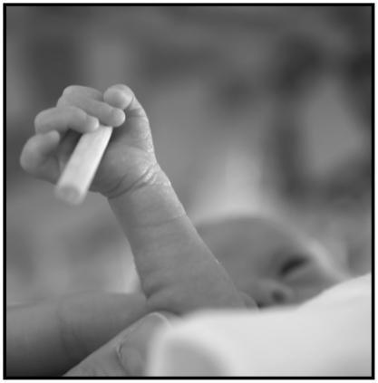
This grayscale clinical photograph depicts the palmar grasp reflex in a preterm infant. The focus is on the infant's hand and forearm as it securely grips a small, smooth cylindrical object. The infant's fingers are flexed around the cylinder, demonstrating the tactile and motor response characteristic of the primitive grasp reflex. The cylinder, approximately 35 mm in length and 6 mm in diameter, is positioned within the palm, with the distal end protruding. In the blurred background, the infant's face is partially visible, indicating the neonatal clinical setting. This image serves as an educational tool for neonatal development, illustrating haptic perception and sensorimotor coordination in premature newborns. It is particularly relevant for pediatric and neonatal research focusing on the maturation of the central nervous system and the evaluation of primitive reflexes as indicators of neurological health.
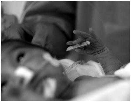
This black and white clinical photograph depicts a preterm neonate in a Neonatal Intensive Care Unit (NICU) setting, illustrating a study on neonatal haptic perception and the palmar grasp reflex. The primary focus is the infant's hand holding a small, light-colored triangular prism stimulus (approximately 35 mm in length). The infant exhibits medical support common in neonatal care, including adhesive hydrocolloid dressings or tapes on the face, likely securing an orogastric or nasogastric tube and respiratory support interfaces, such as CPAP prongs or nasal cannulae. A monitor lead or tubing is also visible near the chest area. This procedural image demonstrates the assessment of somatosensory processing and motor coordination in premature infants. It highlights the clinical relevance of developmental milestones and sensory integration in early neurodevelopmental evaluations.
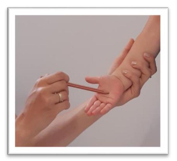
A clinical photograph illustrating the standardized testing procedure for the Palmar reflex (grasp reflex) on a child's right hand. The image depicts an examiner's hand securely stabilizing the child's forearm while keeping the palm supinated and open. Using the other hand, the examiner applies a tactile stimulus to the central palmar surface using a thin, rod-like instrument (spatula). The child's fingers are visible in a neutral to slightly flexed position, awaiting the motor response. This procedure is used in pediatric neurology to assess the integration of primitive reflexes; persistence beyond early infancy can indicate developmental delays or central nervous system dysfunction. The setting appears to be a clinical or therapeutic environment, focusing on developmental milestone screening and neuromotor assessment.
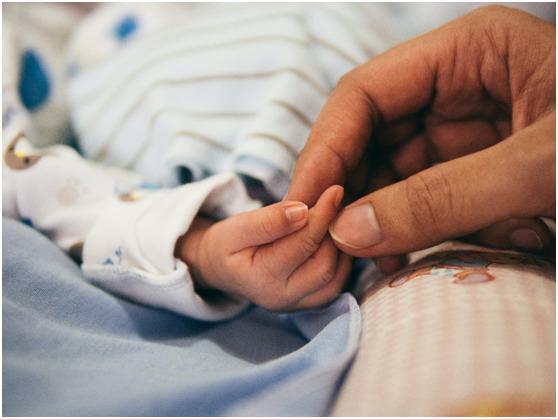
A high-detail clinical photograph capturing the physical interaction between an adult and a neonate, illustrating relative scale and developmental anatomy. The image focuses on the hands, where an adult's index finger and thumb are gently grasping the smaller hand of an infant. This comparison highlights the significant difference in hand size, skin texture, and nail morphology between developmental stages. The infant is shown in a clinical or home care setting, wearing a patterned long-sleeved garment and resting on blue and pink check-patterned bedding. The image demonstrates the palmar grasp reflex or general neonatal bonding, serving as a visual reference for pediatric physical examination, neonatal development, and maternal-infant health education. The lighting is soft and natural, emphasizing the fine skin folds of the infant's hand against the larger, mature hand of the adult.
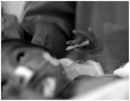
**Imaging Modality:** Clinical photograph, grayscale. **Anatomical Region:** Neonatal patient (head and hand). **Clinical Context:** The image depicts a preterm infant in a Neonatal Intensive Care Unit (NICU) setting. The infant is undergoing clinical observation or sensory assessment. **Key Landmarks and Visual Features:** * **Medical Equipment:** Visible medical interventions include a nasogastric or orogastric tube and adhesive tape applied to the mid-face/cheek area, typical for securing enteral feeding or respiratory support lines. A secondary medical tube is visible near the shoulder area. * **Neonatal Morphology:** The infant exhibits characteristic preterm features, including thin skin with visible wrinkling on the dorsal aspect of the hand and slender digits. * **Sensory Assessment:** The infant’s hand is shown in a grasping posture, holding a small, light-colored triangular prism. This suggests a functional assessment of the palmar grasp reflex or tactile stimulation. * **Positioning:** An adult hand is partially visible in the background, likely providing stability or assisting in the clinical examination. **Diagnostic Relevance:** This image serves as a clinical record of neonatal development, reflexive behavior, and the standard of care/monitoring for premature infants in a critical care environment.
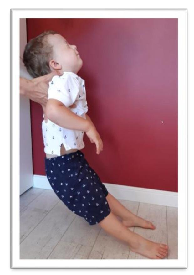
Clinical photograph illustrating the assessment of the Moro reflex in a preschool-aged child. The image depicts a therapist supporting the child's upper torso and head while the child is leaned backward. The child's head is in a position of cervical extension (tilted back) with eyes closed, which is a provocative maneuver to stimulate the vestibular system and trigger the reflex. The upper limbs are observed in a state of abduction or 'throwing out' to the sides, while the lower limbs show knee flexion. This clinical examination is used in pediatric neurology and physiotherapy to assess the integration of primitive reflexes. Persistent, unintegrated Moro reflexes beyond early infancy can indicate developmental delays or neurological maturity issues. The image demonstrates the standard physical therapy test position used to evaluate for residual reflex activity in older children, where compensatory movements like arm flailing or loss of balance are monitored.
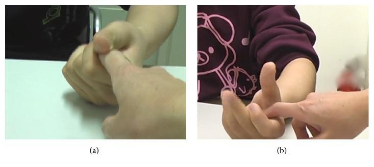
Two-panel clinical photograph demonstrating the evaluation of a grasp reflex in a pediatric patient with hemiplegia. Panel (a) illustrates the patient's hand in a forced, involuntary clenched fist position. An examiner's index finger is inserted into the palm, but the patient displays a pathological grasp reflex, showing an inability to inhibit the flexion or voluntarily open the hand to release the finger. Panel (b) shows the same clinical test after treatment with Botulinum Toxin Type A (BTX-A). In this frame, the patient demonstrates significant improvement in motor control; although the examiner's finger still provides a palmar stimulus, the patient is now able to voluntarily extend the fingers and open the hand. The comparison illustrates the reduction of spasticity and forced grasping following chemodenervation of the flexor muscles (including flexor digitorum superficialis and profundus), facilitating better functional movement and release capability in a neurorehabilitation context.
This clinical photograph captures an intraoperative scene of a surgical drainage procedure on a neonate's right hand. The image focuses on the palmar surface, where a longitudinal incision has been made to access the deep palmar space. A pair of surgical forceps is seen held by a gloved hand, facilitating the evacuation of thick, yellowish-white purulent material (pus) from the abscess site. The infant's hand rests on a sterile white gauze pad, which is positioned over a teal surgical drape. The surrounding skin of the palm and wrist appears slightly erythematous, consistent with localized inflammation secondary to a deep space infection. This visual demonstrates the management of neonatal hand infections, specifically the technique for incision and drainage of a palmar abscess. It serves as an educational example for pediatric surgery, infectious disease, and hand surgery specialties regarding the clinical presentation and surgical intervention of deep tissue neonatal infections.
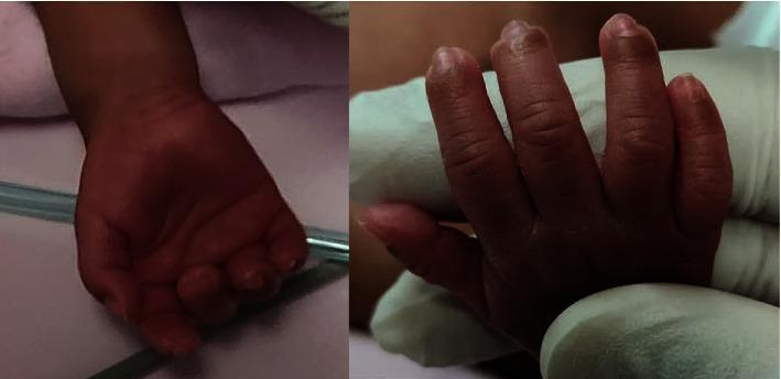
Clinical photograph of a neonate's right hand shown in two views: a palmar view (left) and a dorsal view (right). The hand belongs to an extremely premature infant (24 weeks gestation). The images demonstrate the complete resolution of previously documented digital ischemia. The skin displays a healthy, uniform reddish-pink hue across all digits and the palm, with no residual cyanosis, pallor, or bluish discoloration. The distal phalanges of the second, third, and fourth fingers appear well-perfused with intact skin integrity and no evidence of tissue necrosis, ulceration, or scarring. The soft tissue contour is normal for a preterm infant, showing no signs of significant edema or atrophy. These images illustrate successful recovery and reperfusion in neonatal vascular compromise, potentially following medical interventions like topical nitroglycerin.
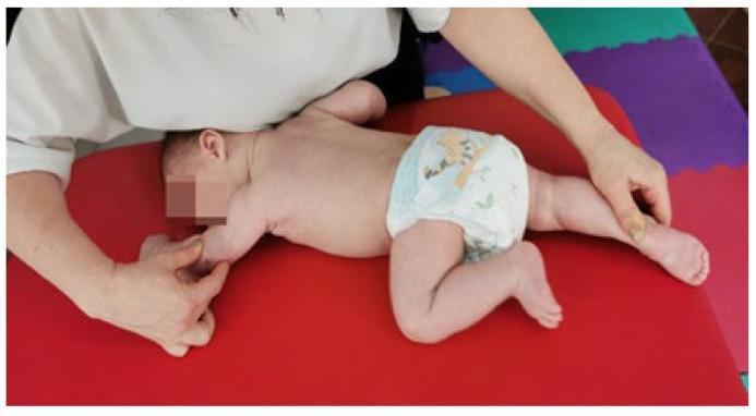
A clinical photograph illustrating a pediatric physical therapy session, specifically demonstrating Vojta therapy and the reflex crawling (Reflexkriechen) technique. The infant is positioned in a prone posture on a stable red treatment surface within a clinical or educational setting. A therapist's hands are visible, applying specific tactile stimuli and directional pressure to trigger global motor patterns. The infant's posture shows asymmetrical limb positioning characteristic of the crawling reflex: one arm is extended while the ipsilateral leg is flexed toward the torso, and the opposite limbs are in a complementary state of extension and flexion. This technique is often used in the rehabilitation of motor development delays and conditions such as muscular torticollis to improve neurological maturation and postural control. The image serves as an educational example of neuro-developmental treatment (NDT) and the assessment of primitive reflexes in infants.
neonatal primitive reflex testing clinical video demonstration YouTube 2023 2024
ATNR asymmetric tonic neck reflex infant assessment stepping reflex newborn
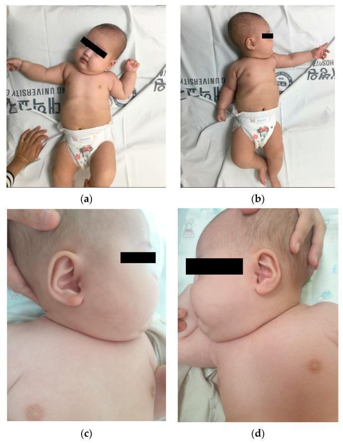
This set of four clinical photographs displays a 2-month-old infant with significant right-sided hemihyperplasia and associated muscular torticollis. Panel (a) and (b) provide full-body views in the supine position, demonstrating marked somatic asymmetry where the right upper and lower extremities appear significantly larger in circumference and volume compared to the contralateral left side. An asymmetric tonic neck reflex is captured in both orientations. Panels (c) and (d) present lateral close-up views of the head and neck. Image (c) shows the right side with visible fullness of the facial and neck soft tissues and a postural tilt, while image (d) provides a comparison of the left side. The visual findings illustrate clinical manifestations of hemihyperplasia, an overgrowth syndrome characterized by the asymmetric enlargement of one or more body parts. The neck positioning is consistent with congenital muscular torticollis, likely secondary to sternocleidomastoid muscle involvement on the hyperplastic side, resulting in a restricted range of motion toward the left.
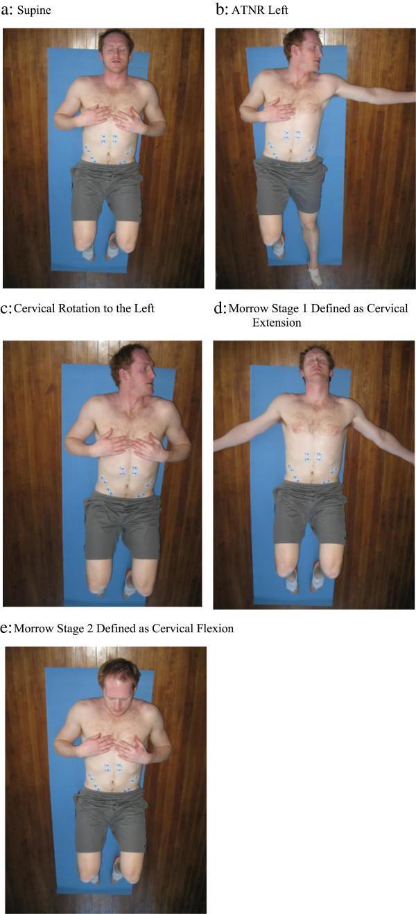
A series of clinical photographs (a-e) showing a male subject in various supine positions used to evaluate abdominal muscle activation in response to primitive reflex stimulation. Six surface electromyography (EMG) electrodes are bilaterally arranged on the abdomen: two over the rectus abdominis (inferior to the xiphoid), two superior to the anterior superior iliac spines (ASIS) for the external obliques, and two medial to the ASIS for the internal obliques. Positions include: (a) Neutral supine. (b) Asymmetric Tonic Neck Reflex (ATNR) Left: Head rotated left, left arm/leg extended, right arm/leg flexed. (c) Cervical Rotation Left: Head rotated left with symmetric limb flexion. (d) Moro Stage 1: Cervical extension with bilateral arm abduction. (e) Moro Stage 2: Cervical flexion with arms resting on the chest. The series demonstrates a kinesiopathology protocol aimed at identifying clinical differences in muscle recruitment patterns, often used in chronic low back pain (CLBP) research to investigate retained primitive reflexes.
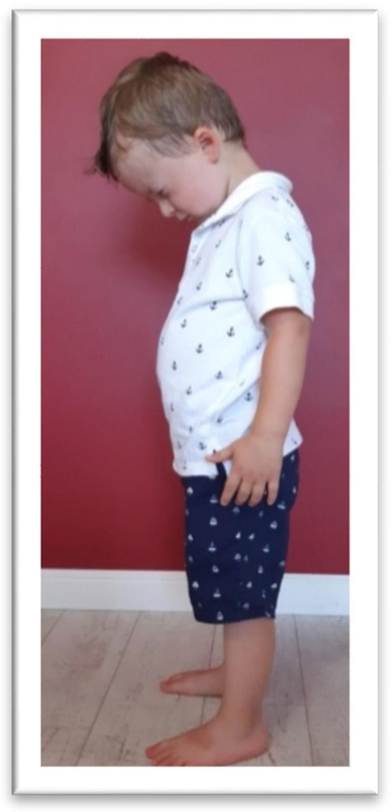
A clinical photograph depicting a pediatric patient in a standing position, demonstrating the assessment for a retained Tonic Labyrinthine Reflex in flexion (TLR FLX). The image illustrates characteristic compensatory motor responses when the child is asked to look downward. Visible findings include significant forward flexion of the head (neck flexion), a rounded or slumped spinal posture (thoracic kyphosis), and slight knee flexion (deflection) rather than maintaining a neutral upright stance. This visual is used in developmental pediatrics and physical therapy to identify primitive reflex persistence, which can impact postural control and motor coordination in preschoolers. The child is viewed from a lateral profile to highlight the sagittal plane deviations of the head, trunk, and lower extremities.
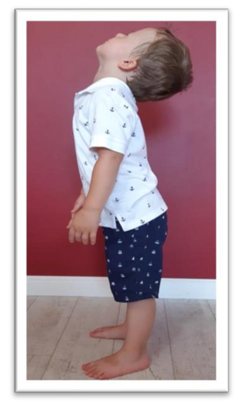
This clinical photograph shows a young child in a standing position, illustrating the examination procedure for the Tonic Labyrinthine Reflex (TLR) in extension. The child is positioned laterally to the camera, demonstrating a significant posterior tilt of the head (neck extension) and an upright torso. The arms hang naturally at the sides, and the feet are flat on the floor, slightly apart. The visual focus is on the body's posture in response to the change in head position, which is used in pediatric neurology and physical therapy to assess the integration of primary reflexes. Incomplete integration of the TLR is suggested if head extension triggers compensatory movements such as total body extension, limb straightening, or a loss of balance. This image serves as an educational reference for clinicians identifying motor reactions related to the vestibular system and brainstem-mediated primitive reflexes during childhood development assessments.
This diagnostic image is a transverse greyscale ultrasound of an infant's neck, focusing on the thyroid gland. The image demonstrates a characteristic morphological alteration where the anterior margin of the thyroid gland exhibits a loss of its typical multi-curved contour, replaced by a single, continuous convex curve that creates a 'bowed' appearance. This structural change results in a subjective impression of gland enlargement or bulkiness, despite objective volumetric measurements (1.75 ml) remaining within normal limits for a newborn. The echotexture of the gland is relatively homogenous and the borders remain smooth. The clinical context involves congenital hypothyroidism, specifically associated with a compound heterozygous DUOX2 mutation, illustrating how altered gland shape can confound subjective visual assessment of thyroid size in neonatal screenings. Key landmarks visible include the thyroid lobes, isthmus, and adjacent vascular structures.
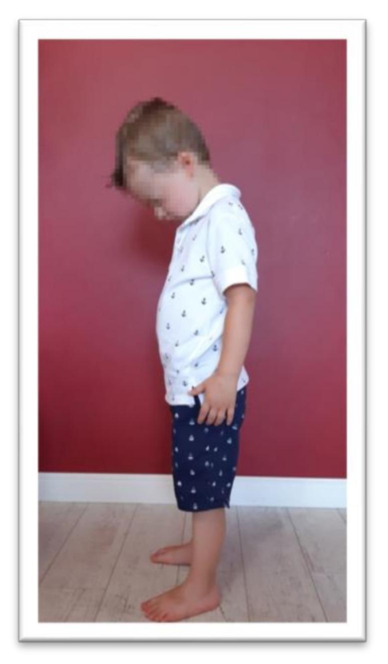
This clinical photograph shows a pediatric patient in a standing position, illustrating the standardized test for Tonic Labyrinthine Reflex (TLR) flexion. The child is depicted in a lateral profile, demonstrating a controlled forward flexion of the head with the chin tucked toward the chest. The trunk remains upright but slightly inclined anteriorly, while the upper and lower extremities are positioned naturally—arms at the sides and feet parallel on the floor. In the context of neurodevelopmental assessment, this position is used to evaluate the integration of primitive reflexes. Clinicians observe for compensatory motor patterns that indicate incomplete reflex integration, such as fist-clenching, involuntary knee deflection, or loss of postural balance triggered by the vestibular stimulus of head flexion. The image serves as an educational reference for pediatric physical therapy and neurological examination protocols in preschool-aged children.
site:youtube.com neonatal reflex examination newborn pediatric physiotherapy
Clinical Images
1. Palmar Grasp Reflex - Preterm Neonate (NICU)
2. Palmar Grasp - Standardised Testing Technique
3. Moro Reflex - Retained in Older Child (Assessment for Integration)
4. Tonic Labyrinthine Reflex (TLR) - Extension Assessment
5. Tonic Labyrinthine Reflex (TLR) - Flexion Assessment
6. ATNR + Moro - Research Protocol (EMG Surface Mapping)
Video Resources
Make a side-by-side comparison poster: normal vs retained reflex patterns
Okay. So now you have given me the card and everything. Now the workshop is going to be a four-hour workshop, and it is going to be very much focused on, um, how to assess these reflexes, then, um, what is normal age and everything, and what if it is retained, what could be the conditions, um, why is it important to assess. Also, what is the importance of these reflexes as to why God has given these reflexes to babies, right? Apart from that, if in, if, uh, other children, if these reflexes are retained, how to in- integrate them and how can they affect their daily functions. Apart from that, I want specifically for cerebral palsy, hypotonia, autism, ADHD, that how these reflexes can affect if, uh, is unintegrated, and, uh, at least 10 times strategies each for every reflex to get integrated. So give me a PDF, a proper PDF of four hours, whatever pages it takes, with all the links and the references.
primitive reflexes cerebral palsy autism ADHD retained integration
primitive reflex integration strategies cerebral palsy autism ADHD hypotonia pediatric physiotherapy 2024