
Tissues
What aspect of tissues are you interested in? · What level of detail do you need?
four basic tissue types epithelial connective muscle nervous histology
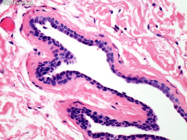
This histology slide is stained with Hematoxylin and Eosin and viewed under light microscopy. It shows branching tubular glands embedded in loose eosinophilic connective tissue stroma. The glands exhibit a well-organized epithelial lining consisting of tall columnar to cuboidal cells with basally oriented nuclei and scant cytoplasm. Lumina are variable in size and shape, ranging from rounded to elongated, with occasional pale secretory material within the lumen. The epithelial cells display minimal cytologic atypia, occasional small nuclear crowding, and no evident mitotic activity in this field. The surrounding stroma is pink and fibrous, with sparse inflammatory cells and few myofibroblasts. Overall architectural pattern is compatible with benign glandular epithelium or non-neoplastic glandular proliferation; differential considerations include normal mucosal glands, simple hyperplasia, or benign adenoma. There is no clear evidence of invasion into the adjacent stroma within the imaged section. This image is valuable for educational purposes in histology and diagnostic pathology, illustrating glandular morphology, lumen formation, epithelial-stromal relationships, and the spectrum between benign and neoplastic glandular processes. It supports teaching of ductal anatomy, tissue architecture, and histopathologic correlation with clinical scenarios. Clinically relevant context includes post-surgical specimens and mapping of glandular margins. This supports comparative pathology training and exam preparation.
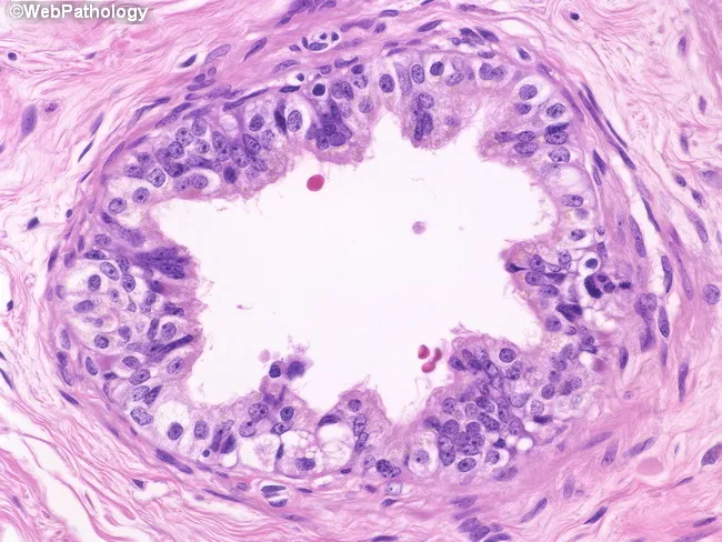
Imaging modality: Light microscopy of a hematoxylin and eosin–stained paraffin-embedded tissue section of efferent ductules from the caput epididymidis. The specimen shows multiple small tubular ducts arising from rete testis and converging toward the proximal epididymis with a centrally placed, undulating lumen. The epithelial lining is pseudostratified columnar, comprising tall ciliated cells interspersed with shorter nonciliated (absorptive) cells and scattered basal cells. A subtle lamina propria contains sparse lymphocytes. The mucosa forms a pronounced border around the lumen, reflecting active resorption of tubular fluid, a primary function of these ducts. The ciliated cells appear to augment luminal transport, while absorptive cells exhibit apical microvilli features. The surrounding smooth muscle and connective tissue provide peristaltic support. Notable features include variable epithelial height and occasional lymphocytes in the mucosa. In normal specimens, the lumen is irregular (undulating) and the epithelium shows mixed cell types without dramatic cytologic atypia. Clinically, this histology is relevant for evaluating obstructive infertility, congenital agenesis, or inflammatory involvement of the male reproductive tract. This image is useful for education, teaching histology of male reproductive ducts, differential diagnosis against epididymal pathologies, and research on sperm transport physiology. High magnification review emphasizes microvascular integrity and epithelial plasticity.
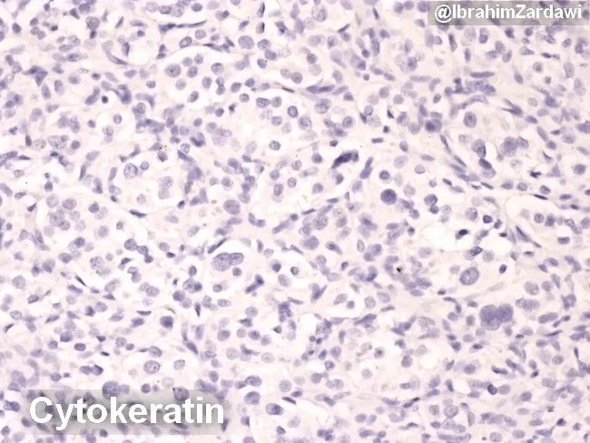
This is a histology image from formalin-fixed paraffin-embedded tissue stained by Cytokeratin immunohistochemistry and counterstained with hematoxylin, viewed under brightfield light microscopy. The tissue section reveals numerous rounded to polygonal cells with oval to eccentric nuclei, inconspicuous cytoplasm, and a diffuse cellular distribution set against a pale connective tissue background. Cytokeratin positivity highlights epithelial components within the specimen, enabling delineation of epithelial lineage amidst possible non-epithelial elements. The staining pattern supports epithelial differentiation and helps distinguish carcinomatous epithelium from lymphoid or mesenchymal neoplasms in diagnostic workflows. Observed features include uniform to mildly pleomorphic nuclear morphology, occasional nucleoli, and a generally cohesive cellular arrangement with minimal mitotic activity suggested by the micrographs. The image is most relevant to pathology and oncology; it is commonly used to confirm epithelial origin in suspected carcinomas, to characterize metastatic disease, or to support grading and staging processes in conjunction with additional markers (CK7, CK20, p40, CK5/6). Clinically, CK IHC informs tumor typing, guides treatment planning (surgery, chemotherapy, radiotherapy), and contributes to prognostic assessment. This slide is a representative educational example for students and clinicians studying cytokeratin-based identification of epithelial tumors. This description emphasizes search-relevant terminology for histology, immunohistochemistry, and epithelial tumors.
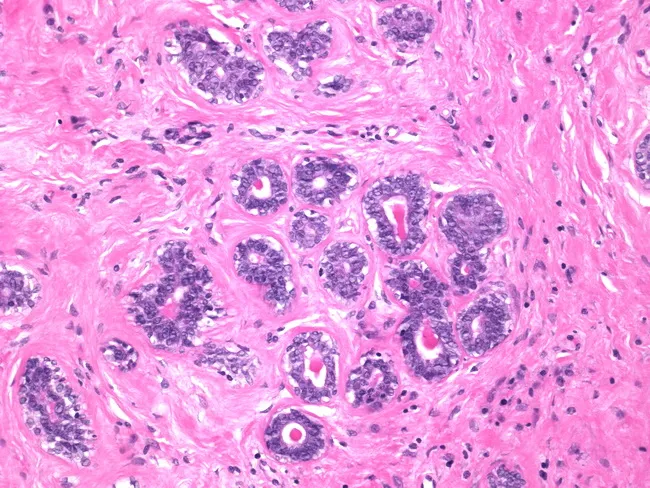
Histology: breast fibroepithelial lesion evaluated by light microscopy after Hematoxylin and Eosin staining. The specimen demonstrates tubular and glandular elements embedded in a fibrous loose stroma. Ducts and glands are lined by a bilayered epithelium composed of cuboidal to low columnar cells with uniform, bland nuclei. A continuous myoepithelial cell layer surrounds the tubules, supporting a benign biphasic architecture typical of a fibroadenoma. The stroma is predominantly loose connective tissue, with sparse to moderate cellularity and no overt stromal overgrowth. No prominent leaf-like stromal projections, mitotic figures, or cytologic atypia are evident within the epithelium or stroma. This combination of ductal epithelial proliferation with intact myoepithelial lining and a nonproliferative stroma favors a benign fibroepithelial lesion rather than a phyllodes tumor. The diagnostic significance rests on stromal characteristics: hypercellular stroma with progressive stromal overgrowth and leaf-like architecture would point toward phyllodes; absence of these features supports fibroadenoma. Clinically, these findings correlate with a small, mobile breast mass commonly seen in young to middle-aged women and may be managed conservatively or with excisional biopsy depending on size, growth, and patient preference. Immunohistochemistry for myoepithelial markers (e.g., p63, smooth muscle actin) can aid confirmation when needed.
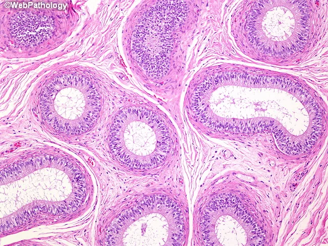
Imaging modality: Light microscopy of a formalin-fixed epididymal tissue section stained with Hematoxylin and Eosin (H&E). Anatomical context: Epididymis of the male reproductive tract, examining tubules that represent the head, body, and tail regions. Core histology: Tubules are lined by tall, pseudostratified columnar epithelium with apical cilia; the epithelial cells include dark-staining columnar cells and basal cells, with occasional clear cells. The lumina are widened and irregular, containing little luminal debris. The tubules sit within a thick tunica muscularis externa composed of multiple concentric smooth muscle layers, which supports peristaltic movement of sperm. Surrounding stroma is relatively loose with fibroelastic connective tissue. The mucosae display intact polarity, with nuclei oriented basally and apical margins bearing dense cilia. This image captures typical epididymal architecture: a specialized tall epithelium, narrow lumens, and a prominent muscular coat essential for sperm maturation, concentration, and propulsion. Pathologic deviations to this pattern would include epithelial desquamation, loss of ciliation, basement membrane disruption, or inflammatory infiltrates. Clinical relevance: normal epididymal histology demonstrates coordinated epithelial activity and smooth muscle contractility; findings aid education in male reproductive pathology, histology teaching, and differential diagnosis of scrotal pathology. In educational databases, this image enhances recognition of epididymal histology.
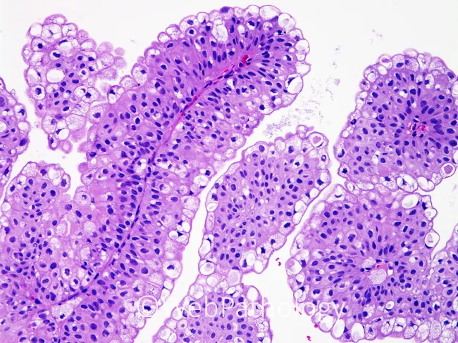
Brightfield hematoxylin and eosin stained histology slide of the small intestinal mucosa. The specimen shows finger-like villi lined by a simple columnar epithelium with scattered goblet cells producing mucin. Enterocytes display basally located nuclei and a dense cytoplasmic border, consistent with healthy absorptive cells. The lamina propria beneath the epithelium contains loose connective tissue and capillaries, and crypts of Lieberkühn appear at the bases of the villi with orderly architecture. Overall mucosal structure is preserved, with villous height and crypt organization within normal limits and no overt inflammatory infiltrate observed in this field. The image emphasizes epithelial surface details, goblet cell distribution, and intercrypt tissue patterns, making it an excellent reference for teaching normal intestinal histology and for contrast with pathological states. The magnification highlights mucosal architecture rather than deeper submucosa. Clinically, this specimen provides a baseline for malabsorption workups, educational comparisons with enteropathies such as celiac disease or infectious enteritis, and a reference standard for histological teaching. It supports differential diagnosis considerations, histologic correlation with serology, and radiologic-pathologic concordance in gastroenterology practice. This image is suitable for medical students, residents, and researchers studying basic histology, mucosal immunology, and nutrient absorption, and it can anchor discussions of normal variant goblet cell distribution across intestinal segments.
stratified squamous epithelium simple columnar epithelium histology microscopy
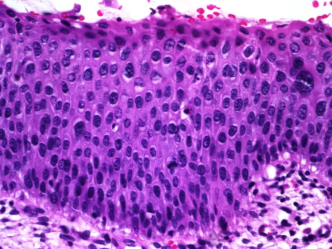
Histology image of mucosal epithelium showing features of squamous epithelial dysplasia on light microscopy. Prepared as a paraffin-embedded hematoxylin and eosin (H&E) stained section and viewed under brightfield microscopy at high magnification (~400x). The epithelium is stratified, with orderly basal cell layer but marked nuclear atypia throughout the lower and mid portions. Nuclear enlargement and hyperchromasia are evident, along with variation in nuclear size (anisokaryosis) and coarse, clumped chromatin. There is partial maturation at the superficial aspect, with surface layer showing more differentiated cells, suggesting some preserved maturation toward the lumen. Occasional mitotic figures are observed in the lower half of the epithelium, indicative of increased proliferative activity. Overall architecture reveals thickened epithelium (acanthosis) with preserved polarity in some cells but dysplastic changes in others. No invasion is evident in this field, though the mitotic activity and cytologic atypia raise concern for preinvasive squamous intraepithelial lesion. The imaging is relevant for diagnosis of epithelial dysplasia or squamous intraepithelial lesion and has clinical significance for risk stratification, biopsy correlation, and surveillance. Potential clinical applications include screening for precancerous lesions in mucosal surfaces, monitoring response to therapy, and educational demonstration of dysplastic cytology for medical students and residents.
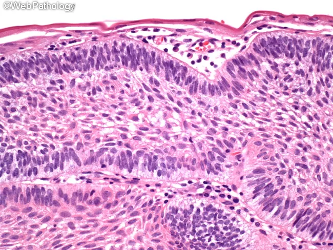
Imaging modality: light microscopy of Hematoxylin and Eosin stained paraffin section of gingival mucosa. Primary subject: peripheral ameloblastoma. Specimen: soft-tissue gingival/alveolar mucosa lesion. Perspective: transverse histologic section of mucosa. The image shows multiple islands and cords of odontogenic epithelium embedded in a dense fibrous stroma beneath the surface stratified squamous epithelium. At the periphery, tall columnar ameloblast-like cells display polarized, basophilic nuclei with reverse polarity; this peripheral palisading is juxtaposed to a central stellate reticulum-like zone. The islands are occasionally elongated and interconnected, with clear demarcation from the surrounding connective tissue. The overlying epithelium may be hyperplastic but lacks invasion into underlying bone. The stromal tissue is fibrous, moderately cellular, with delicate inflammatory cells. These histologic features are characteristic of benign odontogenic tumors confined to the soft tissue, with minimal atypia and low mitotic activity. Clinically, peripheral ameloblastoma presents as a slow-growing gingival mass that mimics reactive lesions; definitive therapy is conservative excision with clear margins. Prognosis is favorable, but local recurrences occur in 15-20% of cases, necessitating re-resection if recurrence is detected. Rare malignant transformation is described in literature. This image demonstrates classic peripheral ameloblastoma histology and serves educational purposes for pathology, oral surgery, and dental education. Educational use.
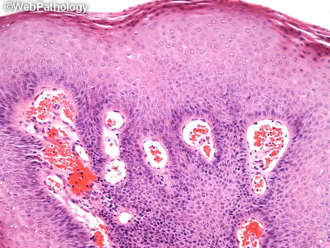
Histology, light microscopy of an oral mucosa biopsy stained with Hematoxylin and Eosin (H&E). The specimen shows a benign squamous papilloma with exophytic papillary projections arising from the surface epithelium. Each papillary frond contains a central fibrovascular core surrounded by keratinizing stratified squamous epithelium, exhibiting mild acanthosis and preserved maturation without dysplasia. The surface is often hyperkeratotic, producing a slightly corrugated or finger-like contour. The underlying lamina propria is fibrous and vascular, with chronic inflammatory infiltrate in keeping with a reactive mucosal lesion. No invasion of the basement membrane is observed, and margins (if excised) are free of tumor. Clinically, this lesion presents as a small, pedunculated or sessile oral growth that is typically benign and curable by simple excision, with rare recurrence. Differential considerations include verruca vulgaris and condyloma acuminatum, which can share papillary architecture but differ in HPV association and distribution. The lesion's benign behavior and lack of malignant transformation make it important to distinguish from squamous cell carcinoma in educational and diagnostic contexts. This image is valuable for education in oral pathology, histopathology teaching, and correlation with clinical oral lesions. This image thus supports educational objectives in surgical pathology, oral medicine, and histology curricula, reinforcing recognition of benign papillomatous lesions.
neuron nerve cell body axon dendrite histology
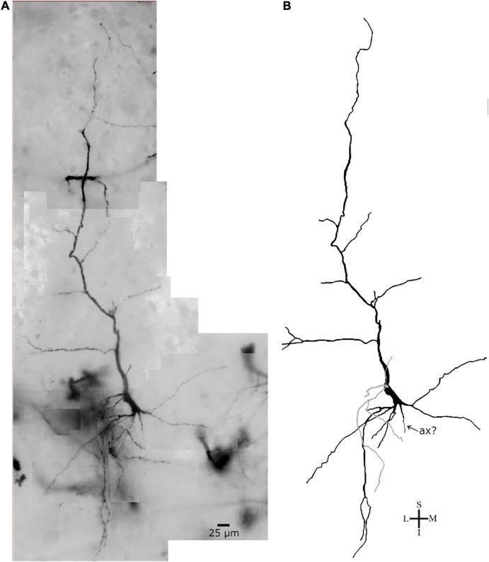
This figure presents a comparative study of a single neuron from inner layer VI of the human precuneus. Panel A displays a brightfield photomicrograph of the neuron prepared using Golgi impregnation, highlighting the cellular morphology against the cortical background. Panel B shows a corresponding two-dimensional digital reconstruction produced with specialized software. The neuron exhibits a characteristic spindle- to rod-shaped cell body (soma). It features two prominent, longitudinally oriented primary dendrites: an ascending (superior) dendrite and a thicker descending (inferior) dendrite, both of which show multiple branching points into secondary and collateral segments. A third, thinner primary dendrite is visible, highlighted in gray in the reconstruction. A process identified as a potential axon hillock (indicated by 'ax?') emerges from the soma, though the distal axon is not visualized. The pial surface is oriented at the top of the frame. This material is used to study neuronal diversity, specifically von Economo-like neurons or modified pyramidal neurons, within human cortical cytoarchitecture. Scale bar = 25 μm.
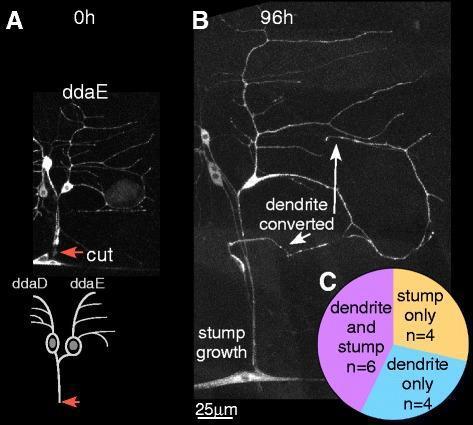
This composite educational graphic illustrates neuronal axon regeneration using Drosophila Class I ddaE sensory neurons as a biological model for neural injury and repair. Panel A presents a 0-hour baseline confocal image and schematic showing the site of distal axotomy (red arrow), located at least 20 μm from the cell body. Panel B displays the same neuron at 96 hours post-injury, demonstrating two simultaneous regenerative mechanisms: 'stump growth' from the original axotomy site and 'dendrite conversion,' where an existing dendrite has transformed into a functional axon. Panel C provides a quantitative comparison chart summarizing the outcomes of 14 experimental cases. The distribution shows that 42.8% (n=6) exhibited regeneration from both the stump and dendrite, 28.6% (n=4) showed stump growth only, and 28.6% (n=4) showed dendrite conversion only. This material demonstrates neural plasticity and the capacity for sensory neurons to specify new axonal processes from dendritic structures following significant distal injury.
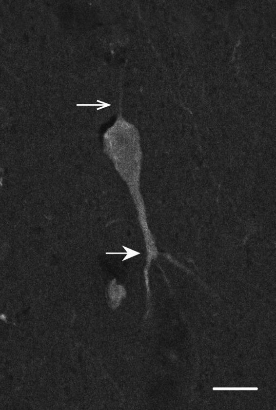
This diagnostic image is a high-magnification fluorescence micrograph showing a single calbindin-D28k-immunoreactive neuron within layer V of the primate cerebral cortex. The cell exhibits a dark, uniform staining pattern against a lighter background, characteristic of inhibitory interneurons. The morphology is defined by a distinct cell body with two primary types of processes. A thin axon (indicated by an open white arrow) extends superiorly toward the pial surface. In contrast, a thick, prominent primary dendrite (indicated by a closed white arrow) descends inferiorly toward the white matter, showing its first branching point at a distance from the soma. This morphological orientation resembles specific infragranular neuron populations. The image includes a 10 µm scale bar in the bottom right corner for dimensional reference. This visual material is used in neuroanatomy and histopathology to demonstrate laminar-specific neuronal morphology and calcium-binding protein expression.
1. Epithelial Tissue
Definition and Key Features
Classification
| Type | Description |
|---|---|
| Simple | Single layer of cells, all touching the basal lamina |
| Stratified | Multiple layers; only the basal layer contacts the basal lamina |
| Pseudostratified | Appears layered (all nuclei at different heights) but is actually one layer - all cells touch the basal lamina |
| Shape | Appearance |
|---|---|
| Squamous | Flat, scale-like |
| Cuboidal | Equal height and width |
| Columnar | Taller than wide |
| Transitional (urothelium) | Special - changes shape with stretching |
- Simple squamous - lung alveoli, blood vessel endothelium
- Simple columnar - GI tract (with goblet cells)
- Pseudostratified columnar ciliated - trachea, bronchi
- Stratified squamous keratinized - epidermis (skin)
- Stratified squamous non-keratinized - oral mucosa, esophagus
- Transitional - urinary bladder, ureters
Surface Modifications
- Microvilli - finger-like projections that increase absorptive surface area (intestinal brush border)
- Stereocilia - long non-motile microvilli (epididymis)
- Cilia - motile projections that move substances along surfaces (trachea)
Glands
- Exocrine glands - secrete onto surfaces via ducts (sweat glands, salivary glands)
- Endocrine glands - ductless; secrete hormones directly into blood (pituitary, thyroid)
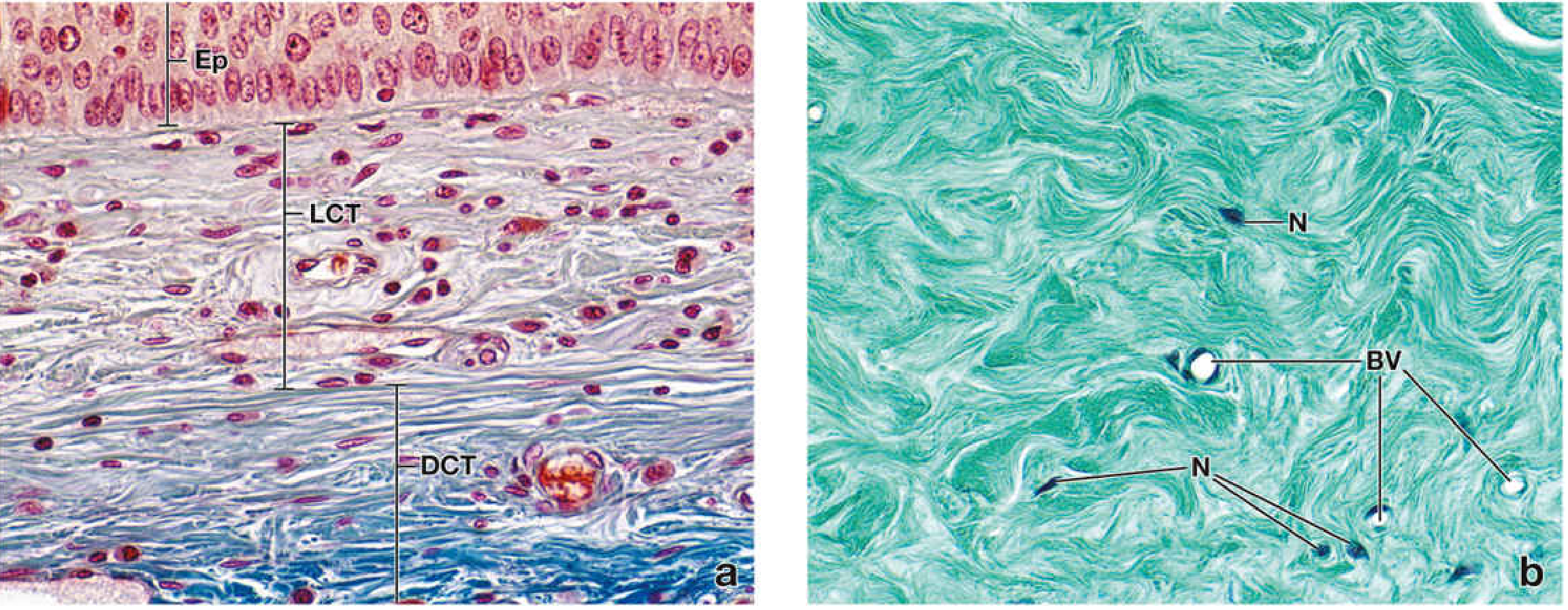
2. Connective Tissue
Definition and Key Features
Connective tissue underlies and supports (structurally and functionally) the other three basic tissues.
- Histology: A Text and Atlas, p. 301
Types of Connective Tissue
- Mesenchyme - loose, gel-like matrix; found in the embryo; gives rise to all other connective tissues
- Mucous (Wharton's jelly) - found in the umbilical cord
| Type | Matrix | Cells | Examples |
|---|---|---|---|
| Loose (areolar) | Loosely arranged collagen + elastin fibers; many cell types | Fibroblasts, macrophages, mast cells, plasma cells | Beneath epithelia; lamina propria |
| Dense regular | Densely packed parallel collagen fibers | Mainly fibroblasts | Tendons, ligaments |
| Dense irregular | Densely packed collagen in random directions | Mainly fibroblasts | Dermis, organ capsules |
| Tissue | ECM character | Key cells |
|---|---|---|
| Bone | Calcified matrix (hydroxyapatite + collagen) | Osteoblasts, osteocytes, osteoclasts |
| Cartilage | Large water-binding proteoglycans (hyaluronan) | Chondrocytes |
| Adipose tissue | Cells packed with lipid; minimal ECM | Adipocytes |
| Blood | Fluid ECM (plasma) | Erythrocytes, leukocytes, platelets |
Cells of Connective Tissue
- Fibroblasts - main cell; synthesize collagen, elastin, and ground substance
- Adipocytes - fat storage
- Mast cells - allergic and inflammatory responses (contain histamine, heparin)
- Macrophages (histiocytes) - phagocytosis
- Plasma cells - antibody production
- Neutrophils, eosinophils, lymphocytes - immune responses
ECM Components
- Collagen fibers - tensile strength (Type I most common)
- Elastin fibers - recoil/elastic properties
- Ground substance - glycosaminoglycans (GAGs) and proteoglycans fill the space
3. Muscle Tissue
Definition and Key Features
The bulk of the cytoplasm consists of the contractile proteins actin and myosin, which form thin and thick myofilaments, respectively.
- Histology: A Text and Atlas, p. 303
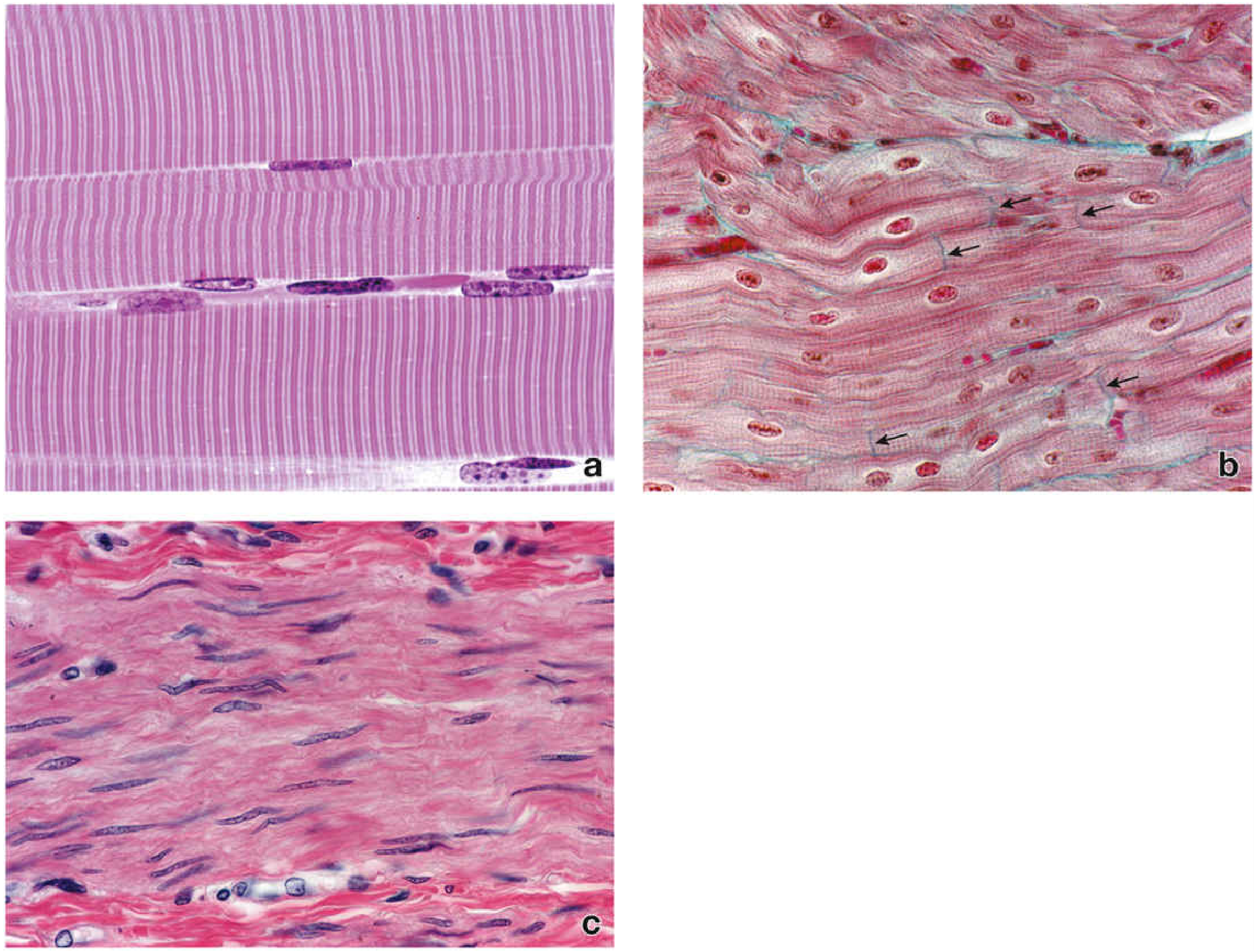
The Three Types of Muscle
| Feature | Skeletal | Cardiac | Smooth |
|---|---|---|---|
| Striations | Yes | Yes | No |
| Nuclei | Multiple, peripheral | 1-2, central | 1, central, elongated |
| Control | Voluntary | Involuntary | Involuntary |
| Cell shape | Long, cylindrical fibers | Branched, shorter cells | Spindle-shaped |
| Intercalated discs | Absent | Present | Absent |
| Location | Attached to skeleton | Heart | Walls of hollow organs (gut, vessels, bladder) |
| Regeneration | Limited (satellite cells) | Very limited | Good |
The Sarcomere (unit of contraction in striated muscle)
- Basic contractile unit bounded by two Z-lines
- I band: thin actin filaments only
- A band: thick myosin (overlaps with actin in middle)
- H zone: myosin only (no actin overlap)
- M line: center of sarcomere
- During contraction: actin slides over myosin, Z-lines move closer; I band and H zone shorten
4. Nervous Tissue
Definition and Key Features
Neurons
- Cell body (soma/perikaryon) - contains the nucleus and organelles; metabolic center
- Axon - single, long process; carries impulses away from the cell body; may be >1 meter long
- Dendrites - multiple, branching processes; carry impulses toward the cell body; receive incoming signals
- Multipolar - one axon + multiple dendrites (most common; motor neurons)
- Bipolar - one axon + one dendrite (retina, olfactory mucosa)
- Unipolar/pseudounipolar - single process that divides (dorsal root ganglion sensory neurons)
Glial Cells (Neuroglia)
| Cell | Function |
|---|---|
| Astrocytes | Structural support; blood-brain barrier formation; regulate extracellular ions |
| Oligodendrocytes | Myelinate CNS axons (each cell wraps multiple axons) |
| Ependymal cells | Line brain ventricles; produce CSF |
| Microglia | Resident immune cells; phagocytosis |
| Cell | Function |
|---|---|
| Schwann cells | Myelinate PNS axons (each cell wraps one axon); enable nerve regeneration |
| Satellite cells | Surround neuronal cell bodies in ganglia |
Organization of the Nervous System
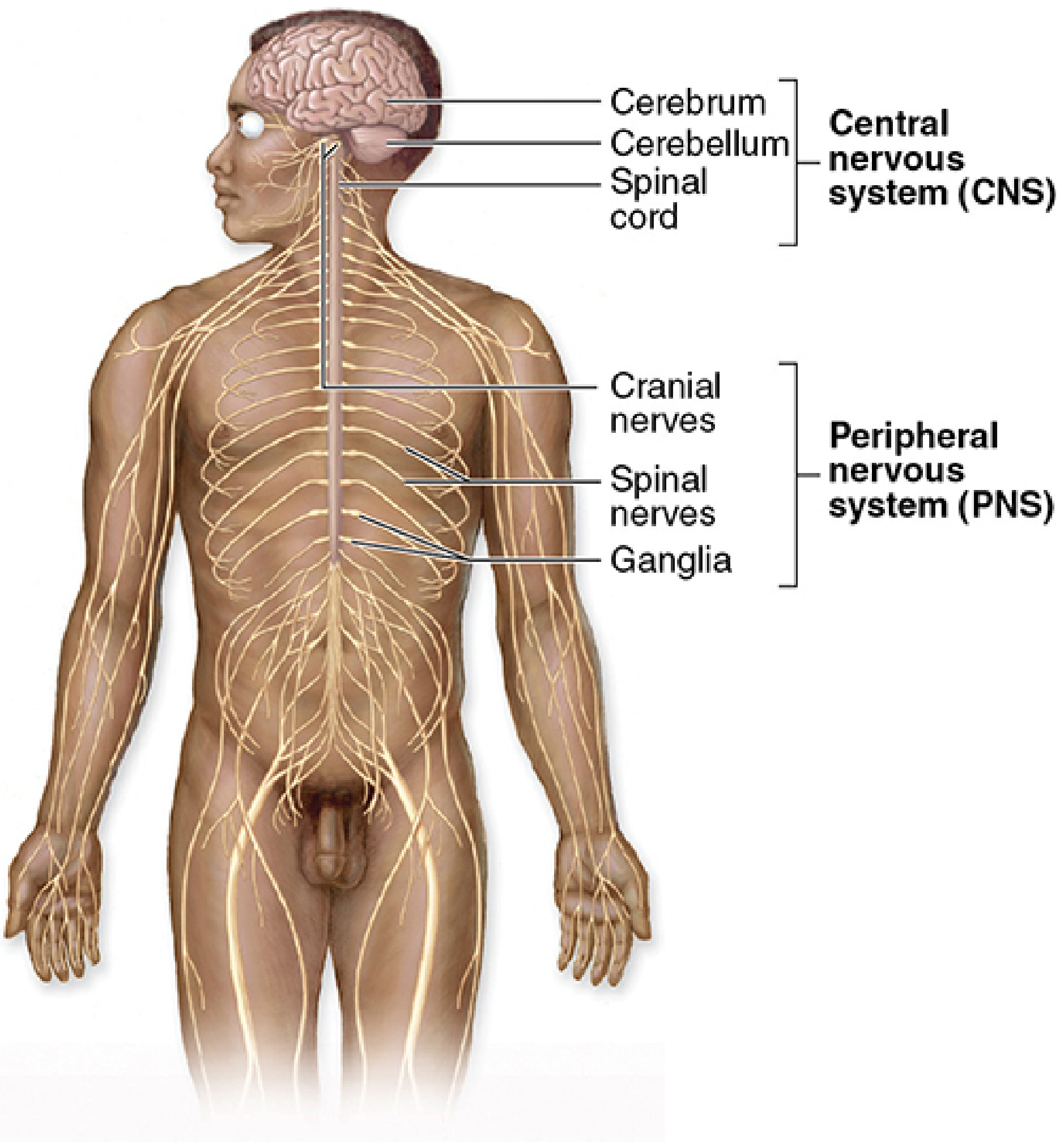
- Somatic nervous system - voluntary control of skeletal muscle
- Autonomic nervous system - involuntary control (sympathetic and parasympathetic)
Synaptic Transmission
- Action potential reaches the axon terminal
- Voltage-gated Ca²+ channels open
- Neurotransmitter vesicles fuse with membrane and release neurotransmitter into the synaptic cleft
- Neurotransmitter binds postsynaptic receptors
- Postsynaptic potential is generated (excitatory or inhibitory)
Histogenesis: Embryonic Origins
| Germ Layer | Tissues Derived |
|---|---|
| Ectoderm (surface) | Epidermis; corneal & lens epithelium; enamel; adenohypophysis; oral & anal mucosa |
| Neuroectoderm | CNS neurons & glia; PNS (Schwann cells, ganglia); melanocytes; adrenal medulla |
| Mesoderm | All connective tissues (bone, cartilage, blood, adipose); skeletal & smooth muscle; cardiac muscle; kidneys; gonads |
| Endoderm | Epithelium of GI, respiratory, urinary tracts; liver; pancreas; thyroid; thymus |
Summary Comparison
| Property | Epithelial | Connective | Muscle | Nervous |
|---|---|---|---|---|
| Hallmark | Close cell apposition; free surface | Extracellular matrix dominates | Contractile proteins (actin/myosin) | Electrical excitability |
| ECM | Minimal (basal lamina only) | Abundant - varies by type | Minimal | Minimal |
| Vascularity | Avascular | Vascular | Vascular | Vascular |
| Renewal | Rapid (mitosis) | Moderate | Limited | Very limited |
| Embryonic origin | Ectoderm, endoderm, mesoderm | Mesoderm (mostly) | Mesoderm | Neuroectoderm |