Image of atopic dermatitis

Searching Images

atopic dermatitis eczema skin

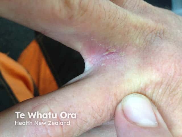
This is a clinical photography image focused on the interdigital web space of the hand, captured using standard digital photography, representing a dermatologic dermatitis in an atopic individual. Modality: Clinical photography; View: close-up interdigital and palmar skin, typically between the fingers, highlighting the first dorsal interosseous and web space. Location: skin of the hand, interdigital fold, palmar aspect; tissue involved: epidermis, with superficial dermal changes. Visual findings: erythematous to violaceous plaques with mild edema, silvery or yellowish scale, fissuring, and subtle maceration in the interdigital clefts; surrounding skin shows dryness and hyperemia. Pattern: localized, symmetric or unilateral involvement of the interdigital spaces, commonly the web spaces between digits 2 and 3 or 3 and 4 in atopic dermatitis. Pathology correlation: clinical diagnosis of eczematous dermatitis (interdigital eczema) in an atopic person; differential includes tinea manuum, allergic contact dermatitis, irritant dermatitis, dyshidrotic eczema. Diagnostic significance: supports atopic dermatitis–associated hand dermatitis; aids in monitoring response to emollients, topical corticosteroids, calcineurin inhibitors, and avoidance of irritants. Clinical utility: educational reference for dermatology trainees, clinicians assessing chronic hand dermatitis, and researchers studying atopic skin disease phenotypes; consider fungal and contact dermatitis testing where indicated. Correlation with patient history and targeted testing strengthens diagnostic accuracy.
This is a clinical photography image focused on the interdigital web space of the hand, captured using standard digital photography, representing a dermatologic dermatitis in an atopic individual. Modality: Clinical photography; View: close-up interdigital and palmar skin, typically between the fingers, highlighting the first dorsal interosseous and web space. Location: skin of the hand, interdigital fold, palmar aspect; tissue involved: epidermis, with superficial dermal changes. Visual findings: erythematous to violaceous plaques with mild edema, silvery or yellowish scale, fissuring, and subtle maceration in the interdigital clefts; surrounding skin shows dryness and hyperemia. Pattern: localized, symmetric or unilateral involvement of the interdigital spaces, commonly the web spaces between digits 2 and 3 or 3 and 4 in atopic dermatitis. Pathology correlation: clinical diagnosis of eczematous dermatitis (interdigital eczema) in an atopic person; differential includes tinea manuum, allergic contact dermatitis, irritant dermatitis, dyshidrotic eczema. Diagnostic significance: supports atopic dermatitis–associated hand dermatitis; aids in monitoring response to emollients, topical corticosteroids, calcineurin inhibitors, and avoidance of irritants. Clinical utility: educational reference for dermatology trainees, clinicians assessing chronic hand dermatitis, and researchers studying atopic skin disease phenotypes; consider fungal and contact dermatitis testing where indicated. Correlation with patient history and targeted testing strengthens diagnostic accuracy.

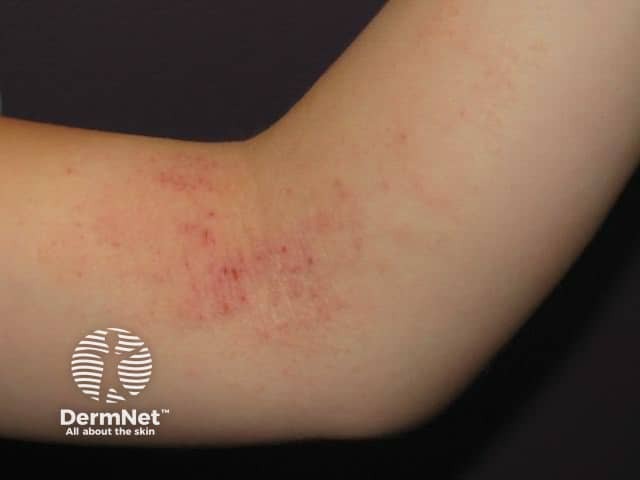
Clinical photography of a pediatric patient highlighting flexural atopic dermatitis in the antecubital region. Modality: Clinical photography (dermatology imaging) captured in color with standard resolution and natural lighting to preserve skin tone. Anatomical context: integumentary system; anterior elbow flexure (antecubital fossa) on the child’s left or unspecified arm. Visual features: erythematous, ill-defined plaques within the flexural creases with mild edema, background skin showing xerosis and secondary excoriations from pruritus; surface may be slightly lichenified and scaly; mild edema and fissuring at the skin folds; distribution is bilateral or unilateral depending on the patient but the image shows a prominent lesion in the flexural area. Pathology: clinical appearance consistent with pediatric atopic dermatitis (atopic eczema) rather than irritant contact dermatitis; features include chronic-relapsing eczema with pruritus, lichenification, and dry skin in flexures. Diagnostic significance: supports a diagnosis of childhood atopic dermatitis with flexural involvement; helps educate on typical morphological pattern and aids in differential diagnosis versus seborrheic dermatitis, contact dermatitis, or psoriasis in pediatric patients. Clinical use: assessment of disease extent, monitoring response to therapy (emollients, topical corticosteroids, calcineurin inhibitors), patient education, and training for students. This image serves as an educational reference for recognizing flexural eczema patterns across age groups and guiding noninvasive management decisions.
Clinical photography of a pediatric patient highlighting flexural atopic dermatitis in the antecubital region. Modality: Clinical photography (dermatology imaging) captured in color with standard resolution and natural lighting to preserve skin tone. Anatomical context: integumentary system; anterior elbow flexure (antecubital fossa) on the child’s left or unspecified arm. Visual features: erythematous, ill-defined plaques within the flexural creases with mild edema, background skin showing xerosis and secondary excoriations from pruritus; surface may be slightly lichenified and scaly; mild edema and fissuring at the skin folds; distribution is bilateral or unilateral depending on the patient but the image shows a prominent lesion in the flexural area. Pathology: clinical appearance consistent with pediatric atopic dermatitis (atopic eczema) rather than irritant contact dermatitis; features include chronic-relapsing eczema with pruritus, lichenification, and dry skin in flexures. Diagnostic significance: supports a diagnosis of childhood atopic dermatitis with flexural involvement; helps educate on typical morphological pattern and aids in differential diagnosis versus seborrheic dermatitis, contact dermatitis, or psoriasis in pediatric patients. Clinical use: assessment of disease extent, monitoring response to therapy (emollients, topical corticosteroids, calcineurin inhibitors), patient education, and training for students. This image serves as an educational reference for recognizing flexural eczema patterns across age groups and guiding noninvasive management decisions.

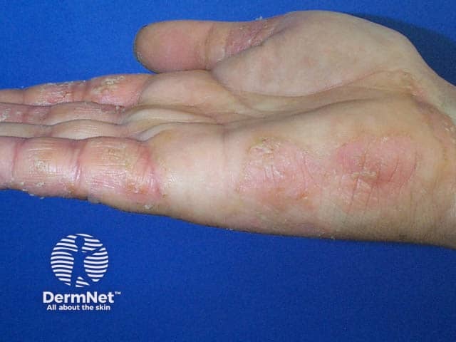
Imaging modality: Clinical photography of the hand. This high-resolution close-up image documents cutaneous changes on the palmar surfaces and digits of a hand, captured from the palmar (volar) aspect. The primary subject is atopic hand eczema (atopic dermatitis affecting the hands), evidenced by xerosis with rough, scaly patches, erythema, and episodic fissuring along the finger creases and interdigital spaces. The skin appears inflamed with mild edema and uniform pink to erythematous coloration in affected areas, while adjacent nonlesional skin remains comparatively normal. The distribution is patchy yet symmetric across involved digits, with involvement of the palmar skin and proximal interdigital folds. The image lacks cross-sectional tissue or histology, so all interpretation is clinically oriented: dermatitis rather than infection, psoriasis, or drug eruption. The visual pattern is characteristic of chronic, atopic-type dermatitis with a tendency toward lichenification with ongoing scratching in exposed areas. Notable diagnostic significance includes identification of hand-specific eczema that complicates daily activities, occupational exposure, and skin barrier dysfunction. This image can support differential diagnosis between atopic dermatitis and irritant/contact dermatitis, guide topical corticosteroid or calcineurin inhibitor therapy, and monitor treatment response. Clinically, correlate with pruritus history, eczema flares, personal or family atopy, and history of irritant exposure. Today.
Imaging modality: Clinical photography of the hand. This high-resolution close-up image documents cutaneous changes on the palmar surfaces and digits of a hand, captured from the palmar (volar) aspect. The primary subject is atopic hand eczema (atopic dermatitis affecting the hands), evidenced by xerosis with rough, scaly patches, erythema, and episodic fissuring along the finger creases and interdigital spaces. The skin appears inflamed with mild edema and uniform pink to erythematous coloration in affected areas, while adjacent nonlesional skin remains comparatively normal. The distribution is patchy yet symmetric across involved digits, with involvement of the palmar skin and proximal interdigital folds. The image lacks cross-sectional tissue or histology, so all interpretation is clinically oriented: dermatitis rather than infection, psoriasis, or drug eruption. The visual pattern is characteristic of chronic, atopic-type dermatitis with a tendency toward lichenification with ongoing scratching in exposed areas. Notable diagnostic significance includes identification of hand-specific eczema that complicates daily activities, occupational exposure, and skin barrier dysfunction. This image can support differential diagnosis between atopic dermatitis and irritant/contact dermatitis, guide topical corticosteroid or calcineurin inhibitor therapy, and monitor treatment response. Clinically, correlate with pruritus history, eczema flares, personal or family atopy, and history of irritant exposure. Today.

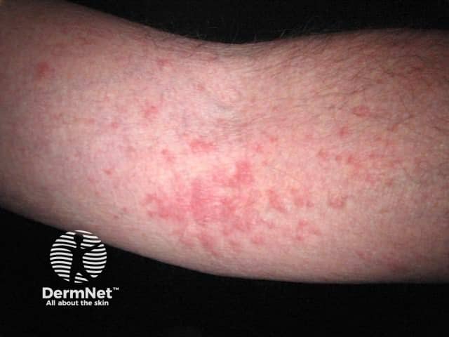
This dermatologic clinical photograph depicts an erythematous, ill-defined patchy to diffuse eruption on the antebrachial forearm consistent with atopic dermatitis (eczema). Acquired with a standard digital camera in close-up view of the skin surface under ambient lighting, it shows diffuse erythema, mild edema, and fine scaling with subtle lichenification in chronic areas. The distribution is localized to the forearm, with no clear involvement of mucosa or nails in this image. The superficial skin changes reflect dermatitis rather than infectious lesions; however, itching and chronic scratching are commonly reported. In epidermal pathology, atopic dermatitis corresponds to spongiotic dermatitis with perivascular lymphocytic infiltrate and potential epidermal hyperplasia in chronic lesions; that histopathology is not directly visible here. Clinically, this image supports diagnosis in the appropriate patient history of pruritus and atopy. Diagnostic significance includes distinguishing eczema from contact dermatitis and psoriasis. Differential considerations include irritant contact dermatitis, allergic contact dermatitis, seborrheic dermatitis, tinea corporis, and nummular eczema. Noting xerosis and barrier dysfunction, management implications involve emollient therapy, topical corticosteroids, and avoidance of irritants. This image is useful for educational demonstrations of typical dermatologic presentations and for image-based search queries related to atopic dermatitis.
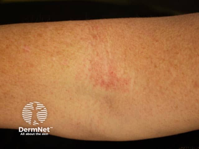
This dermatologic clinical photograph depicts an erythematous, ill-defined patchy to diffuse eruption on the antebrachial forearm consistent with atopic dermatitis (eczema). Acquired with a standard digital camera in close-up view of the skin surface under ambient lighting, it shows diffuse erythema, mild edema, and fine scaling with subtle lichenification in chronic areas. The distribution is localized to the forearm, with no clear involvement of mucosa or nails in this image. The superficial skin changes reflect dermatitis rather than infectious lesions; however, itching and chronic scratching are commonly reported. In epidermal pathology, atopic dermatitis corresponds to spongiotic dermatitis with perivascular lymphocytic infiltrate and potential epidermal hyperplasia in chronic lesions; that histopathology is not directly visible here. Clinically, this image supports diagnosis in the appropriate patient history of pruritus and atopy. Diagnostic significance includes distinguishing eczema from contact dermatitis and psoriasis. Differential considerations include irritant contact dermatitis, allergic contact dermatitis, seborrheic dermatitis, tinea corporis, and nummular eczema. Noting xerosis and barrier dysfunction, management implications involve emollient therapy, topical corticosteroids, and avoidance of irritants. This image is useful for educational demonstrations of typical dermatologic presentations and for image-based search queries related to atopic dermatitis.

Clinical photography of ventral forearm skin showing atopic dermatitis. Imaging modality: Dermatologic clinical photography; close-up, color-balanced capture suitable for visual examination and comparison over time. Anatomical region: antebrachial skin of the forearm; left/right not clearly discernible in the image. Visual features include diffuse erythema with ill-defined, sometimes coalescent plaques, fine scale, mild xerosis, and subtle lichenification. Excoriations from scratching may be present, reflecting pruritus-driven scratching. The appearance is consistent with chronic inflammatory eczema, not acute infection; edema is mild. Distribution appears localized to a distal forearm segment; flexural bias is possible but not required in adults. This pattern aligns with atopic dermatitis or eczema, though differential diagnoses include contact dermatitis, nummular eczema, seborrheic dermatitis, or psoriasis in overlapping cases. When correlated with clinical history (pruritus, atopy, family history, dry skin), the diagnosis of atopic dermatitis is favored. If histology were obtained, findings would typically include spongiotic epidermal changes with perivascular lymphocytic infiltrate. This image is clinically relevant for education, teledermatology triage, and documentation of lesion morphology, severity, and distribution. Potential clinical use cases include baseline documentation, monitoring therapeutic response (emollients, topical corticosteroids, calcineurin inhibitors), and patient counseling on trigger avoidance and skin care.
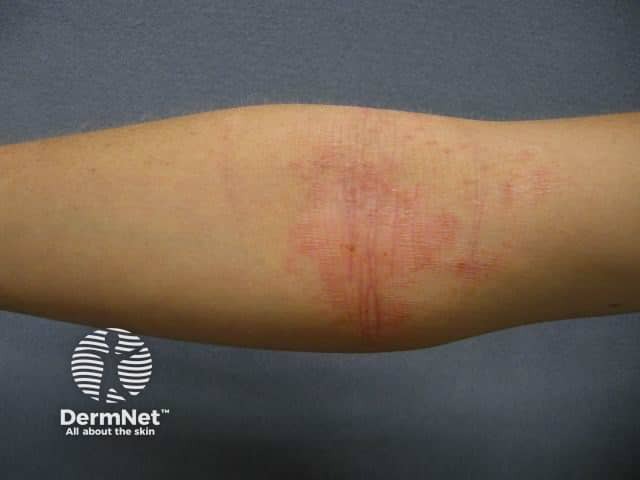
Clinical photography of ventral forearm skin showing atopic dermatitis. Imaging modality: Dermatologic clinical photography; close-up, color-balanced capture suitable for visual examination and comparison over time. Anatomical region: antebrachial skin of the forearm; left/right not clearly discernible in the image. Visual features include diffuse erythema with ill-defined, sometimes coalescent plaques, fine scale, mild xerosis, and subtle lichenification. Excoriations from scratching may be present, reflecting pruritus-driven scratching. The appearance is consistent with chronic inflammatory eczema, not acute infection; edema is mild. Distribution appears localized to a distal forearm segment; flexural bias is possible but not required in adults. This pattern aligns with atopic dermatitis or eczema, though differential diagnoses include contact dermatitis, nummular eczema, seborrheic dermatitis, or psoriasis in overlapping cases. When correlated with clinical history (pruritus, atopy, family history, dry skin), the diagnosis of atopic dermatitis is favored. If histology were obtained, findings would typically include spongiotic epidermal changes with perivascular lymphocytic infiltrate. This image is clinically relevant for education, teledermatology triage, and documentation of lesion morphology, severity, and distribution. Potential clinical use cases include baseline documentation, monitoring therapeutic response (emollients, topical corticosteroids, calcineurin inhibitors), and patient counseling on trigger avoidance and skin care.

This is a clinical dermatology photograph of a forearm, capturing a close-up view of suspected atopic dermatitis (eczema). Modality: clinical photography; imaging technique: standard color photography; surface skin lesion. Anatomically, the image shows the anterior aspect of the forearm (antebrachial region) with integumentary changes. The skin exhibits erythematous patches with mild scale and subtle edema, following linear and patchy distributions along the forearm. There may be faint superficial fissuring or skin cracking in the flexural areas, and some areas appear slightly lichenified due to chronic scratching. The surrounding skin is otherwise normal in tone. These findings are compatible with atopic dermatitis, including the hallmark features of erythema, scaling, and potential lichenification resulting from chronic scratching. Notable differential considerations include irritant or contact dermatitis, nummular eczema, psoriasis, seborrheic dermatitis, or other inflammatory dermatoses; clinical history and distribution help narrow the diagnosis. The diagnostic significance is that this image demonstrates typical eczema morphology: diffuse or regional erythematous plaques, pruritus, and chronicity. This photo is suitable for educational use in dermatology training, primary care assessment, and teledermatology, aiding recognition of acute and chronic atopic dermatitis, disease severity assessment, and treatment monitoring. Clinically correlates with pruritic history and chronic relapsing disease burden in practice.

This is a clinical dermatology photograph of a forearm, capturing a close-up view of suspected atopic dermatitis (eczema). Modality: clinical photography; imaging technique: standard color photography; surface skin lesion. Anatomically, the image shows the anterior aspect of the forearm (antebrachial region) with integumentary changes. The skin exhibits erythematous patches with mild scale and subtle edema, following linear and patchy distributions along the forearm. There may be faint superficial fissuring or skin cracking in the flexural areas, and some areas appear slightly lichenified due to chronic scratching. The surrounding skin is otherwise normal in tone. These findings are compatible with atopic dermatitis, including the hallmark features of erythema, scaling, and potential lichenification resulting from chronic scratching. Notable differential considerations include irritant or contact dermatitis, nummular eczema, psoriasis, seborrheic dermatitis, or other inflammatory dermatoses; clinical history and distribution help narrow the diagnosis. The diagnostic significance is that this image demonstrates typical eczema morphology: diffuse or regional erythematous plaques, pruritus, and chronicity. This photo is suitable for educational use in dermatology training, primary care assessment, and teledermatology, aiding recognition of acute and chronic atopic dermatitis, disease severity assessment, and treatment monitoring. Clinically correlates with pruritic history and chronic relapsing disease burden in practice.

Here are clinical images of atopic dermatitis (eczema) showing characteristic features:
Flexural involvement (classic pediatric presentation) - erythematous, ill-defined plaques in the antecubital fossa with xerosis and lichenification:
Flexural atopic dermatitis
Forearm involvement - diffuse erythema with fine scale and subtle lichenification from chronic scratching:
Forearm atopic dermatitis
Hand eczema - xerosis, erythema, and fissuring at finger creases, a common site in chronic atopic disease:
Hand atopic dermatitis
Key features to note across these images: erythematous, ill-defined plaques; dryness (xerosis); fine scaling; and lichenification (skin thickening) from chronic rubbing/scratching. Distribution is classically flexural (antecubital and popliteal fossae) in children, though hands and forearms are commonly affected in adults. Differentials to keep in mind include contact dermatitis, nummular eczema, seborrheic dermatitis, psoriasis, and tinea, which are distinguished by history of atopy, pruritus pattern, and distribution.
This is a shared conversation. Sign in to Orris to start your own chat.