
Anatomical basis of facial palsy
facial nerve anatomy course branches diagram
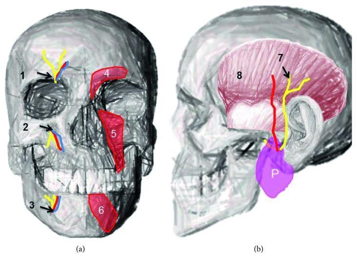
Anatomical diagrams of the human skull illustrating terminal branches of the trigeminal nerve and associated facial structures. Figure (a) is an anterior view highlighting the supraorbital notch (1), infraorbital foramen (2), and mental foramen (3), with accompanying neurovascular bundles shown in yellow (nerves) and blue/red (vessels). The diagram also depicts the overlying musculature: the corrugator supercilii (4), levator labii superioris (5), and depressor labii inferioris (6). Figure (b) provides a lateral view demonstrating the course of the auriculotemporal nerve (7) emerging from the parotid gland (P) and ascending across the temporal region. The temporalis muscle (8) is shown in pink, providing anatomical context for nerve distribution. This educational illustration demonstrates the relationship between bony landmarks, superficial facial muscles, and the sensory nerve exits essential for clinical procedures such as regional nerve blocks and the management of trigeminal neuralgias.
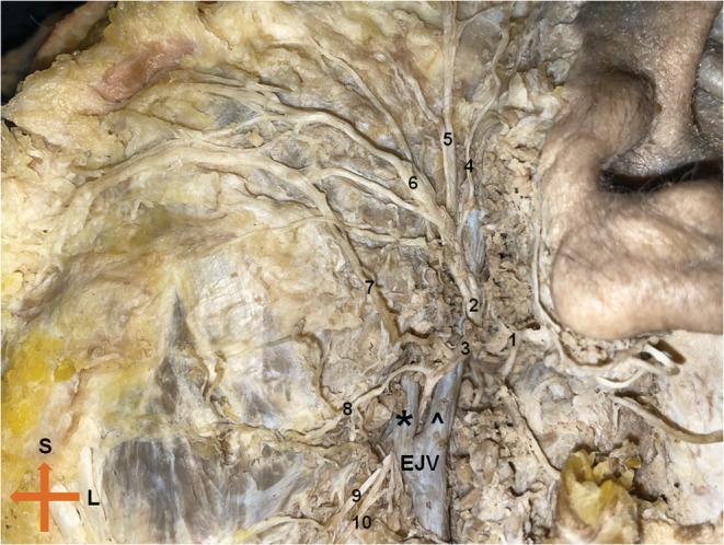
This clinical photograph displays a cadaveric dissection of the left extratemporal facial nerve (Cranial Nerve VII), highlighting its complex branching pattern and vascular relationships. The main trunk bifurcates into the temporofacial and cervicofacial divisions. The superior temporofacial division gives rise to the temporal and zygomatic branches, which course toward the upper face, and a buccal branch. The inferior cervicofacial division exhibits a rare anatomical variation where it bifurcates around the posterior facial vein; the upper branch passes superficially while the lower branch courses deep to the vein. Key anatomical landmarks include the retromandibular vein, the external jugular vein (EJV), and the masseter muscle. Labeled structures include the temporal (4), zygomatic (5), buccal (6), and marginal mandibular (8, 9) branches. This image is an essential educational resource for surgical anatomy, particularly for parotidectomy and maxillofacial procedures, demonstrating the critical proximity of neural branches to venous structures in the parotid region.
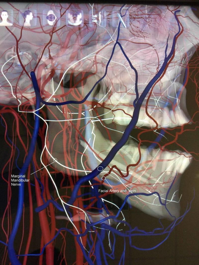
Anatomical diagram and 3D visualization illustrating a rare neurovascular anatomical variation in the maxillofacial region. This lateral view focuses on the right side of the mandible and cervical area, highlighting the spatial relationship between the marginal mandibular nerve (MMN), the facial artery, and the facial vein. The MMN is depicted as a blue tubular structure descending toward the lower border of the mandible. Crucially, the diagram demonstrates an atypical course where the MMN runs deep (medial) to the facial artery and vein (rendered in red), contrasting with the standard anatomical presentation where the nerve typically crosses superficial to these vessels. The visualization overlays these structures on a skeletal model of the skull and mandible, showing the nerve's proximity to the mandibular angle and body. This educational content is relevant for surgical anatomy, particularly in the context of neck dissections, submandibular gland excision, and rhytidectomy, where preserving the MMN is critical to prevent paralysis of the depressor muscles of the lower lip.
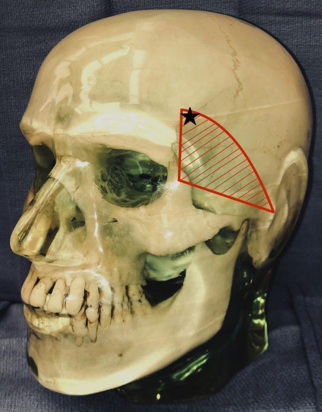
This anatomical diagram utilizes a lateral view of a human skull model to illustrate the surgical vulnerability of the temporal branch of the facial nerve. A red hatched triangular region is overlaid on the temporal fossa, bounded by the zygomatic arch inferiorly, the zygomatic process of the frontal bone anteriorly, and extending toward the superior temporal line. This area represents the danger zone where the temporal branch of the facial nerve is most superficial. A black star is marked within the superior-anterior portion of this shaded region, indicating the specific placement of a Mayfield head clamp pin that resulted in postoperative frontalis muscle palsy. The image serves as a clinical education tool for neurosurgical and orthopedic surgical positioning, highlighting the risk of iatrogenic nerve injury from external fixation devices when applied near the course of the facial nerve branches.
Anatomical Basis of Facial Palsy
1. Overview of the Facial Nerve (CN VII)
| Fiber Type | Function | Nucleus/Origin |
|---|---|---|
| Special visceral efferent (SVE) | Muscles of facial expression, stapedius, stylohyoid, posterior digastric | Motor nucleus (pons) |
| General visceral efferent (GVE) | Lacrimal gland, submandibular/sublingual glands, nasal glands | Superior salivatory nucleus |
| Special visceral afferent (SVA) | Taste - anterior 2/3 tongue (via chorda tympani), palate (via GSPN) | Nucleus tractus solitarius |
| General somatic afferent (GSA) | Skin of EAC, conchal auricle | Geniculate ganglion |
| Visceral afferent | Mucosa of nose, pharynx, palate | Nucleus tractus solitarius |
- Cummings Otolaryngology, p. 2451
2. Course of the Facial Nerve - Segment by Segment
A. Intracranial / Posterior Fossa Segment
- Exits the pons at the pontomedullary junction, lateral to the abducens nucleus
- The facial nerve fasciculus loops around the abducens (VI) nucleus in the floor of the fourth ventricle - this explains why pontine lesions at this level cause ipsilateral CN VI + VII palsy together
- Travels with CN VIII across the cerebellopontine angle (CPA) to enter the IAC
B. Meatal Segment (within the IAC)
- The nerve lacks a fibrous sheath or endoneurium here - surrounded only by a thin arachnoid layer
- This makes it especially vulnerable to compressive lesions (e.g., vestibular schwannoma)
C. Labyrinthine Segment
- The shortest and narrowest segment of the fallopian canal
- Travels superior to the cochlea to reach the geniculate fossa
- The geniculate ganglion contains sensory cell bodies; it is separated from the middle fossa floor by bone that is dehiscent in ~25% of ears
- A watershed vascular zone between vertebrobasilar (AICA labyrinthine branch) and external carotid (petrosal branch of MMA) supplies - making this the most susceptible site for ischemic/inflammatory nerve injury (e.g., Bell's palsy)
- The Greater Superficial Petrosal Nerve (GSPN) exits anteriorly here toward the pterygopalatine ganglion (lacrimation, nasal glands)
D. Tympanic (Horizontal) Segment
- Runs along the medial wall of the middle ear, forming the superior wall of the oval window niche
- Most common site of congenital bony dehiscence (above the oval window in ~40%)
- Vulnerable to injury in suppurative otitis media and cholesteatoma
E. Second Genu and Mastoid (Vertical) Segment
- At the pyramidal eminence, the nerve makes a second bend inferiorly
- The stapedius branch leaves here - damage proximal to this causes hyperacusis
- The chorda tympani branches off distally - carries taste (anterior 2/3 tongue) and parasympathetic fibers to submandibular/sublingual glands
- The space between the mastoid segment and chorda tympani is used surgically to create a posterior tympanotomy (facial recess)
F. Extratemporal Segment
- Exits via the stylomastoid foramen, receiving blood supply from the aponeurosis of the posterior digastric muscle
- Enters the parotid gland and divides into upper (temporofacial) and lower (cervicofacial) trunks
- Five terminal branch groups emerge from the parotid periphery: Temporal, Zygomatic, Buccal, Marginal mandibular, Cervical (mnemonic: To Zanzibar By Motor Car)
- Cummings Otolaryngology, p. 2451-2452; Gray's Anatomy for Students, p. 1052
3. Surgical Landmarks by Segment
| Segment | Key Surgical Landmark |
|---|---|
| Labyrinthine | Vertical crest (Bill's bar) in IAC |
| Geniculate ganglion | Retrograde dissection of GSPN (middle fossa approach) |
| Tympanic | Cochleariform process; supratubal recess |
| Second genu | Oval window |
| Mastoid | Pyramidal eminence; short process of incus; chorda tympani |
| Stylomastoid foramen | Cephalic edge of posterior digastric aponeurosis |
- Cummings Otolaryngology, Table 126.1
4. UMN vs LMN Lesion - The Critical Clinical Distinction
Upper Motor Neuron (UMN) Lesion
- Lesion is above the facial nucleus (above the pons) - usually cortex or internal capsule
- The forehead (frontalis muscle) is SPARED because it receives bilateral cortical representation (both hemispheres send corticobulbar fibers to the facial nucleus serving the upper face)
- Results in contralateral lower face paralysis only
- Emotional/involuntary facial movements may be preserved (the pathway for these travels separately via the basal ganglia)
Lower Motor Neuron (LMN) Lesion
- Lesion at or below the facial nucleus
- ALL ipsilateral facial muscles are paralyzed - upper face including forehead, lower face, orbicularis oculi
- Associated features depend on the precise level (see localization below)
- Kanski's Clinical Ophthalmology, p. 833; Rosen's Emergency Medicine, p. 1533
5. Anatomical Localization of the Lesion
| Site of Lesion | Facial Palsy Pattern | Additional Features |
|---|---|---|
| Cortex/Internal capsule | Contralateral lower face only (UMN) | Contralateral hemiplegia, aphasia |
| Pons (near CN VI nucleus) | Ipsilateral complete LMN facial palsy | Ipsilateral CN VI palsy, contralateral hemiplegia, ipsilateral gaze palsy (PPRF involvement) |
| Cerebellopontine angle | Ipsilateral complete LMN facial palsy | Decreased lacrimation and salivation, hyperacusis, loss of taste (anterior 2/3 tongue), ± deafness/tinnitus (CN VIII) |
| Geniculate ganglion (e.g., Ramsay Hunt) | Ipsilateral complete palsy | Ear pain, vesicles in EAC/ear/face, loss of taste, hyperacusis, decreased lacrimation |
| Between geniculate ganglion and stapedius nerve | Ipsilateral complete palsy | Hyperacusis + loss of taste (no lacrimation deficit) |
| Between stapedius and chorda tympani | Ipsilateral complete palsy | Loss of taste only (no hyperacusis, no lacrimation deficit) |
| Below chorda tympani / at stylomastoid foramen | Ipsilateral motor palsy only | No taste loss, no hyperacusis, no lacrimation deficit |
| Parotid gland | Ipsilateral facial palsy (may be partial/branch-specific) | Expressionless face, sense of numbness |
- Kanski's Clinical Ophthalmology, p. 833-834; Cummings Otolaryngology, p. 2452
6. Clinical Images
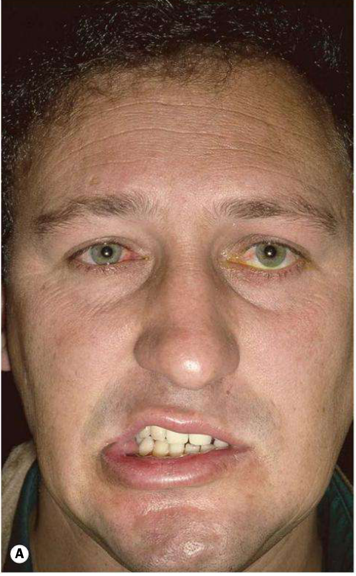
7. Vascular Supply and Vulnerability
- It is the narrowest segment - edema causes compression rapidly
- It is a vascular watershed zone between the vertebrobasilar system (AICA via labyrinthine artery) and external carotid system (petrosal branch of MMA + stylomastoid artery from posterior auricular artery)
- It lacks epineurium and the associated vascular plexus that normally protects against compressive injury
- Cummings Otolaryngology, p. 2452
Summary Diagram of Localization
Cortex/IC → UMN: contralateral lower face only
↓
Pons → LMN: ipsilateral all face + CN VI + gaze palsy
↓
CPA → LMN: ipsilateral all face + ↓tear/saliva + hyperacusis + taste loss + ±deafness
↓
Geniculate ganglion → LMN: same as CPA but with ear pain/vesicles (Ramsay Hunt)
↓
Post-geniculate, pre-stapedius → LMN + hyperacusis + taste loss
↓
Post-stapedius, pre-chorda → LMN + taste loss
↓
Post-chorda / stylomastoid → Pure LMN motor palsy (Bell's palsy typical level)
↓
Parotid → Partial/branch palsy only
Create a summary of Bell's palsy pathophysiology and treatment
"Bell palsy" treatment
Bell's palsy facial nerve inflammation mechanism
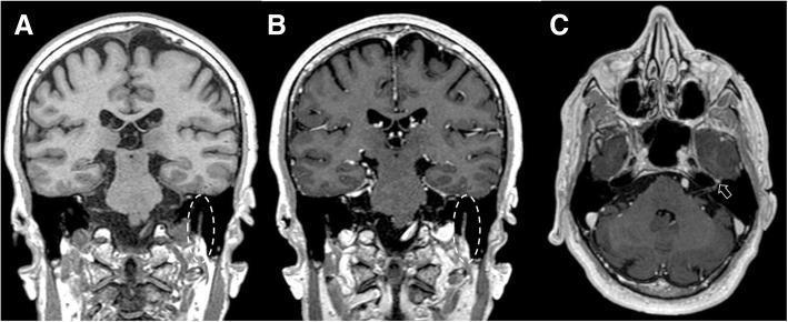
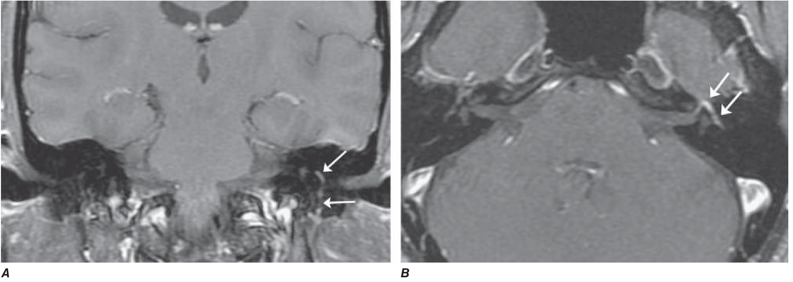
This set of diagnostic MRI images illustrates a clinical case of Bell’s palsy (idiopathic facial palsy). The figure comprises three panels: (a) and (b) are T1-weighted coronal views, while (c) is a T1-weighted axial view. Panels (a) and (b) provide a side-by-side comparison of the left facial nerve pre- and post-gadolinium administration. In the pre-contrast image (a), the nerve within the dotted circle displays normal dimensions and signal intensity. In the post-contrast image (b), the same region exhibits marked contrast enhancement and focal swelling, characteristic of inflammation and blood-nerve barrier disruption. Panel (c) is a post-contrast axial slice showing linear enhancement extending along the facial canal toward the geniculate ganglion (indicated by the arrow), which is located within the petrous portion of the temporal bone. These findings represent the typical radiological manifestation of Bell's palsy, distinguishing it from neoplastic processes by the absence of nodularity. The images serve as an educational resource for identifying cranial nerve VII pathology, relevant for neurology, neuroradiology, and emergency medicine specialties.
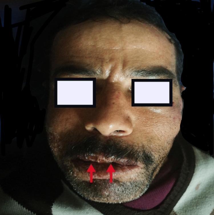
This clinical photograph shows a male patient exhibiting clear signs of left-sided peripheral facial nerve palsy (Bell's palsy) and concurrent erosive cheilitis. The facial asymmetry is characterized by the loss of forehead wrinkles on the left side, drooping of the left eyebrow, and a flattened left nasolabial fold. The mouth is noticeably deviated toward the unaffected right side. On the lips, the image highlights erosive cheilitis, with red arrows pointing to prominent vertical fissures and crusting on the vermilion border. This combination of symptoms—facial paralysis and lip inflammation—is clinically relevant for identifying conditions such as Melkersson-Rosenthal syndrome. The image serves as a high-quality educational resource for recognizing House-Brackmann Stage III facial nerve dysfunction and associated dermatological manifestations in a neurology or dermatology context.
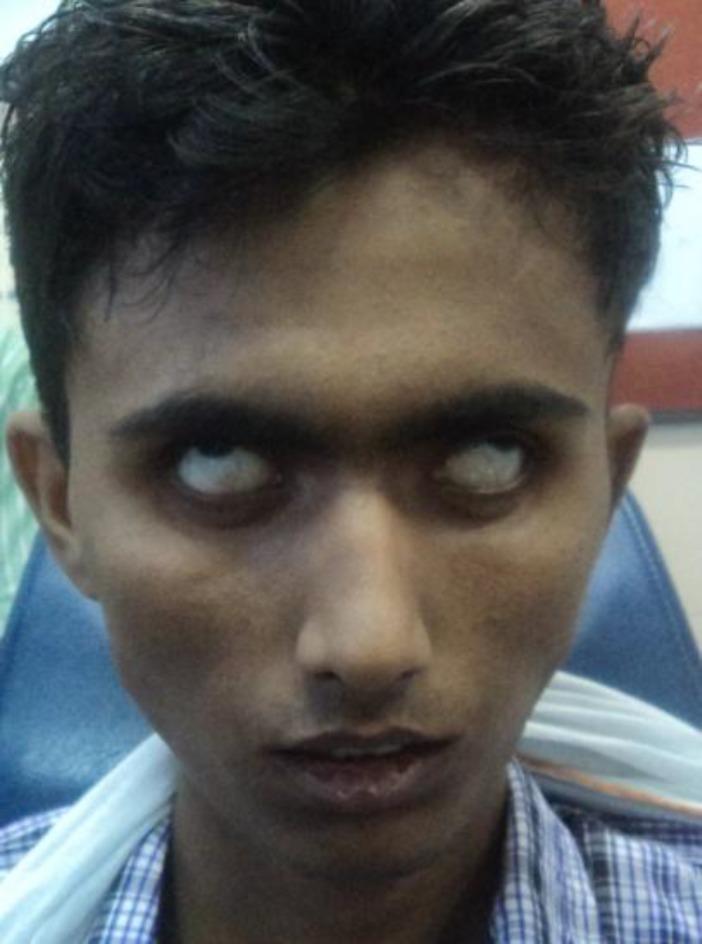
A clinical photograph of a young male patient demonstrating clinical signs of bilateral facial nerve paralysis (Bell's palsy), specifically categorized as House-Brackmann Grade 5 (severe dysfunction). The image highlights a significant lagophthalmos, with a complete inability to close either eyelid voluntarily. The eyes are wide open, and the upward rotation of the eyeballs (Bell's phenomenon) is visible, showing predominantly white sclera. There is a marked loss of facial muscle tone and expression, resulting in a mask-like appearance. The forehead remains smooth with an absence of wrinkling, and there is a lack of nasolabial fold definition bilaterally. This visual demonstrates the severe neurological impact on the muscles of facial expression, including the orbicularis oculi and frontalis muscles, often seen in systemic or bilateral pathologies such as tuberculous otitis media or other severe infectious/inflammatory conditions.
Bell's Palsy: Pathophysiology and Treatment
Definition and Epidemiology
- Incidence: ~25 per 100,000 per year (lifetime risk ~1 in 60)
- Affects men and women roughly equally; occurs at all ages
- Risk factors: Diabetes mellitus, pregnancy (especially the third trimester and first 2 weeks postpartum), possibly hypertension
- Recurrence rate: ~7-8% of cases; average interval between episodes ~10 years
- Harrison's Principles of Internal Medicine 22e, p. 3598; Adams and Victor's Principles of Neurology 12e, p. 1371
Pathophysiology
Proposed Mechanism: Viral Reactivation
- Viral reactivation in the geniculate ganglion (the sensory ganglion of CN VII, lodged within the labyrinthine segment of the fallopian canal)
- Inflammation of the facial nerve - mononuclear cell infiltrate, consistent with an infectious or immune-mediated process
- Edema and swelling of the nerve within the narrow, rigid fallopian canal
- Compression and vascular compromise - the labyrinthine segment is both the narrowest segment and a vascular watershed zone between the vertebrobasilar and external carotid systems
- Axonal injury - ranging from neuropraxia (conduction block, favorable prognosis) to axonotmesis/neurotmesis (axonal degeneration, poorer prognosis)
- HSV-1 DNA was found in endoneurial fluid surrounding the facial nerve in 11/14 surgical decompression cases (Murakami et al., PCR)
- HSV-1 DNA detected in the geniculate ganglion at autopsy in a Bell's palsy patient
- HSV-1 inoculation into mouse ears/tongues reproduced facial paralysis, with virus antigens found in the facial nerve and geniculate ganglion
Note: Despite the viral hypothesis, a Cochrane review found antivirals alone are no more effective than placebo for complete recovery - raising ongoing debate about causation vs. bystander reactivation.
- Harrison's, p. 3598; Adams and Victor's, p. 1371; Scott-Brown's ORL Vol 1, p. 7252-7269
MRI Findings
Clinical Features
| Feature | Details |
|---|---|
| Onset | Rapid - maximal weakness within 48-72 hours (virtually all by 3-4 days) |
| Preceding symptom | Retroauricular (behind the ear) pain, 1-2 days before palsy |
| Facial weakness | Complete ipsilateral LMN palsy - upper + lower face, including forehead and orbicularis oculi |
| Taste loss | Anterior 2/3 tongue (chorda tympani involvement) - present in most, rarely persists beyond 2 weeks |
| Hyperacusis | Stapedius muscle paralysis - distortion/loudness of sound |
| Eye | Lagophthalmos (inability to close eye), Bell's phenomenon (eyeball rolls up on attempted closure), corneal exposure risk |
| CSF | Mild lymphocytosis in a small number of cases |
Prognosis
- 70-80% of patients recover completely within 1-2 months
- 85% achieve near-normal facial function overall
- ~10% have little or incomplete recovery
- Best prognostic signs:
- Incomplete (partial) paralysis at presentation
- Early recovery of motor function within 5-7 days
- Return of taste within the first week
- Poor prognostic sign: EMG showing denervation after 10 days → indicates axonal degeneration → long delay (3+ months) before regeneration, often incomplete, taking 2 years or longer
Sequelae of Incomplete Recovery
- Synkinesis - aberrant reinnervation (e.g., eye closure with jaw movement - "jaw winking"; tearing with salivation - "crocodile tears")
- Facial muscle weakness / contracture
- Persistent lagophthalmos
- Goldman-Cecil Medicine, p. 2978; Harrison's, p. 3598
Treatment
1. Corticosteroids (First-line, High Evidence)
- Increases complete recovery rate from 63% to 83% at 3 months (vs. placebo)
- Mechanism: reduces nerve edema and compression within the fallopian canal
- Supported by landmark RCTs (Sullivan et al., Engstrom et al.) and guidelines
2. Antiviral Agents (Adjunct in Severe Cases)
- Antivirals alone show no independent benefit over placebo (Cochrane review)
- Combination (prednisolone + valacyclovir) may improve outcomes in complete paralysis (Hato et al.) - though other trials did not confirm this benefit
- Dose doubles for suspected VZV (Ramsay Hunt): acyclovir 800 mg 5× daily or valacyclovir 1 g three times daily
3. Eye Care (Essential in All Cases)
- Artificial tears (lubricating eye drops) during the day
- Eye ointment + taping shut at night
- Consider a moisture chamber or protective eyewear during the day
- Goal: prevent exposure keratopathy, corneal ulceration, and permanent visual damage
4. Physical Therapy
5. Surgical Decompression
Treatment Summary Table
| Intervention | Dose/Regimen | Evidence |
|---|---|---|
| Prednisolone | 25 mg BD × 10 days, or 40-60 mg/day × 7-10 days; within 72h | Strong (Grade A RCTs) |
| Acyclovir/Valacyclovir | Add to steroids in severe/complete palsy | Moderate (benefit uncertain, adjunct only) |
| Eye lubrication | Drops by day, ointment + taping by night | Standard of care |
| Physiotherapy | Facial exercises, neuromuscular re-education | Moderate (systematic review evidence) |
| Surgical decompression | Not recommended | No benefit, possible harm |
Differential Diagnosis (Exclusions Before Labeling Bell's Palsy)
| Condition | Distinguishing Features |
|---|---|
| Ramsay Hunt syndrome (VZV) | Ear pain + vesicles in EAC/pinna, worse prognosis |
| Lyme disease | Tick exposure, erythema migrans, can be bilateral; ≥10% of facial palsy in endemic areas |
| HIV seroconversion | CSF pleocytosis, risk factors |
| Sarcoidosis | Often bilateral, systemic features |
| Guillain-Barré syndrome | Bilateral facial palsy, ascending weakness |
| Parotid malignancy | Slow onset, mass palpable |
| Cholesteatoma/Otitis media | Ear discharge, chronic ear disease |
| Stroke | UMN pattern (forehead spared), other focal deficits |
- Harrison's 22e, p. 3598-3599; Adams and Victor's, p. 1372; Goldman-Cecil, p. 2961
Quick Reference Flowchart
Acute unilateral facial palsy
↓
LMN pattern? (forehead involved)
↓ Yes
Vesicles in ear/EAC? → Yes → Ramsay Hunt → antivirals + steroids
↓ No
Tick exposure / endemic area? → Yes → Lyme serology
↓ No
Bilateral? → Yes → Sarcoid, GBS, Lyme
↓ No
Idiopathic = Bell's palsy
↓
Start prednisolone within 72h
+ eye protection
± antivirals if severe/complete
Anatomical basis of wry neck
torticollis wry neck sternocleidomastoid anatomy
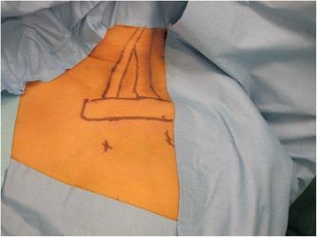
This clinical photograph displays the preoperative preparation of a patient's neck and chest region for arthroscopic release of the sternocleidomastoid (SCM) muscle, a procedure typically used to treat congenital muscular torticollis. The patient is in a supine position, with blue surgical drapes surrounding the sterile field. Anatomical landmarks are clearly delineated on the skin using a dark surgical marker. The markings outline the V-shaped sternal and clavicular heads of the SCM muscle as they converge toward the mastoid process. A horizontal rectangular box marks the superior border of the clavicle. Below the clavicle, two cross-marks indicate the planned sites for surgical access: the anteromedial portal (positioned inferior and medial to the sternoclavicular joint) and the working portal (positioned inferior to the midpoint of the clavicle). This visual serves to educate on surface anatomy palpation and the precise spatial orientation required for minimally invasive cervical muscle release.
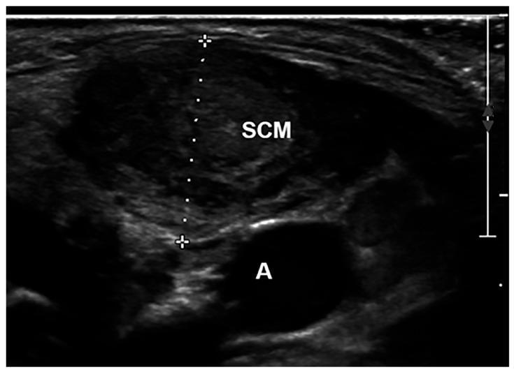
This diagnostic ultrasound image in the transverse plane depicts the anatomy of the neck, specifically focusing on the sternocleidomastoid muscle (SCM) and adjacent structures. The SCM is centrally featured, displaying a heterogeneous, elliptical morphology characterized by internal speckle artifacts typical of muscular tissue. White calipers and a dotted vertical line indicate a measurement of the SCM thickness, spanning the distance between the superficial and deep aponeurosis. Situated inferior and medial to the SCM is a well-defined anechoic (black) circular structure labeled 'A', representing the carotid artery, which serves as a key vascular landmark. The image includes a depth scale on the right margin for quantitative reference. This clinical imaging is representative of diagnostic protocols used in pediatrics to assess musculoskeletal conditions such as congenital muscular torticollis, allowing for the objective measurement of muscle fibrosis or hypertrophy relative to contralateral normal tissue.
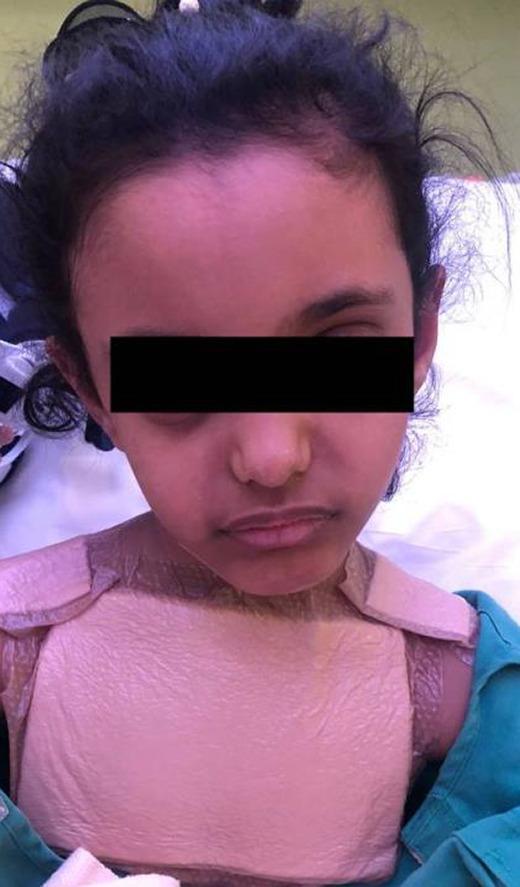
A clinical photograph of a pediatric patient presenting with torticollis (wry neck) and a strained facial expression indicative of clinical discomfort. The image shows a significant lateral tilt and rotation of the head, a common sign of cervical spine instability or muscular spasm. A rigid cervical collar (C-collar) with an anterior thoracic component is applied to the neck and upper chest to provide external stabilization and immobilization of the cervical spine. The clinical context suggests this presentation is associated with Grisel syndrome, a non-traumatic rotatory atlantoaxial subluxation that can occur following inflammatory processes in the head and neck, such as a retropharyngeal abscess. This visual material serves as a teaching tool for recognizing postoperative complications in otolaryngology and the physical manifestations of atlantoaxial instability.
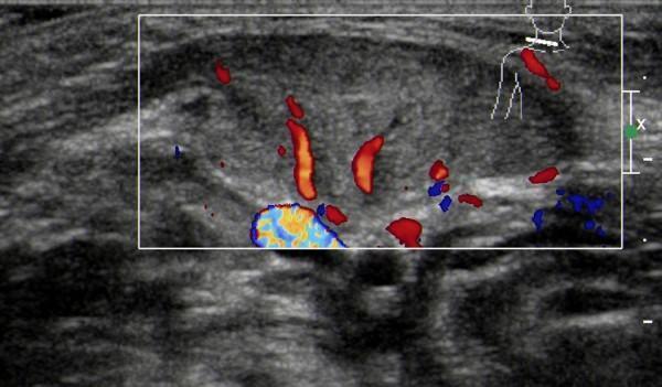
This diagnostic image is a Color Doppler Ultrasound of the neck, specifically targeting the sternocleidomastoid (SCM) muscle in a patient diagnosed with congenital muscular torticollis. The grayscale component of the scan shows a fusiform thickening of the affected SCM muscle with a predominantly hypoechoic internal texture, indicating uneven echo distribution compared to healthy muscle tissue. The muscle boundaries appear relatively distinct in this view. Superimposed Color Doppler signaling reveals increased vascularity within the thickened muscle mass, characterized by short, rod-like or cord-like flow signals in red and blue, representing arterial and venous blood flow. A larger vessel with turbulent flow (mosaic pattern) is visible at the inferior aspect of the ROI. This imaging is characteristic of the early inflammatory or hypervascular stage of muscular torticollis, where ultrasound is utilized to differentiate muscular masses from other cervical pathologies and to guide rehabilitation or surgical treatment planning.
Anatomical Basis of Wry Neck (Torticollis)
Definition
- S. Das Manual on Clinical Surgery 13e, p. 377; Bailey and Love's Surgery 28e, p. 9135
The Central Anatomical Player: The Sternocleidomastoid (SCM)
Attachments
| Head | Origin | Insertion |
|---|---|---|
| Sternal head | Anterior surface of manubrium sterni | Mastoid process of temporal bone and lateral half of superior nuchal line |
| Clavicular head | Medial third of clavicle | Same insertion |
Actions (Bilateral vs Unilateral)
| Contraction | Effect |
|---|---|
| Bilateral | Flexes the neck (when the head is fixed: elevates sternum/ribs in inspiration) |
| Unilateral | Tilts (laterally flexes) the head to the SAME side AND rotates the chin to the OPPOSITE side |
Nerve Supply
- Spinal accessory nerve (CN XI) - motor supply
- C2 and C3 ventral rami - proprioceptive and supplementary motor
Consequence of Pathology
- The head is pulled ipsilaterally (toward the tight SCM) - tilt
- The chin points contralaterally (away from the tight SCM) - rotation
Classification and Anatomical Basis of Each Type
1. Congenital Muscular Torticollis (CMT) - Most Common Type
- Malposition of the fetus in utero / intrauterine constraint
- Intrauterine or perinatal compartment syndrome (venous outflow obstruction → muscle ischemia → fibrosis)
- Birth trauma (forceps delivery, breech presentation)
- Vascular injury / infection
- The "trauma-compartment syndrome" hypothesis is currently most favored: difficult delivery → SCM injury → hematoma → fibrosis
- Head tilts toward the affected (fibrosed) SCM
- Chin rotates away from it
- In untreated or delayed cases: ipsilateral facial underdevelopment (plagiocephaly) - the outer canthus-to-mouth angle is smaller, the eyebrow less arched, the cheek less full - due to restricted muscular pull on the developing facial skeleton
- The fibrosis is most severe in the lower third of the SCM; limited lower-third involvement carries a better prognosis
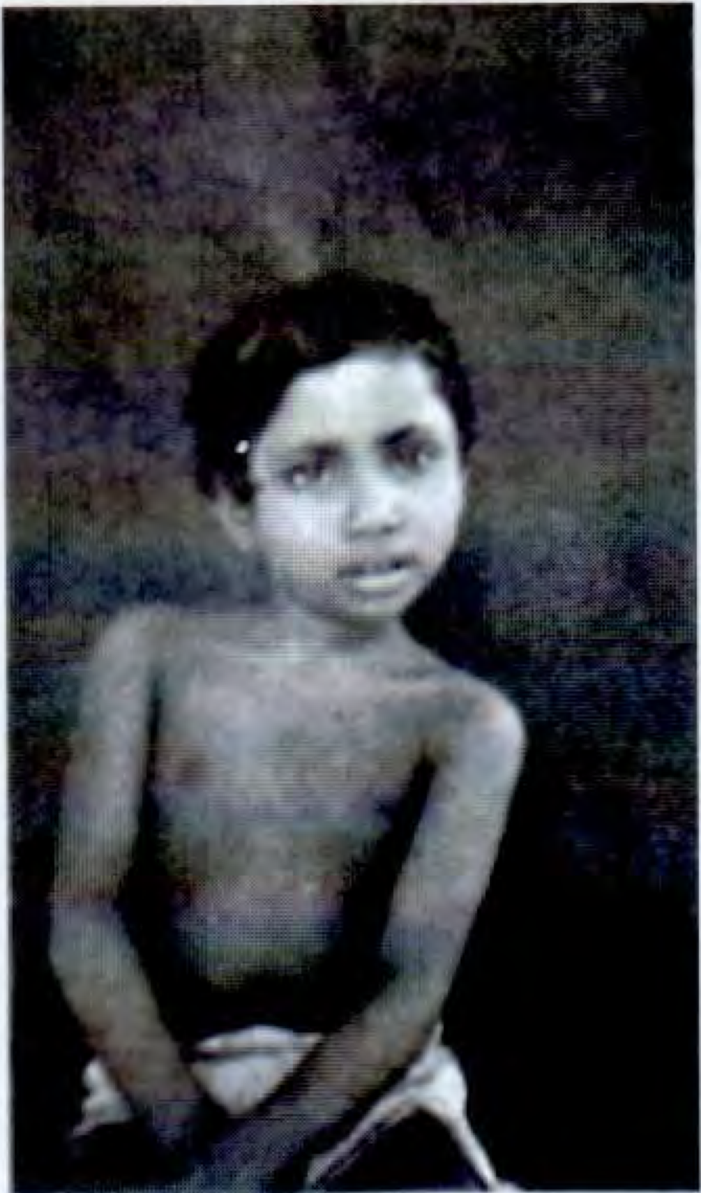
- Campbell's Operative Orthopaedics 15e, p. 78-140; S. Das Manual, p. 377-383
2. Atlantoaxial Rotatory Subluxation (AARS) - Most Common Cause of Acute Acquired Torticollis in Children
- The transverse ligament of the atlas (holds the odontoid peg against the anterior arch of C1)
- Alar ligaments (bilateral, limit rotation)
- Capsular ligaments of the lateral atlantoaxial joints
| Type | Description | Neurological Risk |
|---|---|---|
| I | Rotatory fixation without anterior displacement (most common) | Low |
| II | Rotatory fixation + anterior displacement 3-5 mm (transverse ligament incompetent) | Moderate |
| III | Rotatory fixation + anterior displacement >5 mm (both transverse + alar ligaments) | High |
| IV | Posterior displacement (rare) | High |
- Grisel syndrome - non-traumatic AARS following upper respiratory infection / retropharyngeal inflammation (pharyngeal venous plexus → periarticular C1-C2 ligamentous laxity)
- Post-tonsillectomy / adenoidectomy
- Minor trauma
- Down syndrome (ligamentous laxity)
- Rheumatoid arthritis / JIA
- Campbell's Operative Orthopaedics 15e, p. 1962; Tintinalli's Emergency Medicine
3. Spasmodic (Idiopathic Cervical Dystonia) - Most Common Adult Form
- SCM (primary, causes the tilt and rotation)
- Levator scapulae - elevates the shoulder ipsilaterally (earliest palpable sign)
- Trapezius (upper fibers)
- Posterior cervical muscles bilaterally (by EMG)
- In advanced cases: shoulder girdle, back, face, and limb muscles
- Gestes antagonistes (sensory tricks) - placing a hand on the chin or occiput transiently relieves the spasm; this is pathognomonic of dystonia and reflects abnormal proprioceptive processing
- Spasms worsen with standing and walking, lessen when lying down
- Characteristically jerky or tremulous (vs. fixed in structural torticollis)
- Muscles hypertrophy in chronic cases
- Adams and Victor's Principles of Neurology 12e, p. 110-111
4. Other Anatomical Types
| Type | Anatomical Mechanism |
|---|---|
| Traumatic | Fracture-dislocation of the cervical spine; direct SCM injury |
| Inflammatory / Infective | Inflamed cervical lymph nodes or parapharyngeal abscess compress / irritate the SCM or adjacent tissues; tonsillar/retropharyngeal abscess (Grisel mechanism) |
| Rheumatic | Acute muscular spasm of SCM after exposure to cold/draught - most common benign adult presentation; protective reflex against cervical nerve root irritation |
| Ocular (Compensatory) | Superior oblique palsy (CN IV) → patient tilts head to the opposite side to neutralize diplopia (compensatory head tilt) |
| Bony anomaly | Klippel-Feil syndrome (cervical vertebral fusion), hemivertebrae, os odontoideum |
| Scarring/Contracture | Post-burn contracture, scar tissue involving platysma / cervical fascia |
| Pott's disease | Tuberculous spondylitis of the cervical spine causing muscle spasm / bony instability |
| Posterior fossa tumor | Tonsillar herniation or tumor compressing cervical cord/nerve roots |
| Compensatory (scoliosis) | Head tilt to balance visual axis when thoracic scoliosis creates a tilted shoulder girdle |
Summary of Anatomical Basis by Structure Involved
BRAIN / BASAL GANGLIA
→ Spasmodic (cervical dystonia)
→ Posterior fossa tumor
CERVICAL SPINE (C1-C2)
→ Atlantoaxial rotatory subluxation / Grisel syndrome
→ Klippel-Feil / bony anomaly
→ Pott's disease (TB)
STERNOCLEIDOMASTOID MUSCLE
→ Congenital muscular torticollis (fibrosis)
→ Traumatic / inflammatory SCM injury
→ Rheumatic muscular spasm
CERVICAL NERVES / NERVE ROOTS
→ Cervical disc prolapse / radiculopathy (reflex spasm)
→ Spinal accessory nerve palsy (paradoxical contralateral SCM pull)
ADJACENT STRUCTURES
→ Inflamed lymph nodes, abscess (compressive)
→ Ocular imbalance (compensatory)
→ Scar tissue (contracture)
The Anatomy of the "Cock-Robin" Posture
- A tight/contracted right SCM pulls the mastoid process down and toward the sternum/clavicle
- This laterally flexes (tilts) the head to the right
- The same vector simultaneously rotates the chin to the left (away from the contracted muscle)
- The right sternomastoid is visibly prominent and palpably tight
- The left SCM is relatively elongated and relaxed
- Bailey and Love's 28e, p. 9135-9136; Adams and Victor's, p. 110; Campbell's Operative Orthopaedics 15e, p. 78-135