
Give me a inage showing spinal cord in vertebral column along with intervertebral disc
Reading File
This is a visual/anatomy question. Let me search the medical library for spinal cord and vertebral column images.
Finding Sources
Finding Sources
Reading File
Found exactly what's needed. Let me fetch both the spinal cord diagram and the intervertebral disc image.
Loading Image
Loading Image
Here are two detailed anatomical diagrams from Gray's Anatomy for Students:
Fig. 1 - Vertebral Canal, Spinal Cord, and Spinal Nerves
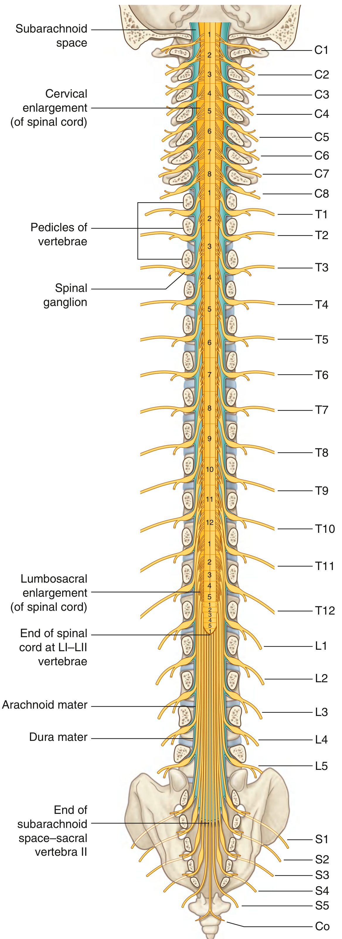
This diagram shows the full length of the spinal cord within the vertebral column, from C1 to the coccyx. Key features labeled:
- Cervical enlargement (for upper limb innervation, C4-T1)
- Lumbosacral enlargement (for lower limb innervation)
- End of spinal cord at LI-LII - the cord does not extend the full length of the canal
- Subarachnoid space, arachnoid mater, and dura mater
- All spinal nerve levels (C1-C8, T1-T12, L1-L5, S1-S5, Co) exiting through intervertebral foramina
- Spinal ganglia at each level
Fig. 2 - Intervertebral Foramen and Intervertebral Disc
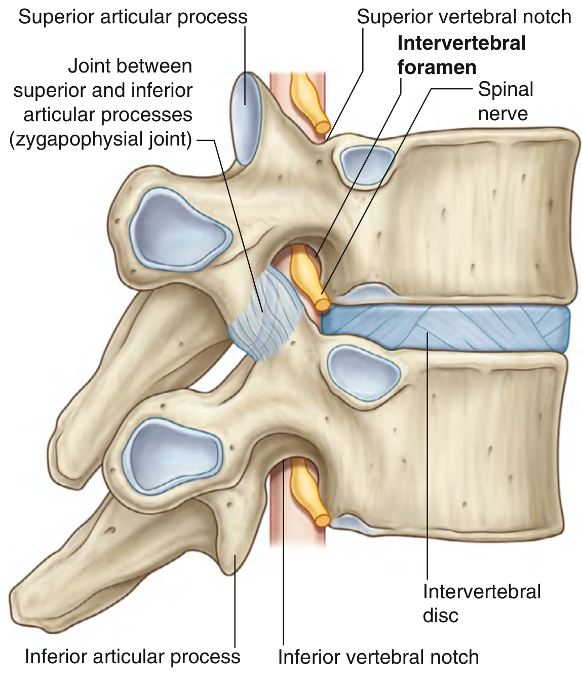
This close-up lateral view shows:
- The intervertebral disc (shown in blue) sitting between two vertebral bodies
- The intervertebral foramen - the opening through which the spinal nerve exits
- The zygapophysial joint (between superior and inferior articular processes)
- Superior/inferior vertebral notches that together form the foramen boundary
- The spinal nerve passing through the foramen, with the disc forming its anterior border
Key anatomical note: The intervertebral disc forms the anterior border of the intervertebral foramen. Disc herniation can therefore compress the exiting spinal nerve at that level, causing radiculopathy. - Gray's Anatomy for Students
This is a shared conversation. Sign in to Orris to start your own chat.