
Thoracic bones
Reading File
The thoracic bones include the thoracic vertebrae, ribs, sternum, and clavicles - forming the bony thorax (chest cage) that protects the heart and lungs.
Searching sources...
Finding Sources
Finding Sources
Finding Sources
Finding Sources
Reading File
Reading File
Reading File
Reading File
Now let me read the key thoracic bones section from Gray's Anatomy for Students, and fetch key diagrams:
Reading File
Loading Image
Now let me get the section on bones of the thorax (ribs and vertebrae) from Gray's:
Reading File
Loading Image
Finding Sources
Reading File
Reading File
Loading Image
Finding Sources
Reading File
Loading Image
Loading Image
I now have comprehensive content from the textbooks. Here is a complete, well-illustrated overview of the thoracic bones:
Thoracic Bones (The Bony Thorax)
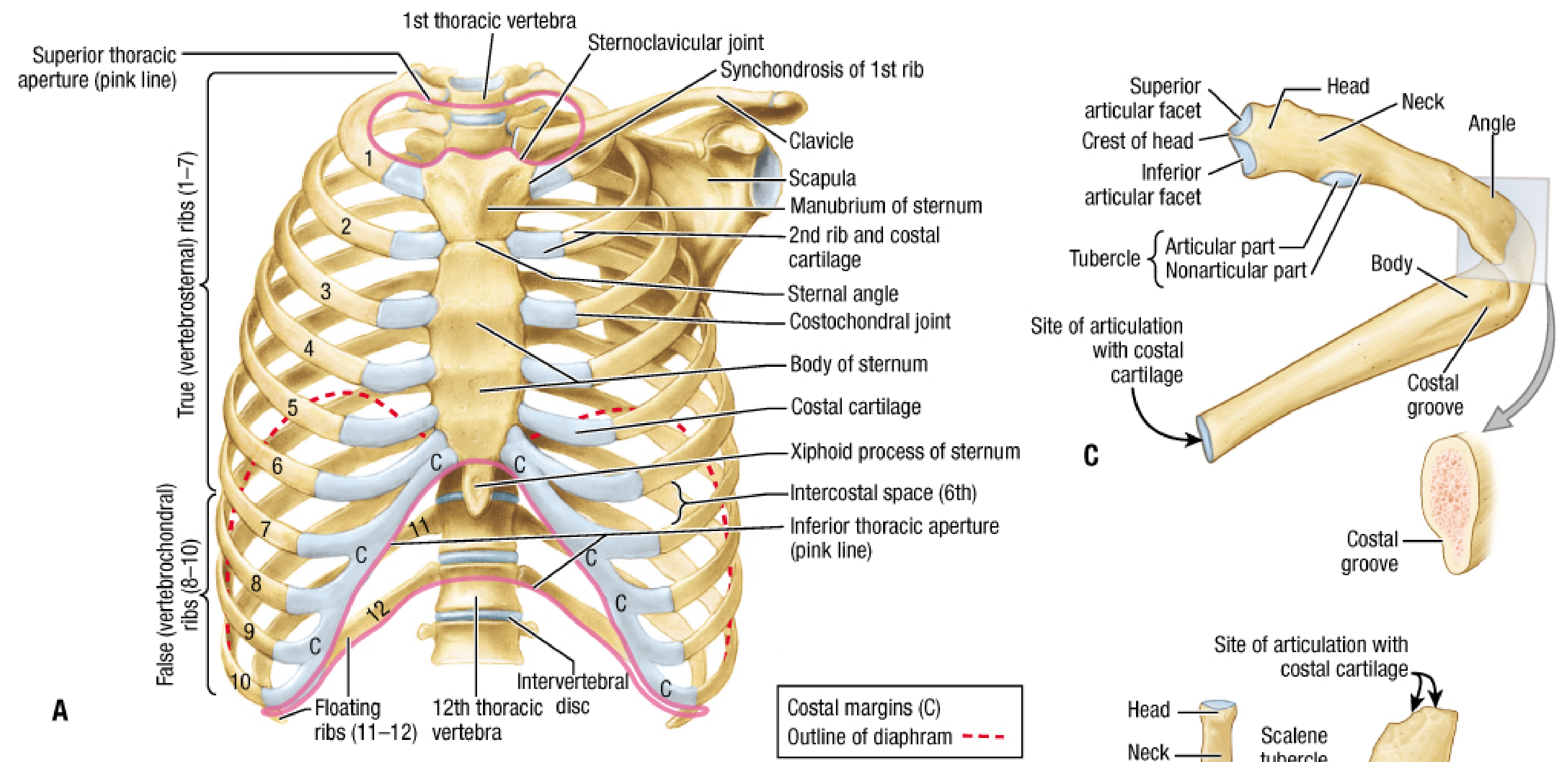
The bony thorax is a cage formed by 12 pairs of ribs with costal cartilages, 12 thoracic vertebrae (T1-T12), and the sternum. It protects the heart, lungs, and great vessels, and plays a key mechanical role in breathing.
(Fischer's Mastery of Surgery, 8th ed. - Skeleton of Thoracic Wall)
1. Overview - Thoracic Cage
2. The Sternum
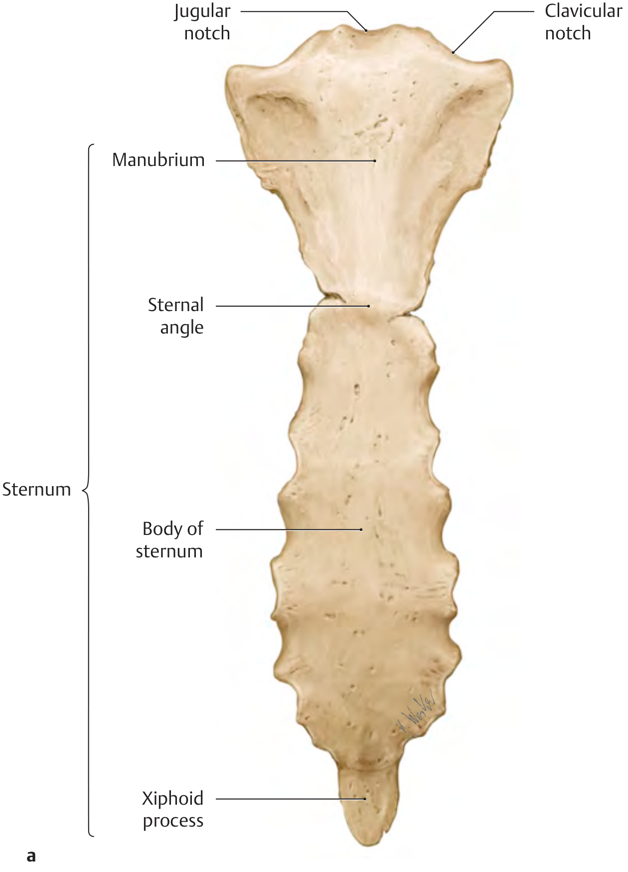
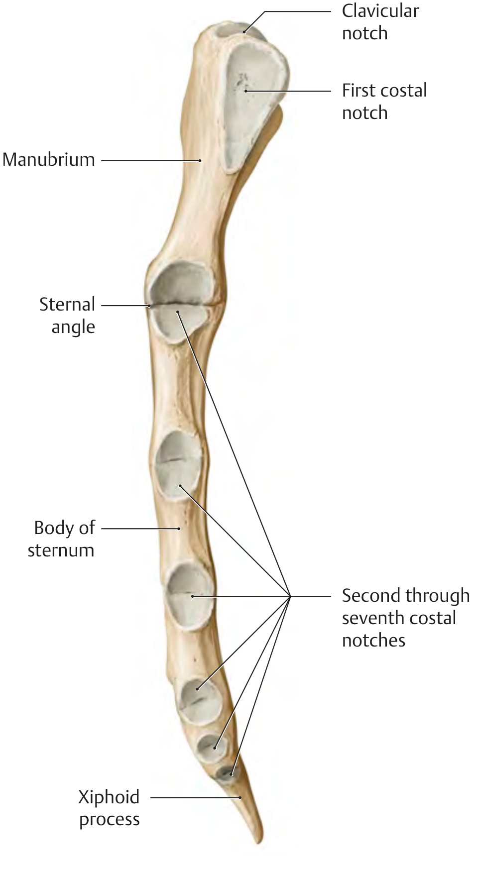
The sternum is a flat bone, slightly convex anteriorly, with costal notches along its lateral borders. It has three parts:
| Part | Key Features |
|---|---|
| Manubrium | Most superior; has jugular notch (top), clavicular notches (sides), and first costal notch |
| Body | Longest part; bears costal notches for ribs 2-7 |
| Xiphoid process | Most inferior; variable, often cartilaginous in adults; no rib attachments |
Sternal angle (angle of Louis): The junction of manubrium and body, where the 2nd costal cartilage articulates. It marks the level of the intervertebral disc between T4/T5, the bifurcation of the trachea, and the aortic arch. It is an important palpable landmark.
The manubrium-sternal and xiphosternal connections are synchondroses that gradually ossify with age.
(General Anatomy and Musculoskeletal System - THIEME Atlas; Gray's Anatomy for Students)
Anterior view:
Lateral view (showing costal notches):
3. The Ribs (12 pairs)
Classification
| Type | Ribs | Description |
|---|---|---|
| True (vertebrosternal) | 1-7 | Costal cartilage connects directly to sternum |
| False (vertebrochondral) | 8-10 | Costal cartilage joins the cartilage above (not directly to sternum) |
| Floating | 11-12 | Ends free in abdominal muscles; no sternal connection |
Parts of a Typical Rib (ribs 3-9)
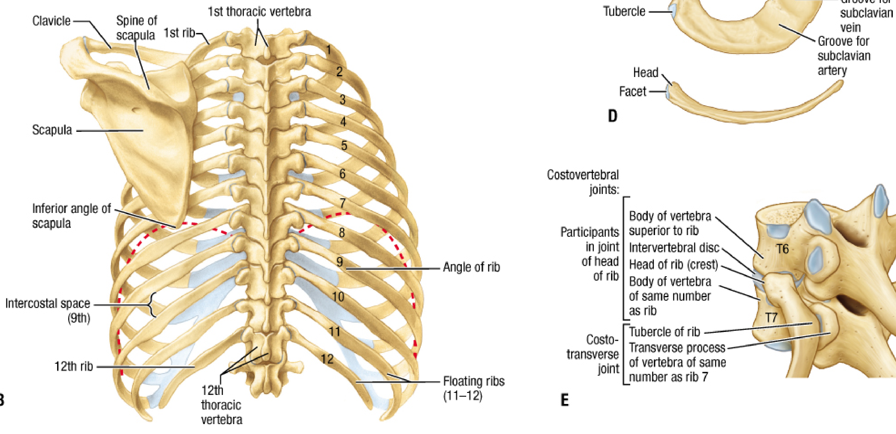
- Head - Has two demi-facets; articulates with the vertebral body of the same number and the vertebral body above (costovertebral joint)
- Neck - Connects head to tubercle
- Tubercle - Has articular part (articulates with transverse process of same-numbered vertebra = costotransverse joint) and a non-articular part
- Body (shaft) - Curved; rounded superiorly, thinner inferiorly forming the costal groove (housing the intercostal vein, artery, and nerve - VAN from top to bottom)
- Angle - Where the body bends sharply anterolaterally
Atypical Ribs
- Rib 1: Broadest and most curved; flat; no costal groove; has grooves for subclavian vessels and a scalene tubercle; head has only ONE facet
- Rib 2: Has two facets on head; tuberosity for serratus anterior on outer surface
- Rib 10: Head has only one facet
- Ribs 11 & 12: No neck, no tubercle, no costal groove (floating)
4. Thoracic Vertebrae (T1-T12)
Thoracic vertebrae are distinctive for their articulation with ribs. Key features:
- Costal facets (demi-facets) on the vertebral body - articulate with rib heads
- Costal facets on transverse processes (T1-T10) - articulate with rib tubercles
- Long, inferiorly directed spinous processes
- Heart-shaped vertebral body
5. Thoracic Apertures
Superior Thoracic Aperture
Bounded by: T1 vertebral body (posterior), medial margins of rib 1 (lateral), manubrium (anterior). Opens obliquely into the neck.
Inferior Thoracic Aperture
Bounded by: T12 (posterior), ribs 11-12 posterolaterally, costal margin (ribs 7-10 cartilages) anterolaterally, xiphoid process (anterior). Closed by the diaphragm.
6. Key Joints of the Thoracic Wall
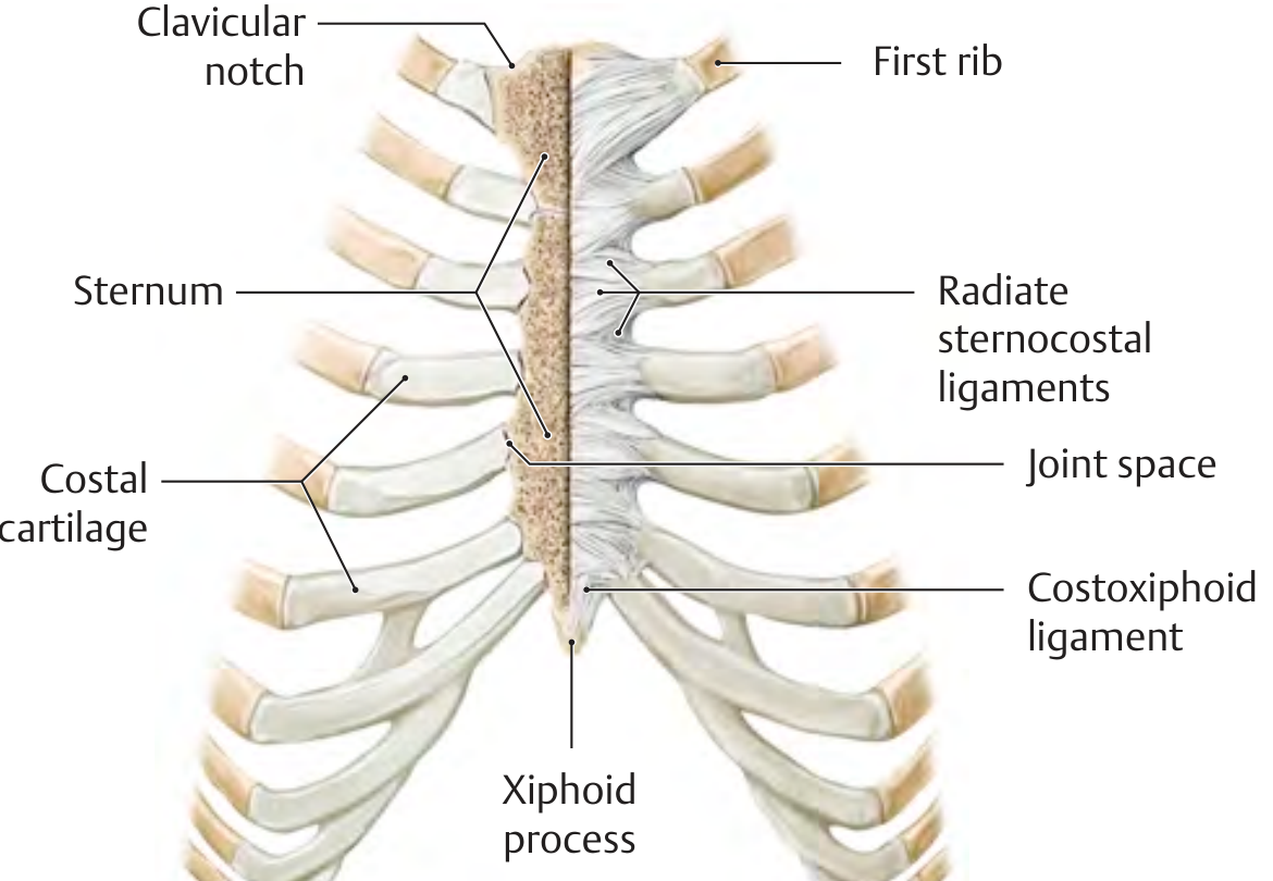
| Joint | Bones | Type |
|---|---|---|
| Sternocostal | Costal cartilages 2-5 + sternum | Synovial (plane) |
| 1st sternocostal | Rib 1 cartilage + manubrium | Synchondrosis |
| Costovertebral | Rib head + vertebral bodies | Synovial |
| Costotransverse | Rib tubercle + transverse process | Synovial (T1-T10) |
| Costochondral | Rib + costal cartilage | Synchondrosis |
| Manubriosternal | Manubrium + body | Secondary cartilaginous (symphysis) |
7. Clinical Pearls
- Sternal bone marrow aspiration: The subcutaneous position of the sternum allows needle insertion through the outer cortex into the medullary cavity to aspirate bone marrow (used in diagnosing leukemia and other blood diseases).
- Flail chest: When ribs are broken in two or more places, a free-floating "flail segment" moves paradoxically inward during inspiration, impairing ventilation. Requires assisted ventilation until healed.
- Rib fractures: Single fractures are painful but rarely dangerous. Ribs 4-9 are most commonly fractured.
- Costal margin: Formed by costal cartilages of ribs 7-10 joining together; clinically used to locate the liver and spleen borders.
(Gray's Anatomy for Students; Fischer's Mastery of Surgery; THIEME General Anatomy)
This is a shared conversation. Sign in to Orris to start your own chat.