
Sternocleidomastoid
Reading File
The sternocleidomastoid (SCM) is a major neck muscle with two heads of origin, running obliquely from the sternum/clavicle to the mastoid process of the skull.
Searching sources...
Finding Sources
Searching the Web
sternocleidomastoid muscle anatomy origin insertion action nerve supply
Reading File
Reading File
Reading File
Loading Image
Finding Sources
Reading File
Finding Sources
Here is a complete overview of the Sternocleidomastoid (SCM) muscle:
Sternocleidomastoid Muscle
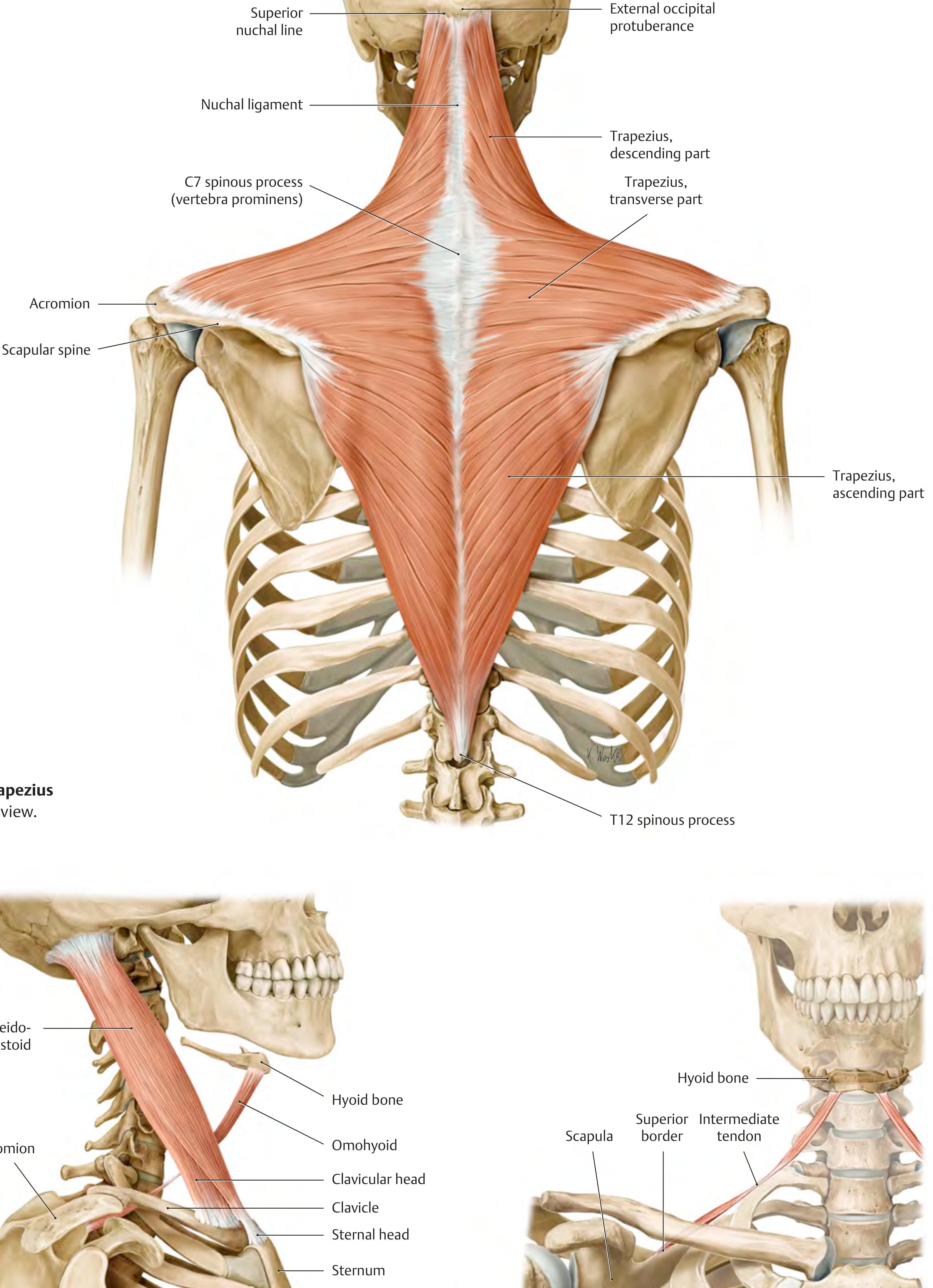
Basic Facts
The SCM is the most prominent muscle in the neck - large, superficial, and visible on inspection. Its name directly describes its attachments: sterno (sternum) + cleido (clavicle) + mastoid (mastoid process).
Origin & Insertion
| Sternal head | Anterior and lateral surface of the manubrium sterni (thick tendon) |
| Clavicular head | Medial third of the clavicle (fan-shaped) |
| Insertion | Lateral surface of the mastoid process of the temporal bone + lateral half of the superior nuchal line of the occipital bone |
The two heads are functionally distinct: the sternal head inserts mainly on the superior nuchal line, and the clavicular head mainly on the mastoid tip.
- Scott-Brown's Otorhinolaryngology Head & Neck Surgery
Actions
| Contraction | Movement |
|---|---|
| Unilateral | Tilts head to the ipsilateral side; rotates head to the contralateral side |
| Bilateral | Flexes the head (anterior flexion of neck); assists in raising the thorax (accessory muscle of respiration) when head is fixed |
Clinical testing of the right SCM: ask the patient to rotate the head to the left against resistance - observe and palpate the right SCM contracting.
- Localization in Clinical Neurology, 8e
Innervation
- Motor: Spinal accessory nerve (CN XI) - passes through the muscle
- Sensory/Proprioception: Ventral rami of C2-C4 (cervical plexus)
Blood Supply (3 segments)
| Segment | Artery |
|---|---|
| Superior third | Occipital artery + posterior auricular artery |
| Middle third | Branch of the superior thyroid artery |
| Inferior third | Suprascapular artery |
The muscle's skin perforators form the basis of the sternocleidomastoid flap used in reconstructive head and neck surgery.
- Scott-Brown's Otorhinolaryngology Head & Neck Surgery
Topographic Relations
The SCM is encased by the investing layer of deep cervical fascia and is a key surgical landmark:
- Divides the neck into the posterior triangle (bounded by SCM, trapezius, and clavicle) and the anterior triangle (bounded by SCM, mandible, and midline)
- Lymph node levels II, III, IV lie deep to it
- Upper part overlies the cervical plexus
- Lower half overlies the carotid sheath and scalenus anterior
- Scott-Brown's Otorhinolaryngology Head & Neck Surgery; Gray's Anatomy for Students
CN XI Lesions - Clinical Significance
Spinal accessory nerve palsy causes:
- Weakness turning the head to the opposite side
- Head rotates slightly toward the unaffected side on attempted flexion (unopposed contralateral SCM)
- Bilateral SCM paresis: weakness of neck flexion, head tends to fall backward
- Localization in Clinical Neurology, 8e
Torticollis
Congenital muscular torticollis: contracture of the SCM causing head tilt toward the affected side and rotation away from it. Often secondary to intrauterine moulding; may present with a palpable "tumour" within the muscle. Most cases resolve with stretching; persistent cases develop facial asymmetry requiring surgical release of the SCM origin and/or insertion.
Acquired torticollis: can be caused by inflammation/infection, ocular problems, atlantoaxial rotatory subluxation, or a posterior fossa tumour.
- Bailey and Love's Short Practice of Surgery, 28e
SCM as Accessory Respiratory Muscle
When the head is fixed, bilateral SCM contraction helps raise the upper thorax, making it an accessory muscle of respiration - clinically visible in respiratory distress (e.g., COPD exacerbations, asthma).
Quick Summary Table
| Feature | Details |
|---|---|
| Origin | Manubrium sterni (sternal head); medial 1/3 of clavicle (clavicular head) |
| Insertion | Mastoid process + superior nuchal line |
| Action (unilateral) | Ipsilateral tilt + contralateral rotation |
| Action (bilateral) | Neck flexion; accessory respiration |
| Nerve | CN XI (motor); C2-C4 (sensory) |
| Blood supply | Occipital / superior thyroid / suprascapular arteries |
| Key role | Neck landmark dividing anterior and posterior triangles; CN XI testing; torticollis |
This is a shared conversation. Sign in to Orris to start your own chat.