
Draw a labelled diagram of spinal cord and arrangement of fibers in spinal cord
Detailed anatomical diagram of spinal cord cross-section showing arrangement of fiber tracts. Show a clean cross-section with: - Central H-shaped gray matter with labeled Posterior horn (dorsal horn), Anterior horn (ventral horn), Lateral horn (intermediolateral column), Central canal, Gray commissure - White matter divided into three funiculi: Posterior funiculus (dorsal column), Lateral funiculus, Anterior funiculus (ventral column) - Ascending tracts labeled: Fasciculus gracilis (medial, posterior column), Fasciculus cuneatus (lateral, posterior column), Lateral spinothalamic tract (pain/temperature), Anterior spinothalamic tract (light touch), Dorsal spinocerebellar tract, Ventral spinocerebellar tract - Descending tracts labeled: Lateral corticospinal tract (posterior lateral funiculus), Anterior corticospinal tract (anterior funiculus), Rubrospinal tract, Vestibulospinal tract, Tectospinal tract, Reticulospinal tract - Color coding: ascending tracts in blue, descending tracts in red, gray matter in gray - Clean educational style with arrows and clear labels
Spinal Cord - Labelled Diagrams
Longitudinal View (Overall Anatomy)
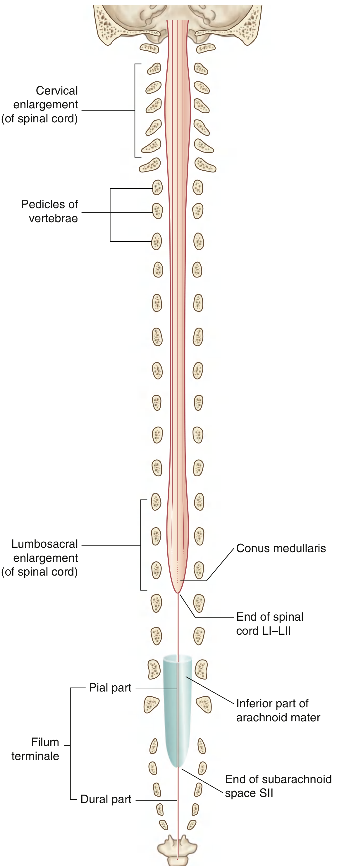
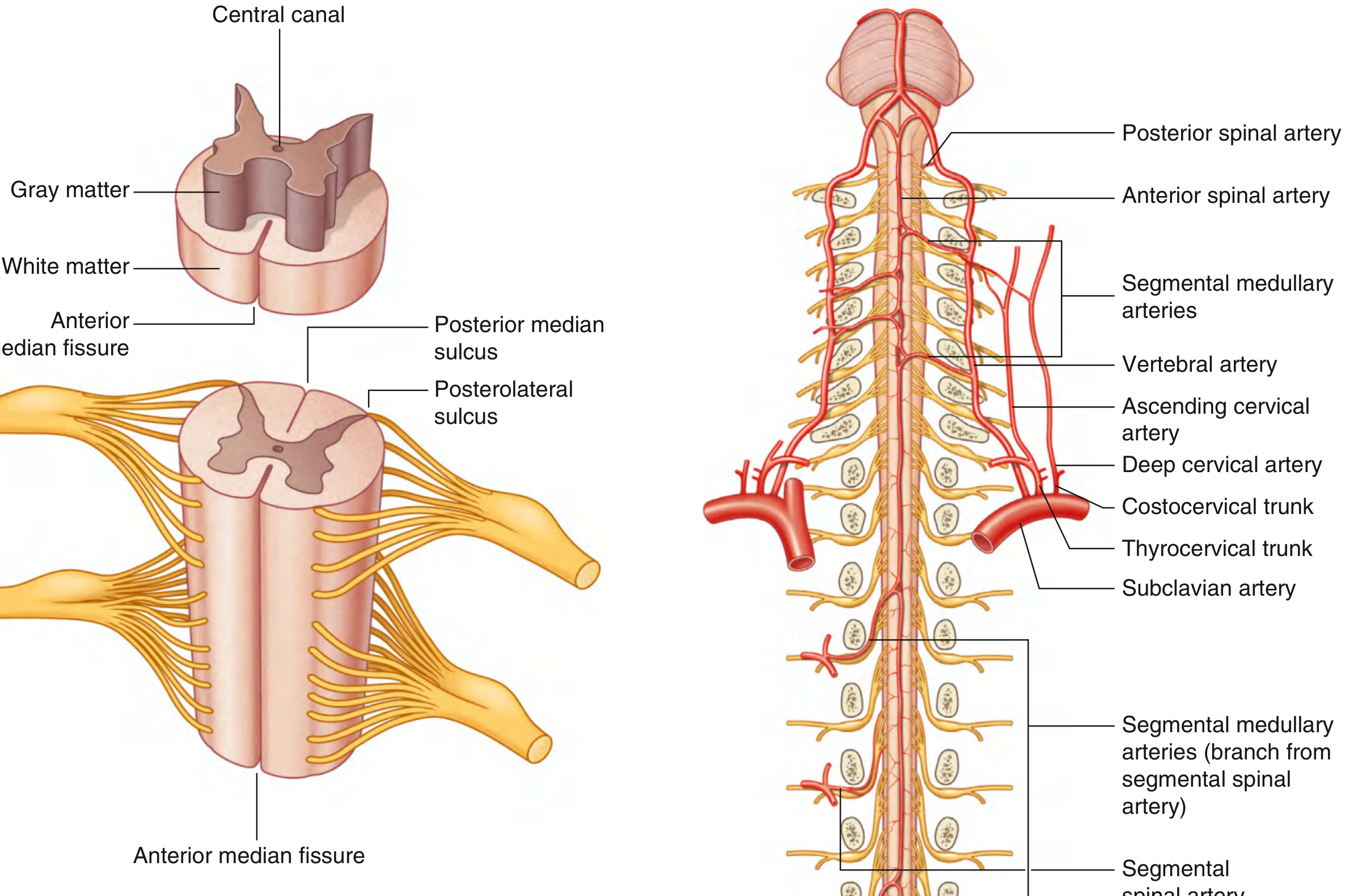
Cross-Section with External Features
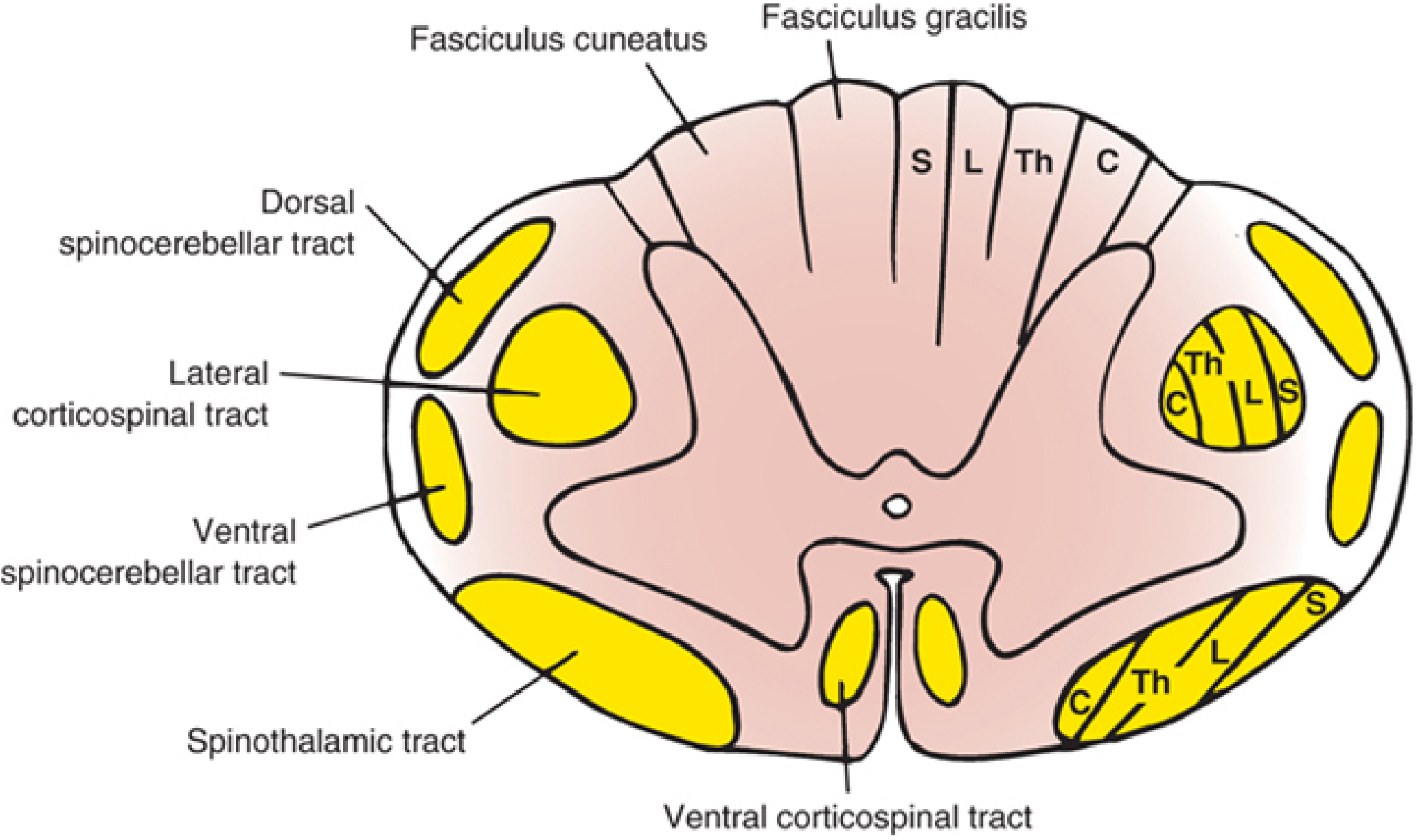
Arrangement of Fiber Tracts (Cross-Section)
Generated Comprehensive Diagram
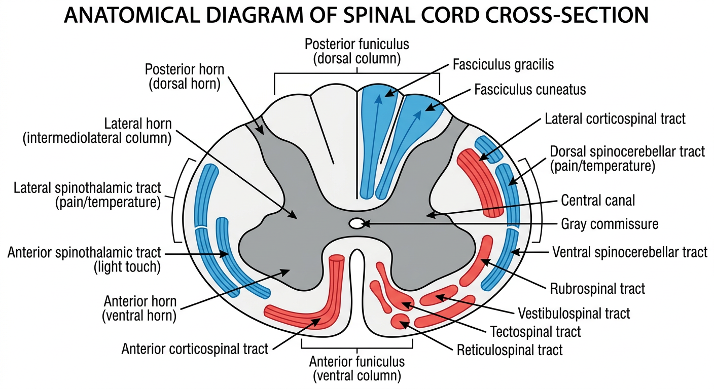
Detailed Notes on Fiber Arrangement
Gray Matter (Central, H-shaped)
| Region | Contents |
|---|---|
| Posterior (dorsal) horn | Receives sensory input; contains laminae I-VI (Rexed); substantia gelatinosa (lamina II) |
| Anterior (ventral) horn | Motor neurons (alpha + gamma); laminae VIII-IX |
| Lateral horn (T1-L2 only) | Intermediolateral column; preganglionic sympathetic neurons |
| Gray commissure | Connects the two halves; contains central canal |
White Matter - Three Funiculi
| Funiculus | Location | Major Tracts |
|---|---|---|
| Posterior (dorsal) | Between dorsomedian and dorsolateral sulci | Fasciculus gracilis, Fasciculus cuneatus |
| Lateral | Between dorsolateral and ventrolateral sulci | Lateral corticospinal, lateral spinothalamic, spinocerebellar tracts |
| Anterior (ventral) | Between ventrolateral sulcus and anterior median fissure | Anterior corticospinal, anterior spinothalamic, vestibulospinal, tectospinal |
Ascending Tracts (Sensory - shown in blue)
- Fasciculus gracilis (medial) - lower limb and lower trunk (T7-sacral); fine touch, proprioception, vibration
- Fasciculus cuneatus (lateral) - upper limb and upper trunk (C1-T6); same modalities
- Somatotopic: sacral fibers most medial, cervical fibers most lateral
- Located in lateral funiculus (anterolateral quadrant)
- Carries pain and temperature (sharp, localized - neospinothalamic)
- Fibers decussate via anterior white commissure over 1-2 segments
- Somatotopic: cervical fibers dorsomedial, sacral fibers ventrolateral
- Located in anterior funiculus
- Carries light touch (crude touch, pressure)
- Also crosses via anterior white commissure
- Dorsal spinocerebellar tract (Clark's column, C8-L2 origin) - unconscious proprioception from lower limb; ipsilateral
- Ventral spinocerebellar tract - lower limb proprioception; crosses twice (effectively ipsilateral)
Descending Tracts (Motor - shown in red)
- Lateral corticospinal tract (in lateral funiculus) - 85% of fibers; crossed; controls voluntary skilled movement of contralateral limbs
- Anterior corticospinal tract (in anterior funiculus) - 15% of fibers; uncrossed (crosses at segmental level); controls axial/proximal muscles
- Rubrospinal tract - lateral funiculus; from red nucleus; controls limb flexors
- Vestibulospinal tract - anterior funiculus; from vestibular nuclei; controls trunk and extensor muscle tone (balance)
- Tectospinal tract - anterior funiculus; from superior colliculus; head-turning reflexes
- Reticulospinal tracts - anterior/lateral funiculi; from reticular formation; modulates tone and reflexes
Clinical Relevance - Tract Lamination
- Central cord syndrome (central lesion) affects arms more than legs (cervical fibers are central)
- Extramedullary compression affects sacral fibers first (peripheral location)