
sprain management
ankle sprain management RICE treatment
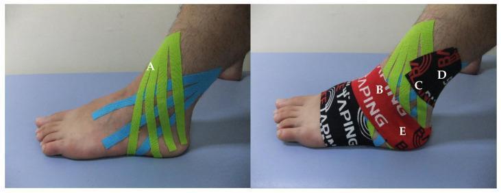
Two-panel clinical photograph demonstrating Ankle Eversion Taping (AET) using kinesiology tape for the treatment of an acute ankle inversion sprain. The left panel (labeled A) shows a fan-shaped application of green and blue kinesiology tape strips originating from the lateral malleolus and extending across the dorsum of the foot, designed to reduce lymphatic edema and swelling. The right panel (labeled B-E) illustrates a multi-step mechanical correction and functional support technique. Step B involves a red 'TAPING' strip applied for posterior talar glide; step C utilizes green tape from the lateral malleolus to the medial instep to induce eversion; step D shows an 80% overlapping layer for mechanical reinforcement; and step E demonstrates a final red strip to support dorsiflexion and talar positioning. The imagery highlights specific physical therapy interventions focused on joint stabilization, biomechanical correction of the subtalar joint, and reduction of localized inflammation in musculoskeletal rehabilitation.
This diagnostic image displays a pair of Megavoltage (MV) portal films, illustrating left lateral (left image) and right lateral (right image) radiation therapy treatment fields for a foot and ankle. The images show the patient's lower extremity positioned inside a radiolucent custom plastic tank. Bony anatomy, including the distal tibia, fibula, and tarsal bones, is discernible through the treatment setup. A calibration reticule, appearing as a grid-like pattern of crosshairs and dots, is superimposed over the images for precise alignment and isocenter verification. The setup involves the use of water-equivalent bolus material (specifically rice packing) to ensure adequate dose buildup at the skin surface while providing immobilization. Distinctive vertical and horizontal lines indicate the edges of the treatment container and the radiation field boundaries. This imaging is used for verification of treatment delivery geometry and anatomical positioning in the management of cutaneous conditions like Mycosis Fungoides or other localized extremity lymphomas.
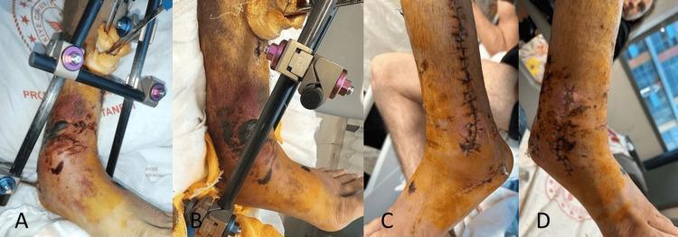
Clinical photographs in a four-panel sequence (A-D) demonstrating the two-stage surgical management of a severe ankle fracture with significant soft tissue injury. Panels A and B (anteromedial views) show the first stage of treatment, featuring a spanning external fixator with metal connecting rods and pins stabilizing the joint. The surrounding soft tissue exhibits marked edema, ecchymosis, and yellowish skin discoloration consistent with high-energy trauma (Oestern and Tscherne Grade 2-3). Panels C (lateral view) and D (medial view) document the second stage after definitive open reduction and internal fixation. Long longitudinal surgical incisions are visible along the anterior and medial distal leg, secured with non-absorbable sutures or staples. While swelling persists in the postoperative phase, the integumentary condition shows stabilization compared to the acute injury phase. This series illustrates the orthopedic protocol of temporary external stabilization to allow soft tissue recovery before definitive internal osteosynthesis in complex ankle fractures.
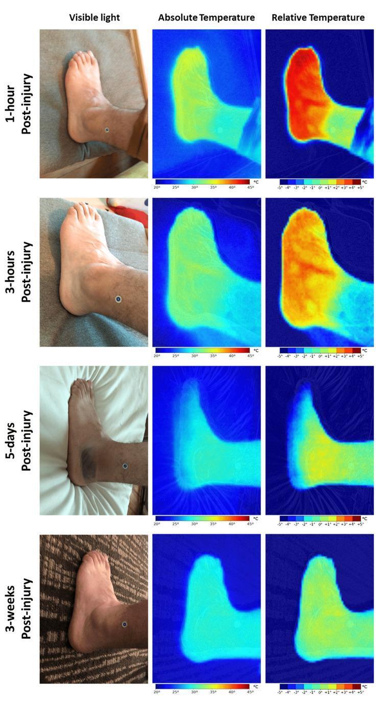
This clinical comparison chart utilizes visible light photography and infrared thermography (IRT) to track the progression and resolution of an acute ankle sprain over four time points: 1-hour, 3-hours, 5-days, and 3-weeks post-injury. The grid layout includes columns for Visible Light, Absolute Temperature (20°C-45°C scale), and Relative Temperature (-5°C to +5°C scale). At 1-hour and 3-hours post-injury, thermograms demonstrate intense hyperthermia (red/orange 'hotspots') across the dorsal and lateral aspect of the foot, indicative of acute inflammatory vasodilation and increased thermal load, despite a relatively normal appearance on visible light images. By day 5, IRT reveals significant cooling (blue/green shifts) as residual inflammation clears, though relative mapping still shows mild elevation. At 3-weeks, the thermal distribution is uniform and baseline temperatures are restored, correlating with clinical resolution. This image serves as a diagnostic educational tool for understanding the physiological timeline of inflammation and the utility of hyperspectral imaging/IRT in detecting subclinical vascular changes during musculoskeletal trauma recovery.
ankle sprain management treatment
Sprain Management
Classification (Grading)
| Grade | Pathology | Clinical Features | Weight-Bearing |
|---|---|---|---|
| I | Microscopic tearing; no complete fiber disruption | Minimal tenderness, small/no effusion | Tolerated immediately or within 24 hrs |
| II | Partial (incomplete) tear of some ligament fibers | Moderate tenderness, hematoma, small-moderate effusion | Unable to bear weight for >24 hrs |
| III | Complete tear of all ligament fibers | Significant tenderness, hematoma, large effusion; positive anterior drawer + talar tilt | Unable to bear weight; positive stress tests |
Acute (Initial) Management: PRICE Protocol
- Protection - avoid re-injury; splint or brace as needed
- Rest - reduce loading on the injured ligament
- Ice - apply for 15-20 min every 1-2 hours to limit swelling and pain
- Compression - elastic/compression bandage; lace-up support is most effective for short-term edema reduction
- Elevation - raise the limb above the level of the heart
A lace-up ankle splint is more effective in short-term edema reduction than semirigid support, elastic bandaging, or taping alone. - Rosen's Emergency Medicine
Grade-Specific Management
Grade I
- PRICE protocol
- Weight-bearing as tolerated
- ROM (range of motion), proprioceptive, and functional exercises as tolerated
- Compression bandage or taping
Grade II
- PRICE protocol
- Air cast or lace-up brace (immobilization)
- Crutches for a few days if needed
- ROM + proprioceptive + functional exercises when tolerated
- A lace-up brace combined with elastic wrapping gives earlier return to function than either alone
Grade III
- PRICE protocol
- Lace-up support or air cast (permits some motion - preferred over rigid cast)
- Crutches until the patient can bear weight without significant pain (varies from a few days to 2-3 weeks)
- Delayed ROM, proprioceptive, and functional exercises
- Prolonged rehabilitation phase
- Surgery is rarely needed initially; delayed operative repair (even years later) yields results equivalent to primary repair
Functional Rehabilitation: 3 Phases
- Phase 1 (0-24 hrs): PRICE protocol - pain/swelling control
- Phase 2 (48-72 hrs onward): Begin active range-of-motion and strengthening exercises
- Phase 3: Endurance training, sport-specific training, and proprioception/balance training
Analgesia
- NSAIDs (e.g., ibuprofen, naproxen) - first-line for pain and inflammation
- Acetaminophen - alternative or adjunct
- Short course of oral opioids (2-3 days) - only if pain is severe; generally not routine
- Corticosteroid injections are not indicated in acute ankle sprain management
Ottawa Ankle Rules (When to X-Ray)
- Bone tenderness at the posterior edge/tip of the lateral or medial malleolus
- Bone tenderness at the base of the 5th metatarsal (navicular for foot rules)
- Inability to bear weight immediately and in the ED (4 steps)
Differential Diagnoses to Exclude
- Fracture of the lateral process of talus or base of 5th metatarsal
- Osteochondral lesion of the talar dome
- Peroneal tendon dislocation
- Syndesmotic ("high ankle") sprain
- Maisonneuve fracture (proximal fibula)
Return to Activity
| Grade | Expected Recovery |
|---|---|
| I | 1-2 weeks |
| II | 2-4 weeks |
| III | 4-8+ weeks; sometimes months |
Special Situations
- High ankle sprain (syndesmotic injury): More serious than lateral sprain; occurs with hyperdorsiflexion + external rotation; significantly prolongs recovery. - Campbell's Operative Orthopaedics
- Medial deltoid sprain: Rare in isolation (<5%); usually associated with lateral malleolar fracture
- Severe acute sprain: A below-knee cast or Aircast produces faster recovery than a tubular bandage alone, though outcomes are equal at 9 months. - Goldman-Cecil Medicine
- Hand/wrist sprains (MCP ligaments): Grade I and II treated nonoperatively with splinting; Grade III UCL tear in the thumb is usually treated surgically (Stener lesion risk). - Rockwood & Green's Fractures
Please write it for medication with dosage for 28 year old
Sprain Medications - 28-Year-Old Adult
Note: A 28-year-old is a healthy adult with no assumed renal, hepatic, GI, or cardiac disease. Full standard adult doses apply. Always confirm no contraindications before prescribing.
1. First-Line: NSAIDs (Anti-inflammatory + Analgesic)
Over-the-Counter Options
| Drug | Dose | Frequency | Max Daily Dose | Notes |
|---|---|---|---|---|
| Ibuprofen | 400-800 mg PO | Every 6-8 hrs | 3200 mg/day | Preferred first choice; take with food |
| Naproxen | 250-500 mg PO | Every 12 hrs | 1500 mg/day | Longer half-life; twice-daily dosing is convenient. Lowest cardiovascular risk among NSAIDs |
| Aspirin | 325-650 mg PO | Every 4-6 hrs | 4000 mg/day | Less preferred due to platelet effects and GI risk |
Prescription NSAIDs (if OTC doses insufficient)
| Drug | Dose | Frequency | Notes |
|---|---|---|---|
| Diclofenac | 50 mg PO | Every 8 hrs | Good for musculoskeletal pain; also available as 75 mg IM |
| Ketorolac | 10 mg PO / 15-30 mg IM or IV | Every 4-6 hrs | Use for severe acute pain only; limit to 5 days max (oral/parenteral combined); rapid onset |
| Celecoxib | 100-200 mg PO | Every 12-24 hrs | COX-2 selective; better GI safety profile |
| Meloxicam | 7.5-15 mg PO | Once daily | COX-2 preferential; convenient once-daily dosing |
| Indomethacin | 25-50 mg PO | Every 6-8 hrs | Effective but higher GI side-effect rate |
2. Second-Line: Acetaminophen (Paracetamol)
| Drug | Dose | Frequency | Max Daily Dose | Notes |
|---|---|---|---|---|
| Acetaminophen | 500-1000 mg PO | Every 4-6 hrs | 4000 mg/day (3000 mg/day if alcohol use) | No anti-inflammatory effect; does not affect platelets; safe on GI tract |
Tip: Alternating acetaminophen and ibuprofen every 3-4 hours (staggered) is a practical strategy to reduce side effects of both while maintaining good analgesia throughout the day. - Tintinalli's EM
3. Topical NSAIDs (Adjunct - Excellent for Sprains)
| Drug | Form | Application | Frequency |
|---|---|---|---|
| Diclofenac gel | 1% topical gel (OTC in US) | Apply up to 4 g to the affected area | Every 6 hrs (4x/day) as needed |
| Ketoprofen gel | 2.5% topical gel | Apply 2-4 g | Every 6-12 hrs |
4. Short-Course Opioids (Reserve for Severe Pain Only)
| Drug | Dose | Frequency | Notes |
|---|---|---|---|
| Tramadol | 50-100 mg PO | Every 4-6 hrs | Max 400 mg/day; weak opioid; avoid in seizure history |
| Codeine + Paracetamol | 30 mg codeine / 500 mg paracetamol | Every 4-6 hrs | Max 8 tablets/day; monitor for constipation |
Recommended Prescription for a Typical Grade II Ankle Sprain (28-year-old)
1. Ibuprofen 400 mg PO three times daily with food × 5 days
   (may increase to 600 mg TID if pain inadequately controlled)

2. Paracetamol 500-1000 mg PO every 6 hours PRN (if pain
   persists between NSAID doses) × 5 days

3. Diclofenac 1% gel - apply 4g to affected area 4 times
   daily × 7-10 days

4. Omeprazole 20 mg PO once daily × 5 days
   (gastric protection while on NSAIDs, especially if taking
   full doses for >3 days)
Gastric Protection (Important)
- Omeprazole 20 mg PO once daily
- Pantoprazole 40 mg PO once daily
What to Avoid in This Patient
| Avoid | Reason |
|---|---|
| Corticosteroid injection | Not indicated for acute sprain management |
| Long-term opioids | Risk of dependence; not appropriate for soft tissue injury |
| High-dose NSAIDs >10 days | GI ulceration risk without added benefit |
| Combining two oral NSAIDs | No added benefit; doubles GI/renal toxicity |
Summary Table
| Priority | Drug | Dose | Route | Frequency |
|---|---|---|---|---|
| 1st | Ibuprofen | 400-800 mg | Oral | Every 6-8 hrs |
| 1st | Naproxen | 250-500 mg | Oral | Every 12 hrs |
| Adjunct | Paracetamol | 500-1000 mg | Oral | Every 4-6 hrs |
| Topical | Diclofenac gel | 4 g | Topical | Every 6 hrs |
| Severe pain | Ketorolac | 15-30 mg | IM/IV | Every 4-6 hrs (max 5 days) |
| If NSAID fails | Tramadol | 50-100 mg | Oral | Every 4-6 hrs (2-3 days max) |