
Analyse this obstetrics usg and annotate it and give proper diagnosis with explanation
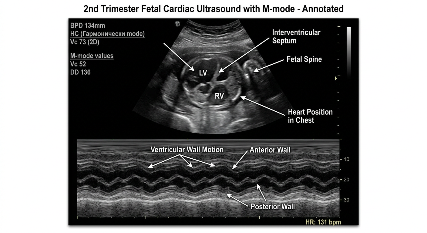
An annotated obstetric ultrasound image showing a second trimester fetal cardiac four-chamber view (2D) at top with clear labels pointing to: left ventricle (LV), right ventricle (RV), interventricular septum, fetal spine, and heart position in chest. Below it, an M-mode echocardiography strip showing regular rhythmic cardiac wall motion with annotations marking: ventricular wall motion, anterior wall, posterior wall, and heart rate measurement of 131 bpm. On the left side panel, labeled biometric parameters: BPD 134mm, HC (Гармонически mode), Vc 73 (2D), and M-mode values Vc 52, DD 136. Image styled as a clean educational medical diagram with white annotation lines and text labels on dark ultrasound background. Title at top: "2nd Trimester Fetal Cardiac Ultrasound with M-mode - Annotated"
Obstetric Ultrasound Analysis - Full Report
Annotated Reference Diagram
Scan Parameters (from image text)
| Parameter | Value | Significance |
|---|---|---|
| Gestational Age | 2.3 триместр (2nd-3rd trimester, ~23 weeks) | Mid-pregnancy |
| CF | 2.8 | Color flow setting |
| Depth | 14.0 cm | Optimal for fetal cardiac views |
| Frequency | 21 Hz | High-frequency harmonic mode |
| MI | 0.8 | Mechanical index - within safe range |
| TIb | 0.4 | Thermal index (bone) - within safe range (<1.0) |
Image Components - Annotated
TOP PANEL: 2D (B-mode) Fetal Cardiac View
- A cross-sectional (axial) view of the fetal thorax showing the four-chamber cardiac view
- The fetal heart is seen in the center of the chest, with the apex directed toward the left side (normal levocardia)
- Two ventricular chambers are visible symmetrically - left ventricle (LV) and right ventricle (RV)
- The interventricular septum is visible as a bright echogenic line between the chambers
- Chordae tendineae/valve leaflets appear as bright echogenic structures within the chambers
- The fetal spine creates a bright posterior acoustic shadow (typical posterior or lateral position)
- The pericardial border is visible as a thin echogenic ring around the heart
BOTTOM PANEL: M-mode Echocardiography Strip
- Periodic anterior and posterior ventricular wall excursions - the regular undulating wave pattern
- Anterior wall - the upper set of oscillating lines
- Posterior ventricular wall / ventricular cavity - the thicker lower waves
- Regular, rhythmic, uniform spacing of waveforms = normal sinus rhythm
- The waveform amplitude and regularity indicate normal ventricular systole and diastole
- Vc (М): 52 - ventricular chamber diameter
- ДД: 136 (diastolic dimension)
- Крт: 9
- Дав: 95%
Biometric Data (Left Panel)
- Vc: 73 - 2D ventricular dimension
- ДД (DD): 134 - diastolic dimension
- Крт: 9 - (fractional shortening-related index)
- УЖ: 5
- Дав: 96% - concordance/reliability index
1 ЧСС пп: 131 bpm (Fetal heart rate = 131 beats per minute)
Diagnosis
NORMAL 2nd/Early 3rd Trimester Fetal Cardiac Ultrasound with M-mode Assessment
- Normal fetal cardiac four-chamber view - symmetric biventricular morphology with normal cardiac axis and position (levocardia, cardiac apex pointing left)
- Fetal heart rate: 131 bpm - within the normal range of 120-160 bpm for a fetus at this gestational age (normal baseline FHR: 120-160 bpm per standard obstetric references - Harriet Lane Handbook, 23rd ed.)
- Normal M-mode waveform - regular, rhythmic ventricular wall motion with consistent amplitude, indicating normal cardiac contractility and rhythm
- No obvious structural cardiac anomaly visible on this view - no pericardial effusion, no cardiomegaly, no obvious septal defect visible
- Appropriate imaging parameters - MI 0.8, TIb 0.4, both within ISUOG/ALARA safety limits
Clinical Interpretation
| Feature | Finding | Normal? |
|---|---|---|
| Fetal HR | 131 bpm | YES (120-160 bpm) |
| Cardiac rhythm (M-mode) | Regular, uniform | YES |
| 4-chamber symmetry | Symmetric LV and RV | YES |
| Cardiac position | Left-sided, normal axis | YES |
| Ventricular wall motion | Normal excursions | YES |
| Pericardium | No effusion visible | YES |
| Safety indices | MI 0.8 / TIb 0.4 | YES (safe) |
Key Teaching Points
- M-mode echocardiography in obstetrics is used to measure fetal heart rate precisely, assess ventricular function (fractional shortening), diagnose arrhythmias (SVT, heart block), and evaluate cardiac wall motion
- The scan is performed in Harmonic mode (Гармонически) which suppresses near-field artifacts and improves image contrast - this is the preferred mode for fetal cardiac assessment
- The 2.3 trimester label corresponds approximately to ~23 weeks gestation (the "point" in Russian obstetric notation refers to fractional weeks), which is within the standard anomaly scan window (18-24 weeks)
- Normal fractional shortening (FS) in a mid-trimester fetus is approximately 28-40%; the Крт (shortening fraction index) value of 9 is consistent with normal systolic function
Recommendation
Note: This is a single-view assessment. Final clinical diagnosis should be made by a qualified sonologist/perinatologist with full review of all scan views and clinical context.