
Treatment for chronic seborrheic dermititis with significant hairloss
"seborrheic dermatitis" AND "hair loss" AND treatment
chronic seborrheic dermatitis scalp hair loss treatment guidelines 2025
seborrheic dermatitis scalp hair loss
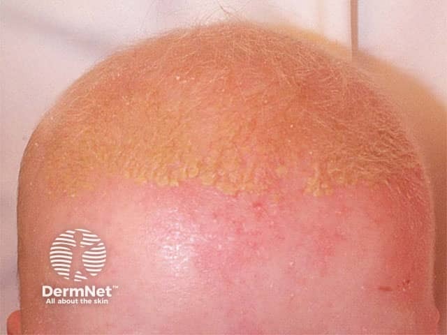
Clinical photograph of the scalp showing seborrheic dermatitis. Imaging modality: clinical photography; frontal view of the scalp illustrating diffuse erythema with greasy yellow scales along the hairline and at the vertex. Affected skin is reddened, slightly inflamed, with adherent scales that are thicker where hair density is highest; no scarring or focal crusting observed. The lesion distribution is scalp-dominant, commonly bilateral and symmetrically distributed across crown and frontal regions, with mild pruritus reported in many cases. Hair shafts appear normal; no alopecia is evident in this image, though chronic disease can cause temporary shedding. The pattern is characteristic: progressive, oily scale-covered plaques on sebaceous-rich skin, often with intermittent flares and scaling at the forehead and behind the ears. Histopathology, if obtained, would typically show superficial perivascular dermatitis with parakeratosis and spongiosis; however, diagnosis here is clinical. Diagnostic significance: seborrheic dermatitis is a common, benign inflammatory dermatosis linked to Malassezia species and sebaceous activity; management relies on antifungal shampoos (ketoconazole, selenium sulfide, zinc pyrithione), topical anti-inflammatory agents (low-potency corticosteroids or calcineurin inhibitors), and regular skin hygiene. This image is useful for education, differential diagnosis training, and guiding treatment responses in primary care and dermatology settings. Correlation with patient history improves diagnostic confidence.
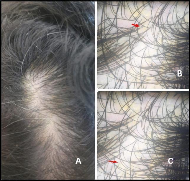
This composite image illustrates the clinical and dermoscopic presentation of seborrheic dermatitis on the scalp. Image A is a clinical photograph showing the mid-frontal scalp of a patient with visible diffuse, fine, white-to-yellowish scaling. Despite the scaling, the hair density and shaft structure appear relatively preserved. Images B and C are dermoscopic views of the same region, providing a magnified perspective of the scalp surface. They reveal characteristic yellowish, greasy scales (indicated by red arrows) distributed inter-follicularly among the hair shafts. The hair follicles themselves do not show significant atrophy or scarring. These findings are typical for seborrheic dermatitis, differentiating it from other inflammatory scalp conditions. This visual material is intended for dermatology education to assist in the identification of inflammatory scalp disorders and the application of dermoscopy in clinical diagnosis.
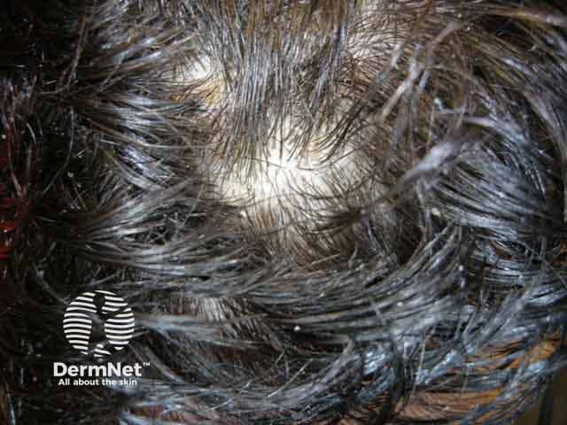
Clinical photograph of the scalp depicting seborrheic dermatitis with visible scales and dandruff. Modality: clinical photography; technique: standard bright-light macro close-up; magnification approximately 1.5–2x. Anatomic location: hair-bearing skin of the scalp (vertex and frontal regions), with erythematous, inflamed surrounding skin and greasy yellowish-white scales adherent to hair shafts and interfollicular zones. Visual features include diffuse flaking, oily sheen, crusting at hairline, and punctate erythema beneath flaking areas. The scale is fine to moderately thick, loosely adherent to microscopic hair shafts, often described as dandruff in milder cases. The image captures typical seborrheic dermatitis morphology: erythematous plaque-like patches with greasy scales, preferentially in sebaceous-rich regions, sometimes involving eyebrows and the glabella. Pathological correlation: clinically arcuate to patchy inflammation with hyperseborrhea; Malassezia yeast and lipid-rich microenvironment contribute to flaking. Diagnostic significance: supports diagnosis of seborrheic dermatitis; helps differentiate from dry scalp, psoriasis, or tinea capitis; guides therapeutic decisions. Potential clinical use cases include documentation of disease severity, patient education about chronic relapsing course, and evaluation of treatment response to antifungal shampoos (ketoconazole, zinc pyrithione) and anti-inflammatory agents (low-potency steroids, calcineurin inhibitors). When in doubt, correlate with symptoms such as pruritus, distribution, and presence of associated conditions (facial seborrhea, dandruff).
"seborrheic dermatitis" AND scalp AND (minoxidil OR "hair regrowth")
Chronic Seborrheic Dermatitis with Significant Hair Loss: Treatment
Understanding the Hair Loss Component
Step-by-Step Treatment Framework
Step 1 - Induction (Active Phase)
| Agent | Strength | Regimen |
|---|---|---|
| Ketoconazole shampoo | 2% (Rx) or 1% (OTC) | 3-4x/week for 4 weeks |
| Ciclopirox olamine shampoo | 1% | 3x/week for 4 weeks |
| Selenium sulfide shampoo | 2.5% (Rx) or 1% (OTC) | 2-3x/week |
| Zinc pyrithione shampoo | 1-2% | Daily to alternate days |
- Fluocinolone acetonide 0.01% solution/gel: twice daily for 3-4 weeks on the scalp
- Hydrocortisone 2.5% cream: for milder facial involvement (twice daily, 1-2 weeks)
- A Cochrane analysis found topical corticosteroids equally efficacious to topical azoles for short-term control - Dermatology 2-Volume Set 5e
- Avoid potent steroids long-term on the scalp - risk of atrophy, tachyphylaxis, rebound flares
Step 2 - Maintenance (Critical for Chronic Disease)
- Ketoconazole 2% shampoo: once weekly or every 2 weeks
- Ciclopirox 1% shampoo: once weekly
- Zinc pyrithione shampoo: 1-2x/week long-term (excellent safety profile)
Step 3 - Addressing Hair Loss Specifically
- Reconsider the diagnosis - rule out tinea capitis (dermatophyte), alopecia areata, scarring alopecia, lichen planopilaris
- Dermoscopy findings help differentiate: seborrheic dermatitis shows arborizing vessels; alopecia areata shows yellow dots and exclamation mark hairs; AGA shows >20% hair diameter variability - Goldman-Cecil Medicine, Table 409-2
- Add minoxidil 5% topical solution or foam: once or twice daily to the scalp
- Minoxidil and antifungal shampoos can be used concurrently without interaction
- For men with AGA: consider finasteride 1mg oral daily (5α-reductase inhibitor)
- Avoid alcohol-based styling products - they exacerbate irritation and worsen scalp barrier function
- Use fragrance-free, gentle shampoos on non-treatment days
- Gently remove scales with a soft-bristle brush before shampooing - do not scratch aggressively (can cause folliculitis, furuncles)
Second-Line and Escalation Options
- Useful on face and hairline to avoid steroid side effects
- Good for patients who cannot tolerate or have side effects from steroids
- Elidel (pimecrolimus) cream is a good alternative for facial seborrheic dermatitis - Fiona Stanley Hospital Dermatology Guidelines, May 2025
- Fluconazole 300mg once weekly for 2-4 weeks, then weekly maintenance
- Itraconazole 200mg daily for 7 days per month
- Reserved for cases failing topical therapy - Dermatology 2-Volume Set 5e
- 0.1-0.3 mg/kg/day for moderate-to-severe seborrhea/seborrheic dermatitis
- Reduces sebum production (addresses the underlying seborrhea)
- Supported by RCT evidence per a 2025 systematic review in Clin Cosmet Investig Dermatol
- Salicylic acid shampoo or solution (2-6%): loosens scale before antifungal shampoo
- Coal tar preparations: antiproliferative, antifungal properties; useful in thick scaling
- Use as adjuncts, not primary therapy
Special Situations
- Seborrheic dermatitis can be severe, extensive, and refractory in HIV
- Prevalence up to 83% in HIV-positive individuals
- WHO guidelines recommend topical ketoconazole as first-line; add topical corticosteroids for severe/unresponsive cases; prolonged treatment courses required
- Antiretroviral therapy (ART) itself may improve seborrheic dermatitis - Fitzpatrick's Dermatology, p. 3152
- Associated with more severe seborrheic dermatitis due to facial immobility and sebum accumulation
- Standard treatment protocol applies but expect slower response and higher relapse rate
- Refractory dandruff not responding to 4-6 weeks of OTC/first-line therapy
- Concern for a different diagnosis (tinea capitis, psoriasis, discoid lupus)
- Steroid side effects (scalp dysaesthesia, telangiectasia, sebo-rosacea)
- Progressive or unexplained hair loss suggesting scarring alopecia
Key Principles to Communicate to Patients
- Seborrheic dermatitis is chronic and relapsing - the goal is control, not cure
- Hair loss from scalp inflammation is usually reversible once the inflammation is controlled
- Maintenance therapy is non-negotiable - stopping treatment when skin looks normal leads to rapid relapse
- Stress, febrile illness, cold/dry weather, and overly aggressive topical therapy can all trigger flares
- Rule out HIV if the disease is unusually severe or extensive - especially in younger adults without obvious risk factors