
Paget disease
Reading File
Paget disease of bone is a chronic metabolic bone disorder characterized by focal areas of disordered bone remodeling - excessive osteoclast activity followed by disorganized new bone formation, producing structurally weak, enlarged bone. Now let me pull the full textbook coverage.
Searching sources...
Finding Sources
Searching PubMed
"Paget disease"[MeSH Terms] AND bone
Excellent sources found. Let me read the most comprehensive ones in parallel - Harrison's (most detailed, 13 paragraphs) and Goldman-Cecil, plus the pathology/pharmacology sections.
Reading File
Reading File
Reading File
I have good content from Harrison's on epidemiology and etiology. Let me now read the remaining Harrison's section (pathology, clinical features, diagnosis, treatment) and also fetch the diagram image.
Reading File
Loading Image
Excellent. I have comprehensive information. Let me now compile the full, structured answer using all the gathered content.
Paget's Disease of Bone
Definition
Paget's disease is a localized bone-remodeling disorder affecting widespread, non-contiguous skeletal areas. The pathologic process begins with overactive osteoclastic bone resorption followed by compensatory, disorganized osteoblastic new bone formation, producing a structurally abnormal mosaic of woven and lamellar bone. Pagetic bone is expanded, less compact, and more vascular - making it susceptible to deformities and fractures. - Harrison's Principles of Internal Medicine, 22nd Ed.
Epidemiology
- Prevalence ~0.5% in the UK and USA; common in Western Europe (Britain, France, Germany) but rare in Scandinavia, Africa, Asia, and the Middle East
- Greater prevalence in males; increases markedly with age
- Autopsy series reveal disease in ~3% of individuals over age 40
- Radiographic prevalence in patients >55 years: 2.5% men, 1.6% women
- For unclear reasons, prevalence and severity have been declining, and age at diagnosis is increasing
- Harrison's, p. 3312; Goldman-Cecil Medicine
Etiology
Both genetic and viral factors are implicated.
Genetic Factors
- Positive family history in 15-25% of patients (raises prevalence 7-10x among first-degree relatives)
- Key gene mutations:
| Gene | Protein | Disease |
|---|---|---|
| SQSTM1 (most common) | Sequestosome-1/p62 | Familial and sporadic Paget's |
| TNFRSF11B | Osteoprotegerin (OPG) | Juvenile Paget's (familial idiopathic hyperphosphatasia) |
| TNFRSF11A | RANK | Familial expansile osteolysis; early-onset Paget's |
| VCP (valosin-containing protein) | - | Inclusion body myopathy + Paget's + frontotemporal dementia (IBMPFD) |
| CSF1 | M-CSF | Increased osteoclast differentiation |
| TM7SF4 | DC-STAMP | Osteoclast fusion |
Viral Factors
-
Paramyxovirus inclusions (measles, RSV, canine distemper virus) found in pagetic osteoclasts
-
Viral mRNA detected in osteoclast precursors
-
Decline in Paget's disease coincides with widespread measles vaccination
-
However, no live virus has been cultured from pagetic bone, and antibody levels against paramyxoviruses are not elevated in affected patients
-
Harrison's Principles of Internal Medicine, 22nd Ed.
Pathophysiology
The RANK/RANKL/OPG axis is central to the pathology:
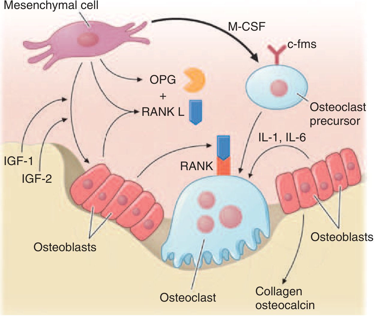
Figure: Factors promoting osteoclast and osteoblast differentiation and function (from Harrison's, 22nd Ed.)
Key cellular abnormalities:
- Pagetic osteoclasts are 10-100x more numerous than normal; may have up to 100 nuclei (vs. 3-5 normally)
- Resorptive surfaces increase sevenfold; erosion rate: 9 μg/day (normal = 1 μg/day)
- Osteoclast precursors are hypersensitive to 1,25(OH)2D3 and hyperresponsive to RANKL
- Marrow stromal cells show increased RANKL expression
- IL-6 is elevated in blood and overexpressed in pagetic osteoclasts
- c-fos (proto-oncogene) and Bcl-2 (antiapoptotic) are overexpressed
Three Phases of Paget's Disease
- Osteolytic phase - prominent bone resorption, hypervascularization; radiographic "blade of grass" or advancing lytic wedge
- Mixed phase - simultaneous resorption and formation; woven bone replaces lamellar bone; fibrous connective tissue replaces marrow
- Sclerotic (osteoblastic) phase - bone formation predominates; "cotton wool" appearance on X-ray; mosaic pattern histologically
Bone mass is normal or increased (not reduced), unless there is vitamin D/calcium deficiency.
Sites Affected
Most common sites (in order):
- Pelvis (most common, ~70%)
- Femur
- Skull
- Tibia
- Lumbar spine
- Cervical spine and clavicle (less common)
Clinical Features
Most patients (~70-80%) are asymptomatic at diagnosis.
Symptoms - Direct Bony Involvement
- Bone pain - deep, aching, worse at rest; most common symptom
- Deformities - bowing of the tibia ("saber shin"), frontal bossing, enlarged skull
- Pathologic fractures - especially transverse "chalk-stick" fractures of the femur or tibia
- Warmth over affected bone due to increased vascularity
Symptoms - Neural Compression
- Hearing loss (most common neurologic complication) - from encroachment on the auditory foramina or cochlear involvement
- Headache and tinnitus (skull involvement)
- Spinal cord/nerve root compression - from vertebral enlargement
- Platybasia (basilar invagination) - can compress the brainstem
- Cranial nerve palsies
Systemic Complications
- High-output cardiac failure - from arteriovenous shunting through hypervascular pagetic bone (rare, requires >35% skeletal involvement)
- Immobilization hypercalcemia - when patients with active disease are immobilized
- Gout and pseudogout - due to hyperuricemia from rapid bone turnover
- Secondary osteoarthritis - from joint deformity (especially hip)
Malignant Transformation
- Osteosarcoma develops in <1% of patients - but in this setting carries a very poor prognosis
- Suspect if: sudden increase in pain, soft tissue mass, rapid rise in ALP
- Less commonly: fibrosarcoma or chondrosarcoma
Diagnosis
Laboratory
| Test | Finding in Active Paget's |
|---|---|
| Serum Alkaline Phosphatase (ALP) | Markedly elevated (best single marker of bone formation activity) |
| Urine hydroxyproline or N-telopeptide | Elevated (bone resorption markers) |
| Serum Ca²⁺, PO₄ | Usually normal |
| Serum PTH | Usually normal |
Radiology
- Plain X-ray - first-line: cortical thickening, bone expansion, coarsened trabecular pattern, "cotton wool" in skull, "blade of grass" lytic front in long bones, "picture frame" vertebra
- Bone scan (technetium-99m) - best for defining extent of skeletal disease; shows intense uptake in pagetic areas
- CT/MRI - useful for suspected complications (nerve compression, sarcomatous change)
Treatment
When to Treat
- Symptomatic disease (pain, neurologic compromise, deformity)
- Pre-operatively for surgery on pagetic bone (to reduce vascularity and bleeding)
- Hypercalcemia of immobilization
- Prophylactic treatment when disease is adjacent to joints, skull base, or weight-bearing bones - but this remains debated
Bisphosphonates (First-Line)
| Drug | Dose | Notes |
|---|---|---|
| Zoledronate (preferred) | 5 mg IV single infusion | Longest remission (months to years); may repeat if ALP remains elevated |
| Risedronate | 30 mg/day × 2 months | Oral; effective; no osteomalacia |
| Alendronate | 40 mg/day × 6 months | Oral; effective; GI side effects at high doses |
| Pamidronate | IV infusion | Used in many countries |
| Etidronate | 5 mg/kg/day | Rarely used now; risk of osteomalacia at higher doses |
Treatment should not exceed 6 months per course but may be repeated after a 6-month break. ALP normalization is the treatment goal. - Katzung's Basic and Clinical Pharmacology, 16th Ed.
Calcitonin (Second-line / Adjunct)
- Subcutaneous or IM: 50-100 MRC units every day or every other day
- Nasal spray: 200-400 units/day
- Effect requires weeks to months; tachyphylaxis (loss of response) develops over time - not due to antibody formation
- Used when bisphosphonates are contraindicated or poorly tolerated
Other Management
- Analgesia (NSAIDs) for bone pain
- Surgery - joint replacement for severe secondary arthritis, osteotomy for deformity, spinal decompression for cord/nerve compression
- Hearing aids for hearing loss (not reversible with treatment)
Summary Table
| Feature | Detail |
|---|---|
| Pathology | Increased osteoclast activity → disorganized bone remodeling |
| Key gene | SQSTM1 (most common) |
| Most affected bone | Pelvis |
| Lab hallmark | Elevated ALP |
| Best extent imaging | Bone scan |
| Most common complication | Hearing loss |
| Most feared complication | Osteosarcoma (<1%) |
| Treatment of choice | Zoledronate 5 mg IV |
Sources: Harrison's Principles of Internal Medicine 22nd Ed., Goldman-Cecil Medicine, Katzung's Basic and Clinical Pharmacology 16th Ed., Firestein & Kelley's Textbook of Rheumatology.
This is a shared conversation. Sign in to Orris to start your own chat.