
Hernia and its examination
inguinal hernia anatomy diagram types
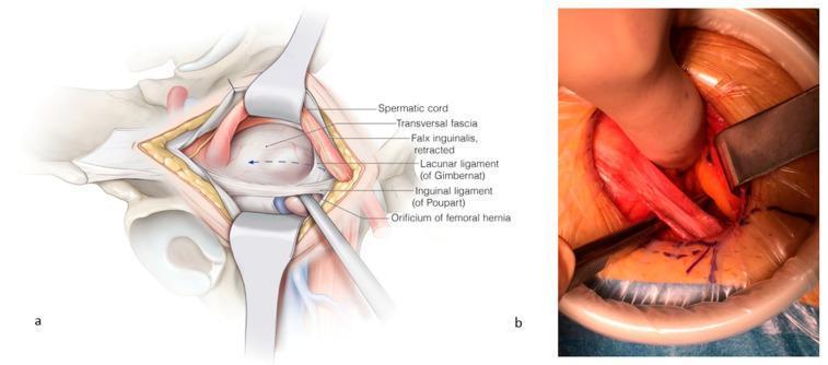
This composite visual contains an anatomical illustration (a) and a corresponding clinical intraoperative photograph (b) detailing the surgical repair of a femoral hernia. Panel (a) provides a labeled anatomical diagram of the right inguinal-femoral region. It depicts the spermatic cord and falx inguinalis retracted cranially, exposing the orificium of the femoral hernia. Key landmarks are identified: the transversalis fascia (opened), the inguinal ligament of Poupart (ventral border), and the lacunar ligament of Gimbernat (medial border). An instrument is shown passing through the femoral orifice to demonstrate the pathology. Panel (b) is a clinical photograph of the surgical field during an open repair. It demonstrates the clinical application of the anatomy shown in panel (a), with retractors maintaining exposure. A surgical instrument is seen incising the lacunar ligament to enlarge the femoral space for mesh placement. The image illustrates the relationship between the inguinal ligament, femoral vessels, and the hernia orifice. The content is intended for intermediate to advanced surgical education, specifically regarding open femoral hernia repair and the anatomy of the femoral canal.
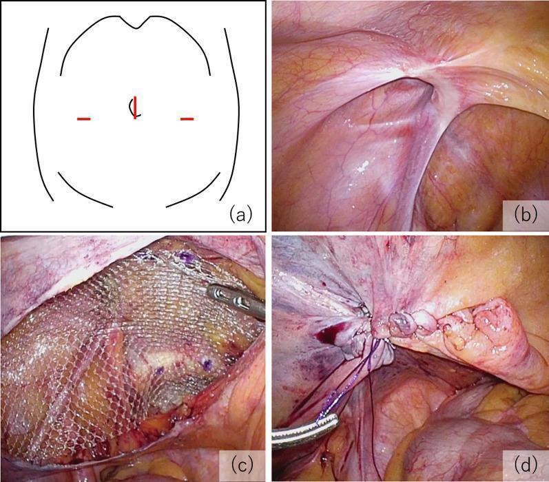
A multi-panel educational figure illustrating the key procedural steps of a Transabdominal Preperitoneal (TAPP) inguinal hernia repair. (a) Schematic diagram of the abdomen showing the laparoscopic port arrangement: a 12 mm umbilical port and two 5 mm lateral ports. (b) Intraoperative laparoscopic photograph showing the internal inguinal anatomy and the hernial orifice, classified as EHS-L2 (European Hernia Society classification for a medium indirect lateral hernia). (c) Surgical view demonstrating mesh fixation; a semi-transparent, macroporous polypropylene mesh is positioned in the preperitoneal space to cover the defect and is secured with visible surgical tacks. (d) Final procedural step showing the closure of the peritoneal flap using continuous suturing to exclude the mesh from the peritoneal cavity, preventing contact with abdominal viscera. The series emphasizes proper anatomical orientation, mesh placement, and peritoneal closure in minimally invasive hernia surgery.
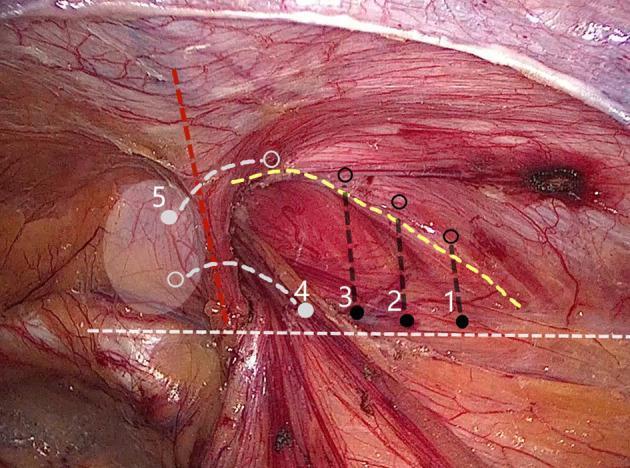
This clinical photograph shows a laparoscopic view of the inguinal region during an internal ring closure procedure, specifically demonstrating a 'five-stitches method' for hernia repair. The surgical field displays the reddish-pink fibrous texture of the conjoined tendon and the iliopubic tract, with prominent epigastric vessels visible. Overlaid on the anatomy are numbered markers (1-5) and colored dashed lines illustrating the suturing technique: black solid dots indicate needle entry points, while open circles represent extraction points. Points 1-3 follow a lateral-to-medial linear path along the iliopubic tract, connected by a dashed yellow line. Points 4 and 5 transition into a U-shaped configuration, marked by a red dashed line, to navigate around the epigastric vessels. A white dashed line indicates the alignment of tissues for approximation. The diagram highlights the clinical strategy of superficial, small-bite suturing intended to avoid underlying neurovascular structures, including the iliac artery and branches of the genitofemoral and ilioinguinal nerves.
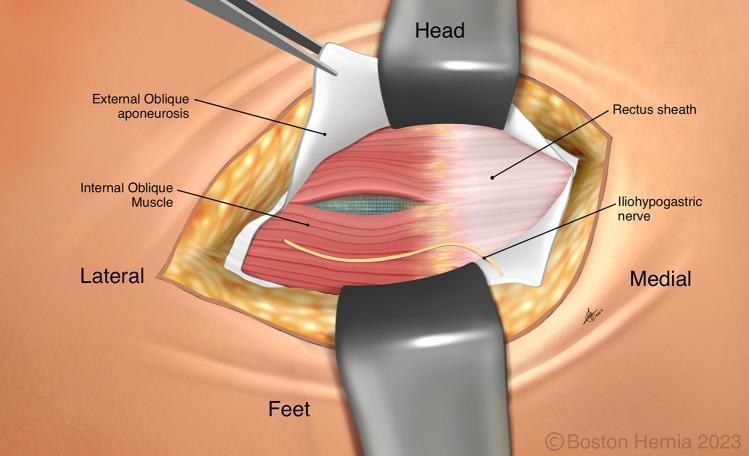
This medical illustration depicts a step in a right inguinal hernia repair (TREPP procedure) showing the abdominal wall layers. The surgical field is oriented with 'Head', 'Feet', 'Lateral', and 'Medial' labels. Retractors and forceps expose the anatomical structures. The external oblique aponeurosis is reflected, revealing the internal oblique muscle. A key feature is the blue surgical mesh visible through a natural split in the fibers of the internal oblique muscle. Medial to this, the rectus sheath is clearly defined. The iliohypogastric nerve is shown coursing superficially across the medial aspect of the internal oblique muscle. The illustration highlights the educational objective of allowing the internal oblique muscles to approximate naturally over the mesh to prevent nerve entrapment or injury by avoiding suturing near the iliohypogastric nerve. This diagram is designed to teach surgical anatomy and mesh placement techniques in preperitoneal hernia repair.
hernia clinical examination cough impulse reducible irreducible
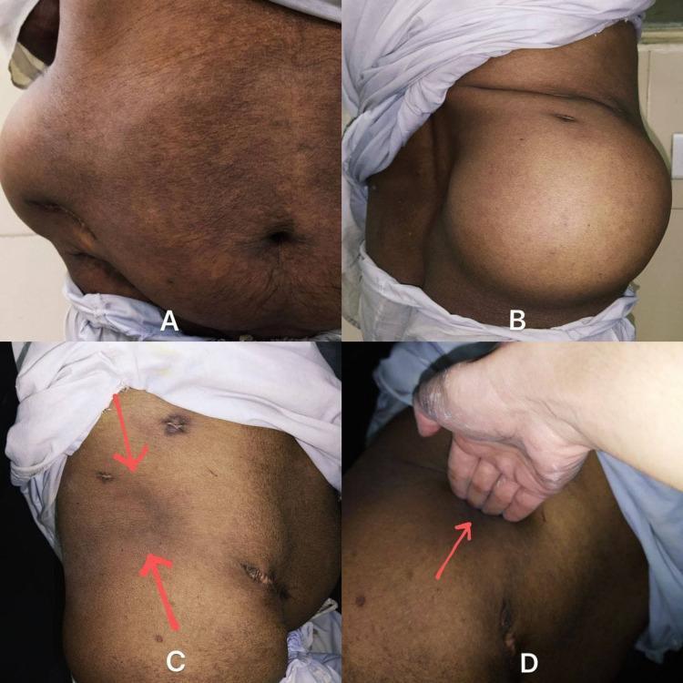
This composite of clinical photographs illustrates the physical examination findings of a right-sided lumbar hernia. Panels A (anterior view) and B (lateral view) demonstrate a large, smooth, and prominent swelling in the right lumbar region that significantly distorts the flank's contour. The overlying skin appears normal with a visible scar, indicative of a previous surgical intervention. Panel C shows the same anatomical region after the manual reduction of the hernia; red arrows indicate the site of the musculoskeletal defect. Panel D demonstrates the clinical technique for assessing the size of the abdominal wall defect, where four fingers are insinuated into the reducible space. This visual series is designed for medical education regarding the diagnosis and physical assessment of lumbar hernias (Petit's or Grynfeltt-Lesshaft hernias), emphasizing key clinical features such as reducibility, the presence of an expansile cough impulse, and the physical palpation of the fascial defect.
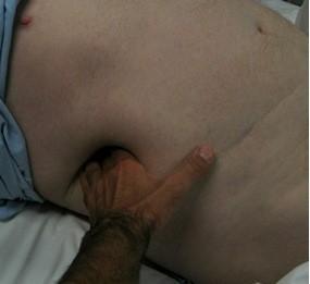
This clinical photograph illustrates a physical examination of a large, reducible Traumatic Transdiaphragmatic Intercostal Hernia (TTIH) in a supine patient. The image shows a significant bulging deformation in the right abdominal and lower thoracic wall region. A clinician's hand is shown applying manual pressure directly onto the herniation, demonstrating its easy reducibility as the abdominal wall yields to the touch. The overlying skin appears intact without signs of acute inflammation or discoloration. This visual focuses on the clinical manifestation of a post-traumatic defect in the abdominal wall and diaphragm, where abdominal viscera (typically including the liver or colon) herniate through the intercostal space. The image is an educational example of hernia assessment techniques and the gross appearance of a chronic traumatic hernia during follow-up.
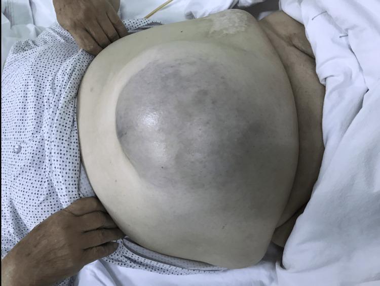
Clinical photograph showing an anterior view of an 80-year-old female patient with a giant, irreducible umbilical hernia. The abdomen is significantly distended, featuring a large, protruding mass approximately 18x18 cm located in the umbilical and infraumbilical regions. The most prominent clinical sign is the diffuse, bluish-purple discoloration of the skin overlying the central aspect of the hernia sac. This cyanotic appearance is highly suggestive of vascular compromise, strangulation, or underlying gangrene of the incarcerated bowel contents. The surrounding abdominal skin is pale but intact, without visible ulceration. This image serves as a critical educational example of an 'acute abdomen' presentation where skin color changes over a hernia site indicate a surgical emergency, necessitating urgent intervention to prevent or address bowel necrosis.
direct indirect inguinal hernia deep ring Hasselbach triangle anatomy
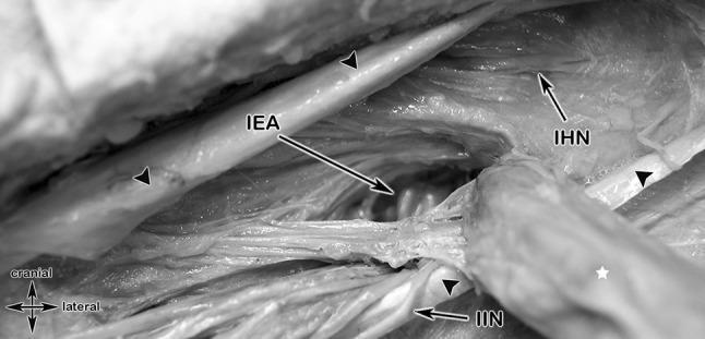
This grayscale clinical photograph of a cadaveric dissection illustrates the surgical anatomy of the inguinal region, specifically highlighting the topographic relationships during an indirect inguinal hernia repair. The dissection reveals the inferior epigastric artery (IEA) running cranially and medially. Lateral and inferior to the IEA, an indirect inguinal hernia is visible, marked with a white star, having protruded through the deep inguinal ring lateral to the epigastric vessels. Key neural structures are identified: the iliohypogastric nerve (IHN) is positioned superomedially to the hernia, while the ilio-inguinal nerve (I-IN) runs inferior and lateral to the IEA, passing toward the superficial inguinal ring. Black arrowheads delineate the reflected edges of the external oblique aponeurosis. The image provides critical educational context regarding the 'triangle of pain' and the importance of identifying these neurovascular structures to avoid iatrogenic injury during herniorrhaphy. The anatomical orientation is provided by a 'cranial/lateral' directional indicator, emphasizing the spatial arrangement of the spermatic cord structures and the transversalis fascia.
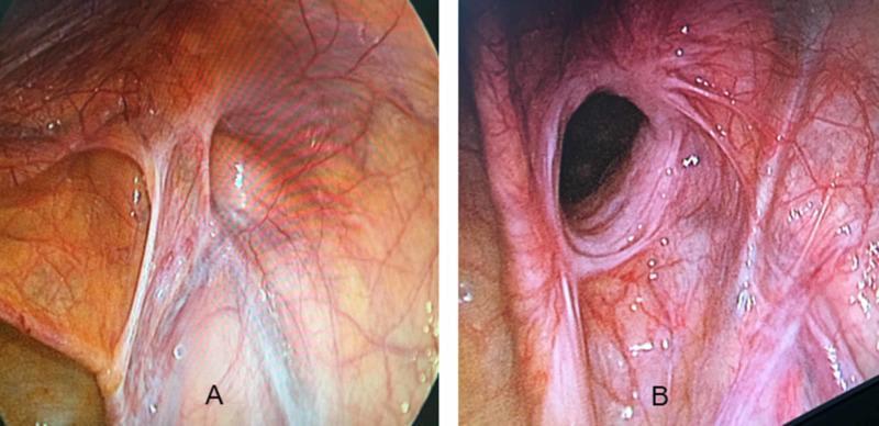
This diagnostic comparison uses laparoscopic (endoscopic) imaging to demonstrate the internal anatomical differences between an indirect and a direct inguinal hernia. Image A displays an indirect inguinal hernia, characterized by a rounded protrusion located lateral to the inferior epigastric vessels. The defect is clearly defined with a localized network of fine vasculature concentrated near the base of the herniated sac. Image B depicts a direct inguinal hernia, presenting as a more diffuse, wide-based bulge through the abdominal wall, located medial to the epigastric vessels in Hesselbach's triangle. The borders of the direct hernia are less distinct than the indirect type, and the overlying peritoneum shows a more prominent, dense, and tortuous vascular network. These images illustrate the surgical perspective during a Transabdominal Preperitoneal (TAPP) repair, emphasizing the importance of anatomical landmarks, specifically the inferior epigastric vessels, in differentiating hernia types based on their exit point relative to the deep inguinal ring.
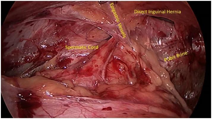
A laparoscopic (endoscopic) surgical view of the posterior inguinal abdominal wall, highlighting key anatomical landmarks for hernia identification. Centrally, the inferior epigastric vessels are visualized, serving as the critical anatomical boundary between direct and indirect inguinal spaces. Medial to these vessels (Hesselbach's triangle), a distinct bulge or protrusion is visible, identified as a direct inguinal hernia. Inferiorly, the whitish, firm structure of the pubic bone is seen, providing a caudal reference point. The spermatic cord is visible laterally, coursing toward the deep inguinal ring. The image demonstrates the laparoscopic perspective of the preperitoneal space, often utilized in TEP (Total Extraperitoneal) or TAPP (Transabdominal Preperitoneal) hernia repair procedures. This clinical photograph provides an educational example of inguinal floor weakness and the topographical relationship between the abdominal wall musculature, vasculature, and pelvic osteology in the context of general surgery and sports medicine.
Hernia - Definition, Types, and Clinical Examination
Definition
- Hernial sac - a protrusion of peritoneum through the defect
- Hernial contents - the viscera inside the sac (omentum, bowel, bladder, ovary, etc.)
- Coverings - layers of abdominal wall that overlie the sac
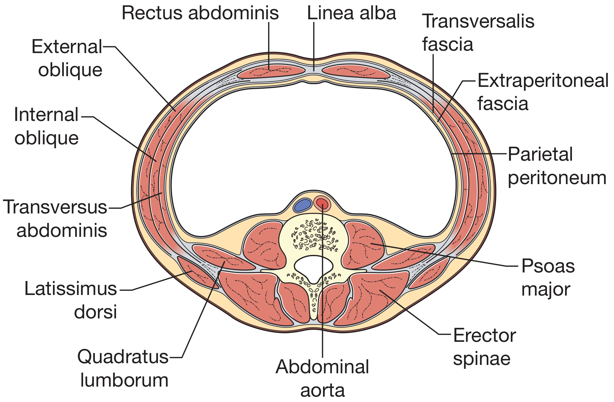
Anatomy of the Abdominal Wall
- Skin and subcutaneous fat
- External oblique (aponeurosis)
- Internal oblique
- Transversus abdominis
- Transversalis fascia
- Extraperitoneal fat
- Parietal peritoneum
- Inguinal canal (indirect & direct hernias)
- Femoral ring
- Umbilicus
- Linea alba (epigastric hernias)
- Surgical scars (incisional hernias)
- Obturator foramen, lumbar triangles, Spigelian line
Classification of Hernias
By Complexity (Clinical States)
| Type | Description |
|---|---|
| Occult | Not detectable clinically |
| Reducible | Swelling appears and disappears spontaneously or on pressure |
| Irreducible | Cannot be replaced back into the abdomen |
| Incarcerated | Irreducible + trapped, at risk of strangulation |
| Obstructed | Bowel within hernia is blocked; no strangulation yet |
| Strangulated | Blood supply to hernia contents is cut off; emergency |
| Infarcted/Gangrenous | Contents are gangrenous; very high mortality |
By Location
| Hernia | Site |
|---|---|
| Inguinal (indirect) | Lateral to inferior epigastric vessels, through deep inguinal ring |
| Inguinal (direct) | Medial to inferior epigastric vessels, through Hesselbach's triangle |
| Femoral | Through femoral ring, below inguinal ligament, medial to femoral vein |
| Umbilical | Through umbilical ring |
| Para-umbilical | Alongside, not through, the umbilicus |
| Epigastric | Through linea alba above umbilicus |
| Incisional | Through a previous surgical scar |
| Spigelian | At the lateral edge of rectus abdominis, through the semilunar line |
| Obturator | Through obturator foramen |
| Lumbar | Through posterior triangles (Petit's or Grynfeltt-Lesshaft) |
| Internal | Bowel entrapment within abdominal cavity (paraduodenal, etc.) |
Special Eponymous Types
- Richter's hernia - only part of the bowel wall is caught in the sac; no bowel obstruction but bowel wall can strangulate and perforate silently. Classic in femoral hernias.
- Littre's hernia - contains Meckel's diverticulum
- Maydl's hernia ("hernia in W") - two loops of bowel in sac with the loop between them inside the abdomen at greatest risk
- Amyand's hernia - appendix in inguinal hernia sac
- Pantaloon hernia - combined direct and indirect hernia straddling inferior epigastric vessels
- Sliding hernia - part of the sac wall is formed by a viscus (e.g., bladder or caecum)
Hesselbach's (Hasselbach's) Triangle
- Lateral border: Inferior epigastric vessels
- Medial border: Lateral edge of rectus abdominis
- Inferior border: Inguinal ligament
Clinical Examination of a Hernia
General Principles - Bailey & Love
- Examine first lying, then standing (hernias enlarge/appear on standing)
- Ask the patient to cough or do the Valsalva manoeuvre to make the hernia visible
- Overlying skin: normally normal colour
- Red/inflamed/discoloured skin over the hernia = contents are strangulating - treat as emergency
- Scars from previous repair
- Reducibility - can it be returned to the abdomen?
- Cough impulse - expansile impulse felt on coughing
- Tenderness - suggests incarceration/strangulation
- Overlying skin colour changes
- Multiple defects and contralateral side (occult contralateral hernias in up to 20%)
- Signs of previous repair
- Scrotal contents for groin hernia
- Place gentle pressure on the lump and ask the patient to cough
- A positive cough impulse = expansile impulse felt = consistent with hernia
- May be absent in irreducible hernias with a tight neck (classic for femoral hernia - often misdiagnosed as lymph node)
- Also present in saphena varix
- Ask the patient to reduce the hernia first (they may be practiced at it)
- If not spontaneously reduced, attempt gentle taxis
- If neither patient nor surgeon can reduce: treatment is more urgent
Inguinal Hernia Examination - Differentiating Direct vs Indirect
- Reduce the hernia (if not already spontaneously reduced)
- Identify bony landmarks: anterior superior iliac spine and pubic tubercle
- Locate the deep inguinal ring - just above the midpoint of the inguinal ligament
- Apply gentle pressure at the deep ring
- Ask the patient to cough:
- Hernia controlled by pressure at deep ring = indirect (lateral)
- Hernia appears medial to pressure = direct (medial)
Note: Even experienced surgeons find it difficult to distinguish direct from indirect with certainty. Final distinction is usually made intraoperatively or at laparoscopy.
- In males, invaginate the scrotal skin with the little finger into the inguinal canal up to the superficial ring
- Ask patient to cough:
- Impulse felt at fingertip = indirect hernia (coming through the deep ring)
- Impulse against the pulp of the finger = direct hernia (pushing straight through the floor)
Inguinal vs Femoral Hernia - Key Differences
| Feature | Inguinal | Femoral |
|---|---|---|
| Position relative to pubic tubercle | Above and medial | Below and lateral |
| Sex | Predominantly male | More common in female (but inguinal still more common in both) |
| Cough impulse | Usually present | Often absent |
| Reducibility | Often reducible | Often irreducible |
| Risk of strangulation | Lower | Higher (tight neck) |
| Richter's hernia | Less common | Classic site |
Groin Swelling - Differential Diagnosis
- Inguinal hernia (indirect or direct)
- Femoral hernia
- Inguinal lymph node / lymphadenopathy
- Saphena varix (has cough impulse, disappears on lying flat, thrill on coughing, associated with varicose veins)
- Femoral artery aneurysm (expansile pulsation)
- Psoas abscess (fluctuant, may extend below inguinal ligament)
- Undescended testis
- Lipoma of cord
- Hydrocele of cord
Complications of Hernia
- Irreducibility - cannot be returned to abdomen
- Obstruction - bowel lumen blocked; vomiting, distension, constipation without blood supply compromise
- Strangulation - compromised blood supply; severe pain, tenderness, vomiting; surgical emergency
- Perforation - gangrenous contents perforate; peritonitis and sepsis
- Infarcted Richter's hernia - partial bowel wall strangulation without obstruction, easily missed
Investigations
- Clinical diagnosis in most cases
- Ultrasound - for occult hernias, confirmation, or scrotal/groin swellings
- CT scan - complex hernias, internal hernias, unclear diagnosis
- Herniography - rarely used; contrast injected into peritoneum
Management Overview
| Approach | Details |
|---|---|
| Conservative (watchful waiting) | Acceptable for minimally symptomatic reducible hernias; patient counselled on strangulation risk |
| Truss/Hernia belt | Temporary; not curative; only in unfit patients |
| Lichtenstein repair | Open, tension-free mesh placement over the inguinal floor |
| Laparoscopic (TEP or TAPP) | Preferred for bilateral, recurrent hernias; faster recovery |
| Emergency surgery | Strangulation, obstruction - irreversible ischaemia may need bowel resection |
Clinical Pearls
- Always examine the contralateral side - occult contralateral hernia present in up to 20% of patients
- Red/blue skin over a hernia = strangulation = emergency
- Femoral hernias in women are frequently missed; any groin lump in a woman below and lateral to the pubic tubercle is femoral until proven otherwise
- A hernia with no upper limit in the scrotum (cannot get above it) is a large inguinal hernia, not a hydrocele
- Richter's hernia can strangulate without bowel obstruction - do not be falsely reassured