
Early stage carcinoma cervix management ,give me a 10mark Drnb surgical oncology answer
"cervical cancer" AND "radical hysterectomy" AND "early stage"
Early Stage Carcinoma Cervix - Surgical Management
Introduction
FIGO 2018 Staging (Early Stages)
| Stage | Definition |
|---|---|
| IA1 | Stromal invasion < 3 mm depth |
| IA2 | Stromal invasion 3-5 mm depth |
| IB1 | Clinically visible lesion ≤ 2 cm |
| IB2 | Clinically visible lesion > 2 cm but ≤ 4 cm |
| IIA1 | Extension to upper 2/3 vagina, tumor ≤ 4 cm |
Surgical Management by Stage
1. Stage IA1 (No LVSI)
- Simple (extrafascial) hysterectomy (Piver-Rutledge type I) is adequate
- Lymph node dissection is NOT required (risk of nodal disease < 1%)
- Fertility-sparing option: Large loop excision of transformation zone (LLETZ/LEEP) or cold knife conization (CKC) with clear margins
2. Stage IA1 (With LVSI) and Stage IA2
- Modified radical hysterectomy (Type II) + pelvic lymph node dissection (PLND)
- Sentinel lymph node (SLN) mapping is an accepted alternative to full PLND for small tumors
- Fertility-sparing option: Radical trachelectomy (vaginal or abdominal) + PLND/SLN mapping
3. Stage IB1 (≤ 2 cm)
- Radical hysterectomy (Type III/Wertheim-Meigs procedure) + bilateral PLND
- This remains the gold standard
- Equally effective as chemoradiation, but surgery avoids long-term radiation morbidity (bowel, bladder, sexual dysfunction)
- Fertility-sparing: Radical trachelectomy is recommended for lesions ≤ 2 cm in patients desiring future fertility (FIGO IA1 with LVSI, IA2, or IB1)
4. Stage IB2 and IIA1
- Radical hysterectomy (Type III) + bilateral PLND ± para-aortic lymph node sampling
- Surgery or concurrent chemoradiation (CCRT) - outcomes are equivalent
- Surgery is preferred in younger patients to preserve ovarian function
- Ovaries can generally be spared in premenopausal patients as ovarian metastasis risk is low
Radical Hysterectomy - Piver-Rutledge-Smith Classification
| Type | Procedure | Indication |
|---|---|---|
| I | Simple extrafascial hysterectomy | IA1 (no LVSI) |
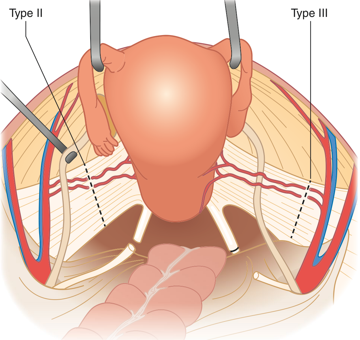
| II | Modified radical - medial 1/2 of cardinal & uterosacral ligaments | IA1+LVSI, IA2 |
| III | Radical - uterine artery at origin, full parametrium, upper 1/3 vagina | IB1, IB2, IIA1 |
| IV | Extended radical - periureteral tissue, superior vesical artery | Recurrent disease |
| V | Exenteration | Recurrent with bladder/rectal involvement |
- Developing paravesical and pararectal pelvic spaces
- Dividing uterine arteries and veins at their origin from internal iliac
- Dissecting the bladder and ureters off the anterior cardinal ligament (ureter "unroofed")
- Dissecting rectum off the posterior cardinal ligament
- Dividing cardinal and uterosacral ligaments fully
- Removing the upper 1-2 cm of vagina with an adequate margin
- Bilateral pelvic lymphadenectomy
Fertility-Sparing Surgery: Radical Trachelectomy
- FIGO stage IA1 with LVSI, IA2, or IB1
- Lesion ≤ 2 cm diameter (most experts' consensus)
- Desire for future fertility
- No lymph node involvement on imaging or SLN biopsy
Pelvic Lymphadenectomy
- External iliac nodes (lateral and medial)
- Obturator nodes (between external iliac vein and obturator nerve)
- Nodes from common iliac bifurcation down to deep circumflex iliac vein
- Accepted alternative for small early-stage tumors
- Avoids full lymphadenectomy morbidity (lymphedema, lymphocyst)
- ICG (indocyanine green) is most commonly used with excellent detection rates
Minimally Invasive vs. Open Surgery - Critical Update
- Minimally invasive radical hysterectomy (laparoscopic/robotic) had significantly worse disease-free survival (91.2% vs. 97.1% at 3 years) and overall survival (93.8% vs. 99% at 3 years) compared to open surgery
- Hazard ratio for recurrence: 3.74 (95% CI 1.63-8.58)
- No difference for tumors < 2 cm
- Open (abdominal) approach is now the standard of care for radical hysterectomy in cervical cancer
- Studies are ongoing to identify patients who may safely undergo minimally invasive surgery
Adjuvant Therapy Post-Surgery
- Positive pelvic lymph nodes
- Positive parametrial margins
- Positive surgical margins
- LVSI + deep stromal invasion + large tumor
- Combination determines need for adjuvant pelvic radiation
Complications of Radical Hysterectomy
| Intraoperative | Postoperative |
|---|---|
| Urinary bladder injury | Vesicovaginal fistula (0.6-4.4%) |
| Ureteral injury | Ureterovaginal fistula |
| Bowel injury | Bladder dysfunction (atonic bladder) |
| Vascular injury | Lymphedema/lymphocyst |
| Ovarian failure (if oophorectomy) |
Summary Algorithm
Early Carcinoma Cervix
        |
        ├── IA1, no LVSI → Simple hysterectomy (or conization if fertility desired)
        |
        ├── IA1+LVSI / IA2 → Type II radical Hx + PLND (or radical trachelectomy)
        |
        ├── IB1 (≤2cm) → Type III radical Hx + PLND (open approach)
        |              [Trachelectomy if fertility-sparing desired]
        |
        └── IB2 / IIA1 → Type III radical Hx + PLND (or CCRT - equivalent)
                        ↓
               Adjuvant CCRT if Peters criteria met
Conclusion
Management of locally advanced cervical cancer
"locally advanced cervical cancer" AND "chemoradiation"
Management of Locally Advanced Carcinoma Cervix
Definition and Staging
| Stage | Definition |
|---|---|
| IB3 | Clinically visible lesion > 4 cm, confined to cervix |
| IIA2 | Vaginal extension (upper 2/3), tumor > 4 cm |
| IIB | Parametrial invasion, not reaching pelvic sidewall |
| IIIA | Extension to lower 1/3 of vagina |
| IIIB | Extension to pelvic sidewall OR hydronephrosis/non-functioning kidney |
| IIIC1 | Pelvic lymph node metastasis (radiologic) |
| IIIC2 | Para-aortic lymph node metastasis (radiologic) |
| IVA | Invasion of bladder or rectal mucosa |
Pre-Treatment Workup
- Clinical examination - bimanual and rectovaginal examination
- MRI pelvis - gold standard for local staging (tumor size, parametrial/vaginal/bladder/rectal involvement)
- PET-CT - for nodal staging (pelvic, para-aortic, supraclavicular) and distant metastasis
- CT chest/abdomen/pelvis if PET unavailable
- Cystoscopy and sigmoidoscopy if bladder or rectal invasion suspected
- Renal function tests (hydronephrosis assessment), CBC, LFTs
Standard of Care: Concurrent Chemoradiation (CCRT)
- GOG 85 (IIB-IVA): Cisplatin + 5-FU + RT vs. hydroxyurea + RT - cisplatin arm had significantly superior PFS and OS
- GOG 120 (IIB-IVA): Weekly cisplatin + RT vs. cisplatin/5-FU/hydroxyurea + RT vs. hydroxyurea + RT - both cisplatin arms were superior
- GOG 123 (Bulky IB): Cisplatin + RT (+ adjuvant hysterectomy) vs. RT alone - 3-year OS 83% vs. 74%
- RTOG/SWOG Intergroup trial: 5-year OS 73% with CCRT vs. 58% with RT alone
CCRT Regimen
| Component | Details |
|---|---|
| Chemotherapy | Cisplatin 40 mg/m² IV weekly × 5-6 cycles (as radiosensitizer) |
| External Beam RT (EBRT) | 45-50.4 Gy in 25-28 fractions to whole pelvis (± para-aortic field) |
| Technique | IMRT (Intensity-Modulated RT) or 3D-CRT to reduce bowel/bladder dose |
| Brachytherapy boost | Intracavitary brachytherapy after EBRT - mandatory |
Brachytherapy - The Non-Negotiable Component
- Brachytherapy inclusion: CSS 64.3% vs. 51.5% without (P < 0.001)
- OS 58.2% vs. 46.2% without (P < 0.001)
- LDR (Low Dose Rate) brachytherapy (classical) - tandem and ovoids/ring
- HDR (High Dose Rate) brachytherapy - now standard; several minutes per fraction
- Total dose to Point A: ~80-90 Gy equivalent (EQD2) combining EBRT + brachytherapy
Extended Field Radiation (EFRT) - Para-Aortic Nodes
- Indicated when para-aortic LN involvement is confirmed (Stage IIIC2) or suspected
- Extends radiation field from pelvis up to L1-L2 (level of inferior mesenteric artery)
- Pelvic CCRT was shown superior to extended-field RT alone (RTOG trial)
- IMRT for para-aortic fields: associated with low GI toxicity (6.5% acute and late)
- Prophylactic EFRT is NOT recommended due to high GI toxicity without proven survival benefit in absence of documented nodal disease
Surgical Staging Before CCRT
- Extraperitoneal laparoscopic lymphadenectomy is preferred (transperitoneal approach has 16-33% radiation enteritis mortality)
- Positive para-aortic nodes: upgrade treatment to extended field CCRT
- PET-CT has largely replaced surgical staging in modern practice, though histologically positive nodes can be missed on imaging
Management of Grossly Positive Para-Aortic Nodes
- Extraperitoneal debulking lymphadenectomy before CCRT improves survival to levels comparable to microscopic nodal disease
- Patients with supraclavicular node involvement (5-30% of para-aortic positive cases) should receive palliative chemotherapy rather than extended CCRT
Neoadjuvant Chemotherapy (NACT) Followed by Surgery
- Paclitaxel + cisplatin or cisplatin + vincristine x 3 cycles, then radical hysterectomy
- The Indian ERCC trial (Gupta et al.) compared NACT + radical surgery vs. CCRT in stage IB2-IIB:
- No superior survival benefit of NACT-surgery over CCRT
- CCRT remains the standard
- NACT + surgery may be considered where radiotherapy resources are limited or in selected stage IIB patients in specialized centers
- A 2025 systematic review and meta-analysis (PMID 40408847) found that induction chemotherapy followed by CCRT improved progression-free and overall survival vs. CCRT alone in LACC
Adjuvant Hysterectomy After CCRT
- GOG 123 included adjuvant hysterectomy after CCRT for bulky IB disease
- No improvement in survival was demonstrated with adjuvant hysterectomy
- Not routinely recommended; CCRT alone is standard
Stage IVA (Bladder/Rectal Invasion)
- Primary CCRT remains standard - may achieve complete response in selected patients
- Bladder/rectal fistula may be palliative issue requiring diversion
- If residual or persistent central disease after CCRT: consider pelvic exenteration in highly selected, fit patients with no distant spread (cures 25-50%)
Recurrent/Persistent Disease After CCRT
Central Pelvic Recurrence (Previously Irradiated)
- Pelvic exenteration - anterior (bladder), posterior (rectum), or total
- Cures 25-50% in carefully selected patients
- Requirements: no pelvic sidewall or distant disease, adequate PS, resectable
- Laterally Extended Endopelvic Resection (LEER) - for selected lateral recurrences
Systemic/Metastatic Recurrence
- Chemotherapy: Paclitaxel + cisplatin/carboplatin ± bevacizumab (GOG 240 trial)
- Bevacizumab (anti-VEGF): Adds ~3.7 months OS benefit (GOG 240, NEJM 2014)
- Pembrolizumab (anti-PD-1): FDA-approved for PD-L1 positive recurrent/metastatic cervical cancer; can be combined with chemotherapy ± bevacizumab
- A 2024 meta-analysis (PMID 39351236) confirmed benefit of chemoradiation + immune checkpoint inhibitors in LACC
Complications of Radiation Therapy
| Acute (during/shortly after RT) | Chronic (months-years later) |
|---|---|
| Radiation proctitis (diarrhea, cramps) | Rectovaginal fistula (< 2%) |
| Radiation cystitis (frequency, bleeding) | Vesicovaginal fistula (1.4-5.3%) |
| Nausea, fatigue | Bowel stricture/obstruction (6.4-8.1%) |
| Hematologic toxicity (with cisplatin) | Sexual dysfunction (vaginal stenosis) |
| Uterine perforation during BT (~9%) | Secondary bladder cancer (2-4x risk) |
Prognosis (5-year OS by Stage - CCRT Era)
| Stage | Approximate 5-yr OS |
|---|---|
| IB3 | 60-70% |
| IIB | 50-65% |
| III | 30-50% |
| IVA | 15-25% |
Summary Algorithm
LACC (FIGO IB3 - IVA)
          |
    PET-CT / MRI staging
          |
    ┌─────┴─────┐
Para-aortic      Pelvic-only
nodes +ve        disease
    |                |
Extended-field   Pelvic CCRT
CCRT (45 Gy)   (45-50 Gy EBRT
                + weekly cisplatin)
          |
    Mandatory intracavitary
    BRACHYTHERAPY boost
    (total EQD2 ~80-90 Gy)
          |
    Assess response (MRI at 3 months)
          |
    ┌─────┴──────┐
Complete        Residual/
response        Recurrence
(surveillance)      |
             Central only → Exenteration
             Metastatic → Chemo ± Bev ± Pembrolizumab
Sentinel lymph node biopsy in cervical cancer Give answer from UpToDate,NCCN guidelines ,devita,berek and hacker and other standard source
"sentinel lymph node" AND "cervical cancer"
NCCN guidelines 2025 sentinel lymph node biopsy cervical cancer recommendations
https://pmc.ncbi.nlm.nih.gov/articles/PMC12399259
PMID: 37696646
PMID: 40555135
PMID: 37586759
SENTIX trial results sentinel lymph node cervical cancer 2024 2025 ultrastaging algorithm
PMID: 40615729
Sentinel Lymph Node Biopsy in Cervical Cancer
Introduction and Rationale
NCCN Guidelines (Version 2.2026) - Current Recommendations
- SLN mapping is now included in the primary treatment recommendations for early-stage cervical cancer (added following SENTIX trial final results, published 2025)
- Recommended for FIGO stage IA1 (with LVSI), IA2, IB1, and IB2 (tumor ≤ 4 cm, clinically node-negative)
- Preferred tracer: ICG (indocyanine green) with near-infrared fluorescence imaging
- Standard bilateral cervical injection at 3 and 9 o'clock positions, both superficially and deeply
- Ultrastaging is a mandatory component of SLN evaluation
- If SLN mapping fails on one side: side-specific lymphadenectomy is required on the non-mapped side
- Any suspicious node must be removed regardless of mapping status
Anatomy: Where are Sentinel Nodes Found?
| Location | Frequency |
|---|---|
| External iliac (medial) | Most common |
| Obturator space (between external iliac vein and obturator nerve) | Common |
| Hypogastric / internal iliac (ventral surface) | Common |
| Common iliac | Less common |
| Presacral / parametrial | Uncommon but important - "unusual" SLNs |
| Para-aortic | Rare - skip metastases possible |
Tracer Techniques
1. Blue Dye (Isosulfan Blue / Patent Blue V)
- Oldest technique; purely visual
- Detection rate: ~60-80%
- Disadvantage: cannot use laparoscopy effectively; anaphylaxis risk (1-2%)
- Injection site per FILMS study protocol: deeply + superficially at 3 and 9 o'clock
2. Technetium-99m Radiocolloid (99mTc)
- Requires preoperative lymphoscintigraphy
- Intraoperative gamma probe detection
- Detection rate: ~80-90%
- Not widely available; radiation handling required
3. Indocyanine Green (ICG) - PREFERRED (NCCN 2026)
- Detected via near-infrared (NIR) fluorescence camera
- Detection rate: 93-100% - highest sensitivity
- Superior to blue dye and comparable to or better than 99mTc
- Works excellently laparoscopically and robotically
- No radiation; minimal allergy risk
- Dose: 1-4 mg diluted in sterile water; 1 mL per quadrant
4. Combination Techniques
- Blue dye + 99mTc has historically been the gold standard (superior to either alone)
- ICG ± blue dye is now preferred in most centers
Injection Technique
- Patient under general or spinal anesthesia
- Inject superficially and deeply at cervical stroma
- Standard: 3 and 9 o'clock (or 12, 3, 6, 9 o'clock quadrants)
- Volume: 0.5-1 mL per injection site; total 2-4 mL
- No prior conization affects detection rates (cold knife cone biopsy does NOT reduce SLN detection)
- Allow 5-10 minutes after injection before starting dissection
The NCCN SLN Algorithm (Cervical Cancer)
Inject tracer (ICG/blue dye/99mTc) into cervix
              ↓
      Bilateral mapping?
     ↙              ↘
   YES                NO (unilateral or failed)
     |                        |
Excise all mapped        Side-specific PLND on
SLNs bilaterally        unmapped/failed side
     |                + excise mapped SLNs
     ↓
Remove ALL suspicious nodes regardless of mapping
              ↓
     PATHOLOGICAL ULTRASTAGING of all SLNs
              ↓
    ┌─────────┴─────────┐
SLN negative           SLN positive
(omit PLND)       (complete treatment/
                   adjuvant therapy)
Pathological Ultrastaging - The Critical Step
- Bisect the node along the longest axis
- H&E staining of all sections
- Cytokeratin immunohistochemistry (IHC) (AE1/AE3 or MNF116) on all levels
- Serial sections at 200-500 μm intervals
Classification of SLN Metastases:
| Category | Size | Clinical significance |
|---|---|---|
| Macrometastasis | > 2 mm | High risk; adjuvant CCRT |
| Micrometastasis | 0.2-2 mm | Intermediate risk; emerging data |
| Isolated Tumor Cells (ITC) | < 0.2 mm | Uncertain; controversial management |
Key Landmark Trials
SENTICOL I & II (France)
- Prospective multicenter studies establishing SLN feasibility in cervical cancer
- SENTICOL II: 206 patients; bilateral detection rate 97%; sensitivity 82%; NPV 97%
- Demonstrated that SLN biopsy with ultrastaging detects low-volume metastases missed by standard H&E
SENTIX Trial (International, NCT02494063) - 2025 Final Results
- 47 centers, 18 countries; FIGO IA1/LVSI+ to IB2; n=731 enrolled, n=594 intention-to-treat
- 12.5% of patients had node-positive disease (81/647)
- 44% of N1 cases detected exclusively by ultrastaging (would have been missed by standard assessment)
- Intraoperative frozen section detected only 56.1% of positive nodes
- At 2 years: recurrence rate 6.1% (within noninferiority threshold of 7%)
- 2-year DFS: 93.3%; 2-year OS: 97.9%
- Conclusion: SLN biopsy without systematic PLND does NOT increase recurrence risk in early-stage cervical cancer
- This trial formed the basis for NCCN v2.2026 adding SLN mapping to primary treatment recommendations
Systematic Reviews on Survival (2023)
- Parpinel et al., IJGC 2023 (PMID 37696646) - Meta-analysis of 6 studies: 5-year DFS 94-98%; 5-year OS 99% after SLN biopsy alone - comparable to full PLND
- Mauro et al., IJGC 2023 (PMID 37586759) - 4 studies, 2226 patients: 3-year recurrence-free survival 93.1% (SLN alone) vs. 92.5% (PLND) - no significant difference (p=0.773)
Performance Characteristics (Berek & Novak, NCCN)
| Parameter | Value |
|---|---|
| Overall SLN detection rate | 80-100% |
| Bilateral detection rate | 85-97% |
| Sensitivity | 65-93% |
| Negative Predictive Value (NPV) | 90-99% |
| False-negative rate | 1-7% |
Predictive Factors for SLN Mapping Failure
| Factor | Odds Ratio | P-value |
|---|---|---|
| Tumor size ≥ 2 cm | OR 1.35 | 0.018 |
| Age ≥ 50 years | OR 2.71 | < 0.001 |
| FIGO stage II-IV | OR 2.11 | 0.009 |
| Pelvic LN metastasis | OR 2.15 | 0.025 |
| Prior NACT | OR 1.44 | 0.010 |
Appropriate Candidates (NCCN 2026 Criteria)
- FIGO stage IA1 with LVSI, IA2, IB1, IB2
- Tumor ≤ 4 cm (strongest evidence for ≤ 2 cm)
- No suspicious nodes on preoperative imaging (MRI/PET-CT)
- No prior pelvic radiation
- No prior pelvic surgery likely to disrupt lymphatic drainage
- Planned radical hysterectomy or trachelectomy
- Tumor > 4 cm
- FIGO stage IIB and beyond
- Clinically suspicious or imaging-positive lymph nodes
- Prior radiotherapy to pelvis
- Failed bilateral mapping without side-specific PLND completion
Advantages of SLN Biopsy Over Full PLND
| Advantage | Clinical Impact |
|---|---|
| Reduced lymphedema | 40% vs. < 5% |
| Reduced lymphocyst formation | Major reduction |
| Shorter operative time | ~30-45 min less |
| Less blood loss | Clinically meaningful |
| Detection of unusual/skip SLNs | Improves staging accuracy |
| Ultrastaging detects ITC/micrometastases | Missed by standard H&E in full PLND |
| Less neurovascular injury | Obturator nerve, iliac vessels |
Current Status and Guidelines Summary
| Guideline Body | Recommendation |
|---|---|
| NCCN v2.2026 | SLN mapping added to primary treatment recommendations; preferred for IA1+LVSI to IB2; ICG preferred; ultrastaging mandatory |
| ESGO 2023 | SLN biopsy acceptable alternative to PLND in early-stage; bilateral mapping required |
| Berek & Novak | Considered for tumors < 2 cm; NPV 90-97%; does not replace PLND if unilateral/failed |
| Sabiston Textbook | SLN mapping an alternative to PLND for small early-stage tumors |
| SENTIX 2025 | SLN biopsy without systematic PLND is noninferior in terms of recurrence |
Conclusion
Nerve sparing radical hysterectomy
"nerve-sparing radical hysterectomy" AND "cervical cancer"
nerve sparing radical hysterectomy technique hypogastric nerve pelvic autonomic anatomy 2024 2025
PMID: 36862911
https://pmc.ncbi.nlm.nih.gov/articles/PMC10566162
"nerve-sparing" AND "radical hysterectomy" AND "bladder" AND "quality of life"
nerve sparing radical hysterectomy Sakuragi Raspagliesi technique steps oncologic outcomes sexual dysfunction
Nerve-Sparing Radical Hysterectomy (NSRH)
Introduction and Historical Background
- Kobayashi (1961): First described preservation of the hypogastric nerve in radical hysterectomy
- Okabayashi radical hysterectomy: The original Japanese technique emphasizing anatomical tissue plane dissection
- Sakuragi (2005): Described a systematic NSRH technique for preserving postsurgical bladder function
- Raspagliesi (2004): European adaptation with anatomic nerve identification
- Fujii (2007): Defined the Nerve-Sparing Okabayashi procedure with functional outcomes
Anatomy of Pelvic Autonomic Nerves - The Surgical Basis
Sympathetic Component
- Originates from T11-L2 spinal segments
- Forms the Superior Hypogastric Plexus (SHP) below the aortic bifurcation at the sacral promontory
- SHP divides into left and right Hypogastric Nerves (HN) which descend along the lateral pelvic wall
- Functions: bladder neck continence, orgasm, ejaculation (in males), uterine/vaginal tone
Parasympathetic Component
- Originates from S2, S3, S4 sacral nerve roots
- Forms Pelvic Splanchnic Nerves (PSN) - also called "nervi erigentes"
- Runs along the pelvic sidewall within the cardinal ligament (deep uterine vein region)
- Functions: detrusor contraction (bladder voiding), rectal motility, vaginal lubrication, clitoral engorgement
Inferior Hypogastric Plexus (IHP) = Pelvic Plexus
- Formed by convergence of: Hypogastric Nerve (sympathetic) + Pelvic Splanchnic Nerves (parasympathetic) + Sacral Sympathetic Trunk
- Located in the retroperitoneal space lateral to the rectum, vagina, and cervix
- Situated within the "pars nervosa" of the parametrium (posterior-lateral portion)
- Gives rise to branches to: bladder (vesical branches), uterus, vagina, and rectum
The Three Critical Nerve Groups at Risk in Radical Hysterectomy
| Nerve Structure | Location | Injured When |
|---|---|---|
| Hypogastric nerve | Posterior parametrium / uterosacral ligament | Uterosacral ligament is divided |
| Pelvic splanchnic nerve (PSN) | Deep within cardinal ligament near deep uterine vein | Deep uterine vein ligated, cardinal ligament divided |
| Bladder branches of IHP | Vesico-uterine ligament / paracolpium | Paracolpium and upper vagina divided |
Parametrium: Surgical Anatomy (Okabayashi Division)
| Part | Contents | Radical resection required? |
|---|---|---|
| Pars vasculosa (vascular part) | Uterine artery/vein, lymphatics, loose areolar tissue | YES - must be resected |
| Pars nervosa (nervous part) | IHP, bladder branches, PSN branches | NO - must be preserved |
Indications for NSRH
- FIGO 2018 stage IA2, IB1 (< 2 cm) - strongest evidence
- Stage IB2, IIA1 in selected cases (tumor ≤ 4 cm, no parametrial involvement)
- Clinically negative lymph nodes on preoperative MRI/PET-CT
- No parametrial invasion on imaging
- Good performance status; younger patients (quality of life priority)
- Bulky tumors > 4 cm
- Imaging-suspicious parametrial involvement
- Lymph node metastases on preoperative imaging
- Prior pelvic surgery altering anatomy
Surgical Technique - Step-by-Step
Step 1: Retroperitoneal Development and Nerve Identification
- Open the retroperitoneum bilaterally
- Identify the superior hypogastric plexus at the sacral promontory (below aortic bifurcation)
- Trace the hypogastric nerves bilaterally as they descend along the pelvic sidewall (medial to the internal iliac artery)
- Gentle blunt dissection exposes the nerve as a "stretched string" lateral to the rectum
Step 2: Medial Rectal Space Development
- Develop the mesorectal plane (rectovaginal space)
- This allows skeletonization of the uterosacral ligament, separating its nerve component from the ligament itself
- The hypogastric nerve is traced down to the IHP, preserved medially, while the uterosacral ligament is divided only at its uterine insertion
Step 3: Paravesical and Pararectal Space Development
- Standard development of paravesical space (medial to obliterated umbilical artery)
- Standard pararectal space (medial to hypogastric vessels)
- These spaces define the lateral limits of the parametrium
Step 4: Uterine Artery Ligation - Selective
- Uterine artery ligated at its origin from the internal iliac artery (standard)
- The deep uterine vein complex (where PSN runs) is carefully identified
- PSN is identified BENEATH the deep uterine veins and preserved while the veins are ligated above the nerve
Step 5: Cardinal Ligament Division - The Critical Step
- The cardinal ligament is divided in two phases:
- Pars vasculosa: ligated and divided (uterine artery, lymphatic tissue)
- Pars nervosa: the IHP and its bladder branches are lateralized and preserved
- The bladder branches of IHP run within the vesico-uterine ligament medial to the ureter
- After "unroofing" the ureter, the bladder pillars are divided while staying medial to the nerve branches
Step 6: Vesico-uterine Ligament (Bladder Pillar) Division
- The vesico-uterine ligament (anterior parametrium) is divided in two layers:
- Ventral layer: divided close to the bladder
- Dorsal layer: contains bladder branches of IHP - preserved by sharp dissection and lateral retraction
- This is the most technically demanding step
Step 7: Uterosacral Ligament Division
- The uterosacral ligament contains the upper portion of the IHP
- The ligament is divided at its cervical/vaginal insertion
- The hypogastric nerve running along its posterior surface is dissected free and preserved
Step 8: Vaginal Resection and Specimen Removal
- Standard 1-2 cm vaginal cuff removed
- Specimen checked for adequacy of parametrial margins
The "T-Shaped Nerve Plane" Concept
Superior Hypogastric Plexus (sacral promontory)
              ↓
      Hypogastric Nerve (descends on pelvic wall)
              ↓
    Inferior Hypogastric Plexus (lateral to cervix)
       ↙ (horizontal bar of "T")
  Pelvic Splanchnic     Bladder Branches
  Nerves (S2-S4)        (to vesical plexus)
Classification / Types of NSRH
| Type | Description | Indication |
|---|---|---|
| Type A NSRH | Hypogastric nerve preservation only | Minimal; Stage IA2 |
| Type B NSRH | HN + PSN preservation | Standard; IB1 |
| Type C NSRH | Complete HN + PSN + bladder branches | Selected IB2 |
- Type C1: Nerve-sparing radical hysterectomy (preserves autonomic nerves)
- Type C2: Classical non-nerve-sparing radical hysterectomy
Functional Outcomes: Evidence Summary
Bladder Dysfunction
| Outcome | Classical RH | NSRH |
|---|---|---|
| Urinary retention (short-term) | 20-50% | 5-15% |
| Long-term voiding dysfunction | 15-30% | 3-8% |
| Time to catheter removal | 7-14 days | 3-5 days |
| Need for self-catheterization | 8-12% | 1-3% |
- NSRH associated with significantly less urinary retention vs. classical RH
- HR 1.78 (95% CI 1.37-2.31, p < 0.001) in favour of NSRH
- HR 2.49 (95% CI 1.43-4.33, p = 0.001) in subgroup analysis
Sexual Function
- Nerve-sparing RH associated with significantly improved sexual function vs. standard RH (Berek & Novak, 4th ed.)
- Improved vaginal lubrication, arousal, and orgasm due to preservation of parasympathetic fibers
- A meta-analysis of NSRH for cervical cancer: no worsening of prognosis + improved sexual function vs. non-nerve-sparing surgery
Anorectal Function
- Reduced constipation (sympathetic preservation maintains rectal tone)
- Less defecatory urgency and straining
- Improved anorectal transit times
Oncologic Outcomes: Is Radicality Compromised?
- Parametrial clearance is maintained: provided the correct tissue planes are respected, pars vasculosa resection is complete
- No significant difference in local recurrence rates between NSRH and classical RH in multiple retrospective series
- 5-year OS and DFS are equivalent (Sakuragi 2005; Fujii 2007; Yin 2018)
- Berek & Novak (Robotic NSRH): "No differences with recurrence and survival" compared to non-nerve-sparing robotic RH
- 2024 systematic review (PMID 38438182 - Viveros-Carreño et al.): NSRH is one component of the overall de-escalation strategy - oncologic safety maintained
Minimally Invasive NSRH
- Robotic NSRH technique has been described and validated (Berek & Novak)
- Robotic platform advantages: 3D magnified visualization, tremor filtration, precise dissection in narrow pelvic spaces
- Comparison with open robotic NSRH: No differences in operating time, blood loss, complications, hospital stay
- However: The LACC trial caveat applies - open approach remains standard of care for radical hysterectomy in cervical cancer; MIS NSRH should be performed in specialized centers with careful patient selection (tumors < 2 cm)
Complications and Learning Curve
- Longer operative time (+30-90 minutes vs. standard RH)
- Steeper learning curve: requires detailed retroperitoneal anatomy training
- Risk of inadequate nerve dissection (nerve preserved anatomically but functionally disrupted by thermal spread)
- Para-rectal hematoma if hypogastric nerve dissection is overzealous
- Ureteral fistula (< 2%)
- Lymphocyst, lymphedema
- Bladder injury
Summary Comparison Table
| Feature | Classical RH (Type C2) | NSRH (Type C1) |
|---|---|---|
| Parametrial resection | Full (pars vasculosa + nervosa) | Pars vasculosa only |
| Bladder dysfunction | 20-50% | 5-15% |
| Sexual dysfunction | Common (40-60%) | Significantly reduced |
| Rectal dysfunction | 15-30% | Reduced |
| Oncologic outcome | Reference standard | Equivalent for IA2-IB1 |
| Operative time | Shorter | +30-90 min |
| Recommended approach | Open | Open (MIS at expert centers only) |
Conclusion
Radical hysterectomy
"radical hysterectomy" AND "cervical cancer" AND "outcomes"
PMID: 38416430
Radical Hysterectomy
Definition and Historical Background
- 1895 - Ernst Wertheim (Vienna): Pioneered the abdominal radical hysterectomy with selective lymph node removal; less extensive than modern Type III
- 1944 - Joe Vincent Meigs (Boston): Standardized the radical hysterectomy with systematic bilateral pelvic lymphadenectomy - the operation most performed in the USA
- Okabayashi (Japan): Described a more extensive parametrial dissection technique
- 2008 - Querleu & Morrow: Published the modern anatomical classification (updated 2017) now widely used in Europe and internationally
Indications
- FIGO 2018 Stage IA2 (stromal invasion 3-5 mm) - Type II
- FIGO 2018 Stage IB1 (≤ 2 cm) - Type III (gold standard)
- FIGO 2018 Stage IB2 (2-4 cm) - Type III (surgery or CCRT - equivalent outcomes)
- FIGO 2018 Stage IIA1 (< 4 cm vaginal extension) - Type III
- Adenocarcinoma - preferred over radiotherapy to avoid bowel complications
- Younger patients - to preserve ovarian function (ovaries conserved if premenopausal)
- Patients who are unlikely to need adjuvant radiation (to avoid combined modality morbidity)
- Stage IIB and beyond (parametrial invasion)
- Bulky tumors > 4 cm (relative - high risk of needing adjuvant RT)
- Positive para-aortic nodes (CCRT preferred)
- Poor surgical candidate (medical comorbidities)
- Obesity or severe pelvic adhesions (relative)
Classification Systems
1. Piver-Rutledge-Smith Classification (1974) - Most Widely Used
| Type | Name | Extent of Resection | Uterine Artery Ligation | Indication |
|---|---|---|---|---|
| I | Simple/Extrafascial | Uterus within pubovesicocervical fascia | At uterus | Stage IA1 (no LVSI) |
| II | Modified Radical (Wertheim) | Medial 1/2 cardinal + uterosacral ligaments; upper vaginal cuff | At ureter level | Stage IA1+LVSI, IA2 |
| III | Radical (Meigs) | Full cardinal + uterosacral ligaments to pelvic wall; upper 1/3 vagina | At internal iliac origin | Stage IB1, IB2, IIA1 |
| IV | Extended Radical | Periureteral tissue, superior vesical artery; 3/4 vagina | At origin | Rare: recurrent disease |
| V | Partial Exenteration | Distal ureter + bladder portion resected | - | Rare: tumor involving ureter/bladder |
2. Querleu-Morrow Classification (2008, Updated 2017) - European Standard
| Type | Parametrial Resection | Key Feature |
|---|---|---|
| A | Minimal parametrectomy | Close to cervix; ureter identified but not unroofed |
| B1 | Transection at ureter | Ureter unroofed; partial parametrium |
| B2 | As B1 + paracervical node dissection | - |
| C1 | Complete parametrectomy + nerve-sparing | Autonomic nerves preserved |
| C2 | Complete parametrectomy - classical | No nerve sparing |
| D1 | Laterally extended - at pelvic sidewall | Lateral parametrium to hypogastric vessels |
| D2 | As D1 + hypogastric vessels resected | Exenterative |
Surgical Anatomy: Pelvic Spaces
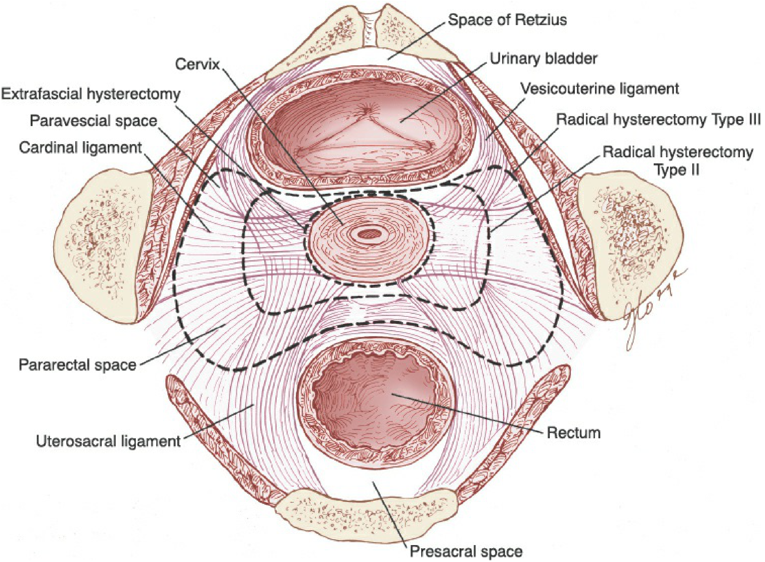
| Space | Medial Border | Lateral Border | Anterior/Posterior |
|---|---|---|---|
| Paravesical | Obliterated umbilical artery (bladder) | Obturator internus muscle | Pubic symphysis (ant) / Cardinal ligament (post) |
| Pararectal | Rectum | Hypogastric artery | Cardinal ligament (ant) / Sacrum (post) |
| Space of Retzius | Bladder | Pubic bones | Anterior |
| Presacral | Rectum | Sacrum | Posterior |
Operative Steps - Type III Radical Hysterectomy (Meigs)
Pre-Operative Preparation
- Bowel preparation (low residue diet, mechanical prep if needed)
- DVT prophylaxis (compression stockings + LMWH)
- Foley/suprapubic catheter placement
- Ureteral stenting (optional, in difficult cases)
- Informed consent: fistula risk, bladder dysfunction, lymphedema
Incision
- Midline infraumbilical incision (most common) - provides excellent exposure
- Low transverse incision (Maylard or Cherney): requires division of rectus muscles - superior lateral pelvic exposure, preferred for wider pelvis
Step 1: Abdominal Exploration
- Inspect peritoneal surfaces, liver, omentum for metastases
- Palpate para-aortic lymph nodes transperitoneally
- Inspect ovaries and fallopian tubes
- Palpate cardinal ligaments for lateral tumor extension
- Palpate the cervix between thumb and fingers to assess extent
Step 2: Retroperitoneal Entry and Lymphadenectomy
- Open round ligament at pelvic sidewall bilaterally
- Develop paravesical and pararectal spaces with blunt and sharp dissection
- Pelvic lymphadenectomy (bilateral):
- External iliac nodes (lateral and medial chains from common iliac bifurcation to deep circumflex iliac vein)
- Obturator nodes (between external iliac vein and obturator nerve; beware aberrant obturator vein in 10%)
- Internal iliac nodes
- Hemoclips applied distally to reduce lymphocyst
- Drains NOT used (increase complications without benefit)
- Inspect/palpate para-aortic nodes; send frozen section if suspicious
- If frozen section confirms nodal metastases: consider abandoning RH in favour of CCRT
Step 3: Para-Aortic Node Assessment
- Indicated if: bulky cervical tumor, grossly positive pelvic nodes, suspicious imaging
- Peritoneum incised medial to ureter, over right common iliac artery
- Nodes removed up to level of inferior mesenteric artery
Step 4: Bladder Mobilization
- Critical early step - done before uterine artery ligation
- Peritoneum over vesicouterine fold incised
- Bladder mobilized off anterior cervix and upper vagina
- Tumor extension into bladder base (undetectable by cystoscopy) may require abandonment
- Must mobilize to lower 1/3 of vagina for adequate margin
Step 5: Uterine Artery Dissection and Ligation
- Superior vesical artery identified and preserved
- Uterine artery isolated where it arises from the superior vesical artery or directly from internal iliac
- In Type III: ligated at its origin from the internal iliac artery (sacrificing the ureteral branch)
- In Type II: ligated at the level of the ureter (preserving ureteral branch)
- Uterine vein ligated; occasionally passes under ureter
Step 6: Ureteral Dissection ("Unroofing")
- Ureter identified on medial peritoneal flap at level of uterosacral ligament
- Traced into the vesicouterine ligament tunnel (ureteral tunnel)
- Consistent arterial branch from uterine artery to ureter: sacrificed in Type III, preserved in Type II
- Ureter "unroofed" from the vesicouterine ligament using a right-angle clamp
- This is the most critical step for avoiding ureteral injury
Step 7: Posterior Dissection
- Peritoneum across the cul-de-sac (rectouterine pouch) incised
- Rectum rolled free from the uterosacral ligaments
- Type III: Uterosacral ligaments divided midway to the sacrum
- Type II: Uterosacral ligaments divided near the rectum
- Cardinal ligament isolated from rectum
Step 8: Cardinal Ligament Division
- Type III: Surgical clamp placed at lateral pelvic sidewall - full parametrial resection
- Type II: Clamp placed at level of the ureteral bed (midportion)
- Specimen-side clamp maintains traction to ensure complete parametrial excision
- Right-angle clamp placed across paravaginal tissues
Step 9: Vaginal Resection and Specimen Removal
- Vagina entered anteriorly with adequate margin of proximal vagina (at least 1-2 cm cuff)
- More vagina excised if colposcopy showed lower disease
- Vaginal edge: sutured hemostatic and either left open with pelvic drain, or closed with suction drain
Step 10: Closure
- Pelvic peritonization (optional - some leave open)
- Suprapubic catheter placed (preferred for postoperative bladder management)
- Abdominal closure in layers
Surgical Approach: Open vs. Minimally Invasive
The LACC Trial (NEJM 2018) - Practice-Changing Evidence
| Outcome | MIS | Open |
|---|---|---|
| 4.5-year DFS | 86.0% | 96.5% |
| 3-year DFS | 91.2% | 97.1% |
| 3-year OS | 93.8% | 99.0% |
| HR for recurrence | 3.74 (95% CI 1.63-8.58) | Reference |
De-escalation: SHAPE Trial (NEJM 2024) - Critical Update
- Low-risk cervical cancer: stage IB1 (2009 FIGO), tumor ≤ 2 cm, limited stromal invasion
- Compared simple hysterectomy vs. radical hysterectomy + LN assessment
- Results: 3-year pelvic recurrence: 2.17% (RH) vs. 2.52% (SH) - noninferiority confirmed (difference 0.35%, 90% CI -1.62 to 2.32)
- Urinary retention: RH 11% vs. SH 0.6% (p < 0.001) - dramatically lower with simple hysterectomy
- Implication: For low-risk stage IB1 (≤ 2 cm, limited stromal invasion, node-negative), simple hysterectomy is now a valid alternative to radical hysterectomy with equivalent oncologic outcomes and significantly less morbidity
Postoperative Management
- Suprapubic catheter for bladder drainage (preferred over urethral)
- Cystometrography at 3-4 weeks postoperative
- Criteria for catheter removal: sense bladder fullness + initiate voiding + residual urine < 75-100 mL
- DVT prophylaxis continued postoperatively
- Early ambulation, nutrition
- Wound care and monitoring for febrile morbidity
Adjuvant Therapy Post-Radical Hysterectomy
High-Risk Features (Peters/GOG 109 Criteria) - Any ONE = Concurrent CCRT
- Positive pelvic lymph nodes
- Positive parametrial margin
- Positive surgical margins
Intermediate-Risk Features (Sedlis Criteria) - Combination = Pelvic RT
- Lymphovascular space invasion (LVSI) + large tumor
- Deep stromal invasion (outer 1/3)
- Tumor size > 4 cm
Complications
Acute Complications (Berek & Novak, p. 2248)
| Complication | Incidence |
|---|---|
| Ureterovaginal fistula | 1-2% |
| Vesicovaginal fistula | 1% |
| Blood loss (average) | ~0.8 L |
| Pulmonary embolism | 1-2% |
| Small bowel obstruction | 1% |
| Febrile morbidity | 25-50% |
| - Pulmonary infection | 10% |
| - Pelvic cellulitis | 7% |
| - UTI | 6% |
Subacute Complications
- For first days postoperatively: decreased bladder volume, increased filling pressure, diminished sensation, inability to initiate voiding
- Suprapubic catheter preferred for management
- Patients continue voiding trials at home; catheter discontinued when residual < 75-100 mL
- Cause uncertain; routine drains do NOT reduce incidence
- Can cause ureteral obstruction, venous obstruction, thrombosis
- Treatment: percutaneous drainage; if failed, surgical marsupialisation with omentum interposition
Chronic Complications
- Most common chronic effect - occurs in ~3% regardless of drainage method
- Likely due to bladder denervation
- Management: voiding every 4-6 hours, Credé's maneuver, intermittent self-catheterization
- Uncommon in absence of adjuvant RT or recurrent disease
- With lymphocyst: treat lymphocyst first
- After RT: ureteral stenting
- Without clear cause: suspect recurrent cancer - CT + FNA, then stenting if negative
Oncologic Outcomes
| Stage | 5-Year Survival (Surgery) |
|---|---|
| IA2 | > 95% |
| IB1 (≤ 2 cm) | 88-95% |
| IB2 (2-4 cm) | 75-88% |
| IIA1 | 70-80% |
- Both modalities achieve equivalent 5-year survival (~85%) for Stage I disease
- Surgery preferred in younger women: ovarian conservation, avoids late radiation morbidity (bowel/bladder fibrosis, sexual dysfunction, secondary malignancy)
- Radiation preferred: poor surgical candidates, locally advanced, parametrial involvement
Advantages of Surgery Over Radiotherapy (Berek & Novak)
| Advantage | Details |
|---|---|
| Ovarian conservation | No radiation-induced menopause in premenopausal patients |
| Vaginal function | Surgical shortening reversible with activity; no radiation fibrosis/atrophy |
| Repairable complications | Surgical injuries repaired; radiation fibrosis/fistula difficult to treat |
| Accurate pathologic staging | Lymph nodes, margins, parametrial status confirmed histologically |
| Avoids bowel/bladder fibrosis | Chronic radiation complications in 8% of RT patients |
| Sexual function | Better preservation than radiation |