
Balint syndrome
- Simultanagnosia - inability to perceive more than one object at a time despite intact primary vision
- Optic ataxia - impaired visually guided reaching/grasping (misreaching under visual guidance)
- Ocular apraxia (psychic paralysis of gaze) - inability to voluntarily direct gaze toward objects in the periphery
Balint syndrome clinical features causes treatment 2024
Balint syndrome visual cortex parieto-occipital lesion
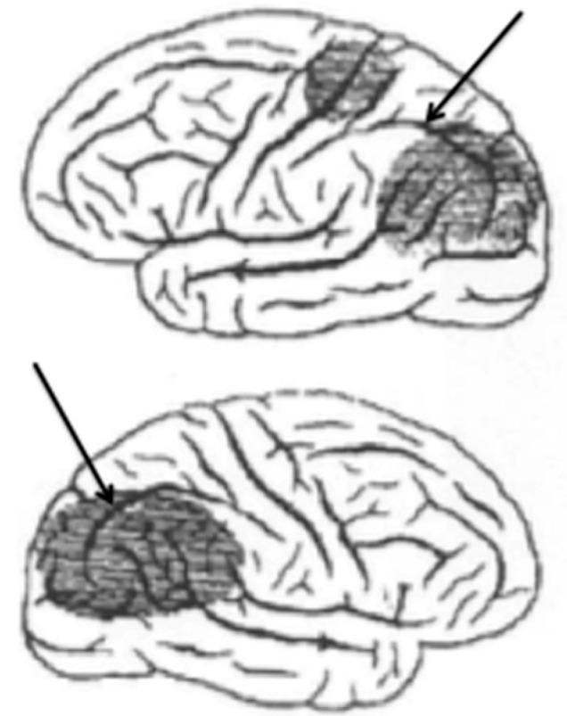
This medical illustration consists of two lateral-view drawings of a human brain, depicting bilateral cortical lesions associated with Bálint syndrome. Both diagrams are oriented with the anterior frontal lobe to the left and the posterior occipital lobe to the right. Darkly shaded regions highlight areas of tissue damage within the parietal lobes. The upper drawing displays two distinct patches of damage: a smaller anterior parietal lesion and a larger posterior parietal-occipital lesion. The lower drawing shows a more singular, large concentrated area of damage in the posterior parietal region. In both illustrations, black arrows point specifically to the intraparietal sulcus, which serves as a landmark dividing the parietal lobe into superior and inferior lobules. The diagram illustrates the pathophysiology of Bálint syndrome, specifically showing bilateral damage to the dorsal visual stream. This content is intended for intermediate to advanced medical students and neuroscience professionals studying neuroanatomy and clinical neurology syndromes related to visual processing and spatial orientation.
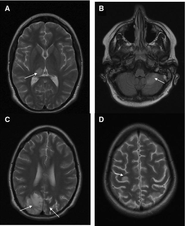
This diagnostic image set consists of four axial brain MRI scans (A, B, C, D) demonstrating the progression of neuropathology associated with POLG mutations and Alpers-Huttenlocher syndrome. Panel A (axial T2-weighted) shows a circumscribed hyperintense lesion in the right posterior thalamus. Panel B (axial FLAIR) reveals high signal intensity lesions in the left cerebellar hemisphere. Panel C (axial T2-weighted), taken two months later, illustrates bilateral, asymmetric parieto-occipital lesions involving both the cortical ribbon and subcortical white matter, with more extensive involvement on the right. Panel D (axial T2-weighted), from a follow-up three months later, shows new hyperintense focal lesions in the cortex (indicated by an arrow). The sequence demonstrates the characteristic migratory and fluctuating nature of brain lesions in POLG-related mitochondrial disease, often presenting as stroke-like episodes or focal status epilepticus. This visual record is significant for illustrating the evolution of metabolic-induced encephalopathy and cortical-subcortical signal changes over a five-month clinical course.
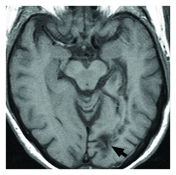
**Modality:** Magnetic Resonance Imaging (MRI) **Sequence:** Axial T1-weighted image **Anatomical Region:** Brain; specifically the posterior supratentorial compartment at the level of the midbrain and occipital lobes. **Observed Pathology:** There is a focal area of T1-hypointensity located in the left parieto-occipital region, consistent with a localized cortical and subcortical cerebral infarction. The lesion is demarcated by a black arrow, highlighting its involvement within the visual processing pathways or associated association fibers. **Visual Features:** - **Signal Intensity:** Low signal intensity (hypointense) on T1-weighted imaging, suggesting tissue necrosis or edema characteristic of an established or subacute infarct. - **Distribution:** Unilateral, localized to the left posterior cortex near the midline and parieto-occipital junction. - **Landmarks:** The midbrain (mesencephalon) is visible centrally with normal morphology; the lateral ventricles are partially visualized anterior to the lesion. **Diagnostic Significance:** The location of the infarct in the left parieto-occipital lobe is clinically significant for potential visual field deficits (e.g., right homonymous hemianopsia) or visuospatial neglect, which are critical factors in assessing functional safety and neurological impairment.
Balint syndrome
Balint Syndrome
The Classical Triad
1. Simultanagnosia (the core deficit)
- The inability to perceive more than one object or visual stimulus at a time, even though individual components can be identified
- Patients "miss the forest for the trees" - they may describe small isolated fragments of a complex scene while having no awareness of the whole
- Example: a patient shown a table lamp may look at its circular base and call it an ashtray
- Objects being fixated may "vanish suddenly," likely because the patient cannot compute a return to the original fixation point after saccadic displacements
- Movement and distracting stimuli greatly worsen the difficulties
- Can be thought of as a deficit in visual-spatial binding
2. Optic Ataxia
- Impaired ability to reach for or point to objects in space under visual guidance
- Key distinguishing feature: coordination using proprioceptive or auditory cues is intact; once an object is touched, the patient can perform smooth movements along its surface
- Results from disruption of transmission of visual information for directing motor acts from occipital cortex to premotor areas - i.e., disruption of the dorsal visual stream ("Where/How" pathway)
3. Oculomotor Apraxia (Psychic Paralysis of Gaze)
- Difficulty directing the eyes voluntarily away from central fixation toward peripheral targets
- The patient cannot make purposeful saccades to objects of interest, despite intact extraocular muscle function
- Kanski's Ophthalmology describes it as "ocular apraxia" - inability to direct gaze voluntarily on command
Pathophysiology
- Visuomotor guidance (reaching, grasping)
- Spatial attention and oculomotor scanning
- Integration of central foveal information with peripheral visual information
- Integration of egocentric (self-centered) and allocentric (object-centered) spatial coordinates
Etiology / Causes
| Cause | Notes |
|---|---|
| Bilateral MCA-PCA watershed infarcts | Most common; hypotension, cardiac arrest, carotid stenosis |
| Posterior cortical atrophy (PCA) | Atypical Alzheimer's disease variant; progressive course; simultanagnosia may predominate |
| Bilateral PCA occlusions | Stroke in the posterior circulation |
| Bilateral hemorrhage | Trauma, hypertension |
| Tumors | Bilateral parieto-occipital involvement |
| Prion disease (CJD) | Rapidly progressive |
| Lewy body disease, FTLD (corticobasal type) | Neurodegenerative |
| Hypoglycemia / sagittal sinus thrombosis | Reversible causes |
Associated Features
- Dressing apraxia: inability to align the body axis with garment axis
- Construction apraxia: severe difficulty copying simple line drawings (worse with right hemisphere lesions)
- Route-finding impairments: inability to orient self with respect to external landmarks
- Visual field defects: inferior quadrant cuts may be present, but are not invariant
- Optic allesthesia: false localization of objects in visual space (isolated feature)
- Cerebral akinetopsia: inability to perceive moving objects (isolated feature)
Bedside Diagnosis
-
Modified letter cancellation test (simultanagnosia): Mix large (7.5-10 cm) and small (2.5 cm) target letters. Patients with simultanagnosia characteristically miss the larger targets - because identifying them requires integrating visual information across multiple fixation points. This also rules out poor acuity as the cause.
-
Complex picture interpretation: Ask the patient to interpret the "Cookie Theft" picture from the Boston Diagnostic Aphasia Examination (BDAE) or the NIH Stroke Scale picture. Patients can describe isolated fragments but cannot integrate the whole scene.
-
Visually guided reaching test: Ask the patient to reach and touch a visible object. Optic ataxia produces gross misreaching under visual guidance.
Differentiation from Related Syndromes
| Feature | Balint Syndrome | Cortical Blindness | Hemispatial Neglect |
|---|---|---|---|
| Lesion laterality | Bilateral parieto-occipital | Bilateral occipital (V1) | Typically right hemisphere |
| Object recognition | Fragmented, one at a time | Absent | Intact for attended side |
| Reaching | Optic ataxia | N/A | Normal |
| Gaze | Oculomotor apraxia | Normal | Ipsilateral deviation |
| Anosognosia | Sometimes | Common (Anton's) | Common |
Treatment and Prognosis
- Stroke: standard acute stroke management; rehabilitation for visually guided activities
- Posterior cortical atrophy (AD): acetylcholinesterase inhibitors may partially help cognitive symptoms; progressive course
- Reversible causes (hypoglycemia, infection): treating the cause may lead to significant recovery
- Rehabilitation: compensatory strategies (e.g., systematic scanning training, environmental modifications) are more effective than restorative attempts; studies on rehabilitation are limited