
Anatomy of NASAL SEPTUM ENY 4TH YR MBBS DETAILED
nasal septum anatomy diagram bones cartilage
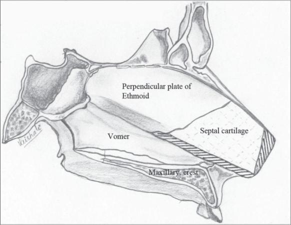
This medical anatomical diagram illustrates the surgical anatomy of the nasal septum, specifically focusing on modifications made during septal correction in cleft lip rhinoplasty. The diagram depicts the septal cartilage in its anatomical relationship to the perpendicular plate of the ethmoid (superior-posterior), the vomer (inferior-posterior), and the maxillary crest (inferior). The septal cartilage is shown as a quadrangular structure with surgical alterations: cross-hatched shading along the inferior and anterior borders represents slivers of excised cartilage to prevent buckling and enhance nasal tip projection. Additionally, the main body of the cartilage features fine vertical scoring lines on its concave (non-cleft) surface, a technique used to nullify the bow-string effect and achieve a flail, midline-correctable state. The illustration serves as a guide for reconstructive surgeons to understand the resection sites and cartilage weakening maneuvers necessary to correct septal deviation and improve nasal symmetry in pediatric or adult cleft patients.
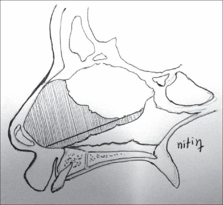
This medical anatomical diagram provides a sagittal cross-section of the human nasal region, specifically illustrating the surgical site for a septal cartilage graft harvest. Key landmarks include the nasal bones superiorly, the anterior nasal spine, and the alveolar ridge and hard palate inferiorly. The central focus is the cartilaginous nasal septum. A diagonal shaded area indicates the precise donor site for harvesting septal cartilage. This area encompasses the lower and anterior portions of the quadrangular cartilage, extending posteriorly along the floor of the nasal cavity towards the vomerine attachment but leaving a superior and anterior 'L-strut' for structural support. The diagram is designed for educational use in plastic and reconstructive surgery, specifically for procedures such as cleft lip rhinoplasty or secondary rhinoplasty where autologous cartilage is required for columellar struts or tip projection.
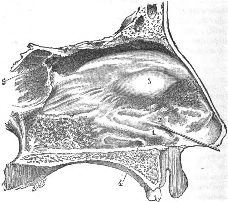
This medical anatomical diagram illustrates a sagittal section of the human nasal septum, highlighting specialized structures of the nasal fossa. Key landmarks are numerically labeled: (1) indicates the 'bead' or Jacobsonian swelling, formed by Jacobson's cartilage in the anteroinferior region; (2) shows the orifice of the Jacobson's canal (vomeronasal organ) with a fine stylet inserted to demonstrate patency; (3) identifies the tubercle of the septum located superior to the bead; (4) marks the nasopalatine infundibulum, the mucosal entrance to the nasopalatine canal; and (5) points to the orifice of the sphenoidal sinus in the posterosuperior aspect. The diagram also visualizes the frontal sinus (6) and the surrounding bony and cartilaginous framework. This illustration is primarily used in otorhinolaryngology and anatomy education to detail the location and morphology of the vestigial vomeronasal organ and associated septal landmarks in adult humans.
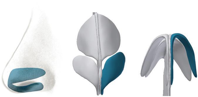
This medical anatomical diagram illustrates the cartilaginous framework of the human nose across three perspectives, specifically highlighting the greater alar cartilages (lower lateral cartilages) in blue. Left (Lateral View): A profile illustration showing the alar cartilage situated in the caudal portion of the nose, demonstrating its role in providing structural support to the nasal tip and the lateral wall of the vestibule. Center (Anterior View): A diagnostic-style reconstruction (derived from CT imaging) showing the relationship between the paired alar cartilages and the central vertical nasal septum. It highlights the symmetry and spatial orientation of the medial and lateral crura. Right (Inferior View): A basal perspective emphasizing the arched, wing-like morphology of the alar cartilages. This view demonstrates how the curvature of the medial and lateral crura forms the characteristic 'McDonald’s arches' shape, defining the contour of the nostrils and the projection of the nasal tip. The diagram serves as an educational resource for plastic surgery and otolaryngology, detailing the essential anatomy required for nasal reconstruction and rhinoplasty.
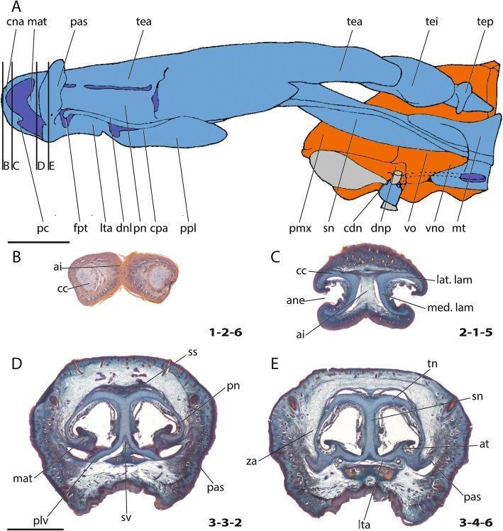
This medical anatomical diagram and series of histological cross-sections illustrate the comparative anatomy and developmental morphology of the cartilaginous nasal capsule. Figure A provides a lateral schematic view where cartilage is color-coded in blue, bone in orange, and soft tissue in gray, depicting key structures such as the cupula nasi anterior (cna), tectum nasi (tea, tei, tep), and the vomeronasal organ (vno). Figures B through E present corresponding histological transverse sections. Section B shows the anterior wall of the cartilago cupularis (cc) and the area internarica (ai). Section C identifies the lateral (lat. lam) and medial (med. lam) laminae surrounding the apertura nasi externa (ane). Section D details the region posterior to the fenestra narina, highlighting the marginoturbinale (mat), paries nasi (pn), and sulcus supraseptalis (ss). Section E demonstrates the anterior margin of the zona annularis (za), featuring the tectum nasi (tn), septum nasi (sn), atrioturbinale (at), and lamina transversalis anterior (lta). These visuals are essential for understanding the embryological development and structural complexity of the mammalian chondrocranium and respiratory intake architecture.
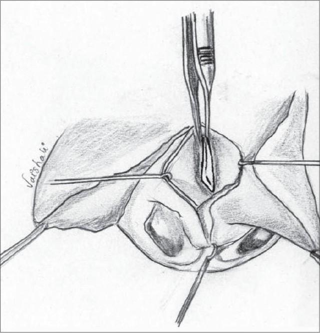
Anatomical diagram and surgical illustration detailing a key step in primary nasal correction for cleft lip or septoplasty. The drawing depicts the surgical field of the nasal septum from an anterior view. It illustrates the septal cartilage being meticulously dissected and freed from the surrounding mucoperichondrium on both sides. Additional separation is shown at the junctions with the vomer and the perpendicular plate of the ethmoid. A surgical scalpel is positioned centrally to perform precise dissection or scoring of the cartilage. Retraction sutures are visible on the lateral edges of the surgical site to maintain exposure of the deep structures. This procedure is clinically relevant for addressing septal deviation and repositioning the nasal midline during reconstructive surgery, such as the Millard procedure for unilateral cleft lip. The illustration serves as an educational tool for plastic surgery and otolaryngology trainees to understand the planes of dissection required for nasal septal mobilization.
nasal septum blood supply Kiesselbach Little's area vascular
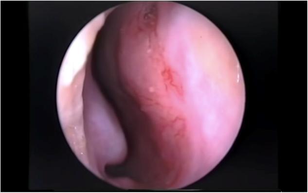
This clinical endoscopic photograph captures the internal anatomy of the nasal cavity, specifically focusing on the anterior nasal septum. The image illustrates the 'Little's area,' which houses the Kiesselbach's plexus, a highly vascularized region where the sphenopalatine, greater palatine, superior labial, and anterior ethmoidal arteries anastomose. The septal mucosa appears pinkish-red and moist. Visible on the surface is a network of fine, branching, and interconnected superficial red blood vessels (telangiectasia). The density of these vessels in this specific anatomical location is the primary clinical source for approximately 80% of anterior epistaxis cases. The image demonstrates the characteristic vascular fragility of this region, which is susceptible to environmental dryness, digital trauma, and inflammation. This visual is intended for ENT specialists and emergency medicine providers to understand the pathophysiology of nosebleeds and identify potential sites for chemical cauterization or topical treatment.
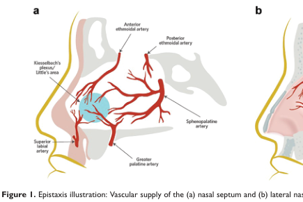
Summary : This illustration shows the vascular supply of the nasal septum, highlighting the main arteries involved in epistaxis (nosebleeds) and their anatomical locations. illustration: # Scene Overview : • Main subject is the vascular anatomy of the nasal septum (left side of the nose in sagittal section). • Perspective is a lateral cross-section, focusing on the internal nasal structures. • Colour palette includes red for arteries, yellow for outline, and blue shading for a specific area (Kiesselbach’s plexus/Little’s area). # Technical Details : • No scale bar or magnification indicated. • Anatomical labels are present for key arteries and regions. # Spatial Relationships : • The following arteries are labeled and shown branching within the nasal septum: • Anterior ethmoidal artery (superior, anterior region) • Posterior ethmoidal artery (superior, posterior region) • Sphenopalatine artery (posterior, central region) • Greater palatine artery (inferior, posterior region) • Superior labial artery (inferior, anterior region) • Kiesselbach’s plexus/Little’s area is highlighted in blue, located anteriorly on the septum, where several arteries converge. # Analysis : • The illustration emphasizes the convergence of multiple arteries at Kiesselbach’s plexus/Little’s area, a common site for anterior nosebleeds (epistaxis). • The vascular supply is shown as dense and interconnected, especially in the anterior septal region, explaining the clinical significance of this area in nasal bleeding. • The lateral nasal wall (panel b) is only partially visible and not labeled in this crop.
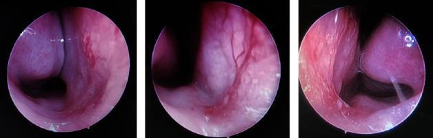
Three clinical endoscopic views of the internal nasal cavity in a patient with allergic rhinitis and epistaxis. The images demonstrate classic mucosal changes including generalized hypervascularity, vascular dilatation, and tortuous superficial blood vessels along the nasal septum and turbinates. The nasal mucosa appears erythematous and edematous, with areas of significant pale swelling (cobblestone appearance) characteristic of allergic inflammation. The inferior turbinates show signs of hypertrophy, causing narrowing of the nasal passage. Visible signs of recent or potential epistaxis are indicated by the prominent, engorged vessels in Kiesselbach’s plexus area and along the anterior septal wall. These endoscopic findings illustrate the chronic inflammatory state of the nasal airway in pediatric or adult allergic patients, highlighting how mucosal fragility and vascular congestion contribute to frequent nosebleeds.
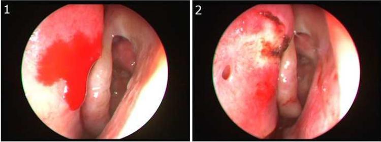
This clinical image consists of two side-by-side endoscopic views of the left nasal cavity, illustrating the identification and management of posterior epistaxis at the S-point. View 1 (left) shows active arterial bleeding with bright red blood pooling on the superior portion of the nasal septum, located above the axilla of the middle turbinate. This specific anatomical site, known as Stamm's S-point, is associated with vascular supply from the anterior ethmoidal artery (AEA). View 2 (right) demonstrates the same anatomical region post-intervention. The active hemorrhage has ceased following localized cauterization, characterized by a visible area of dark, charred tissue and mucosal erythema at the previous bleeding site. This comparison highlights a critical diagnostic location for non-anterior epistaxis and the efficacy of targeted endoscopic cautery. The material is intended for advanced medical education in otorhinolaryngology, focusing on the management of severe nasal hemorrhage.
Anatomy of the Nasal Septum - Detailed (ENT, 4th Year MBBS)
Overview & Functions
- Separation of the nasal airway into two distinct chambers
- Structural support of the nasal dorsum and tip
- Maintenance of the shape of the columella
- Forms part of the internal nasal valve
- Contributes to humidification, filtration and warming of inspired air
Composition - Three Parts
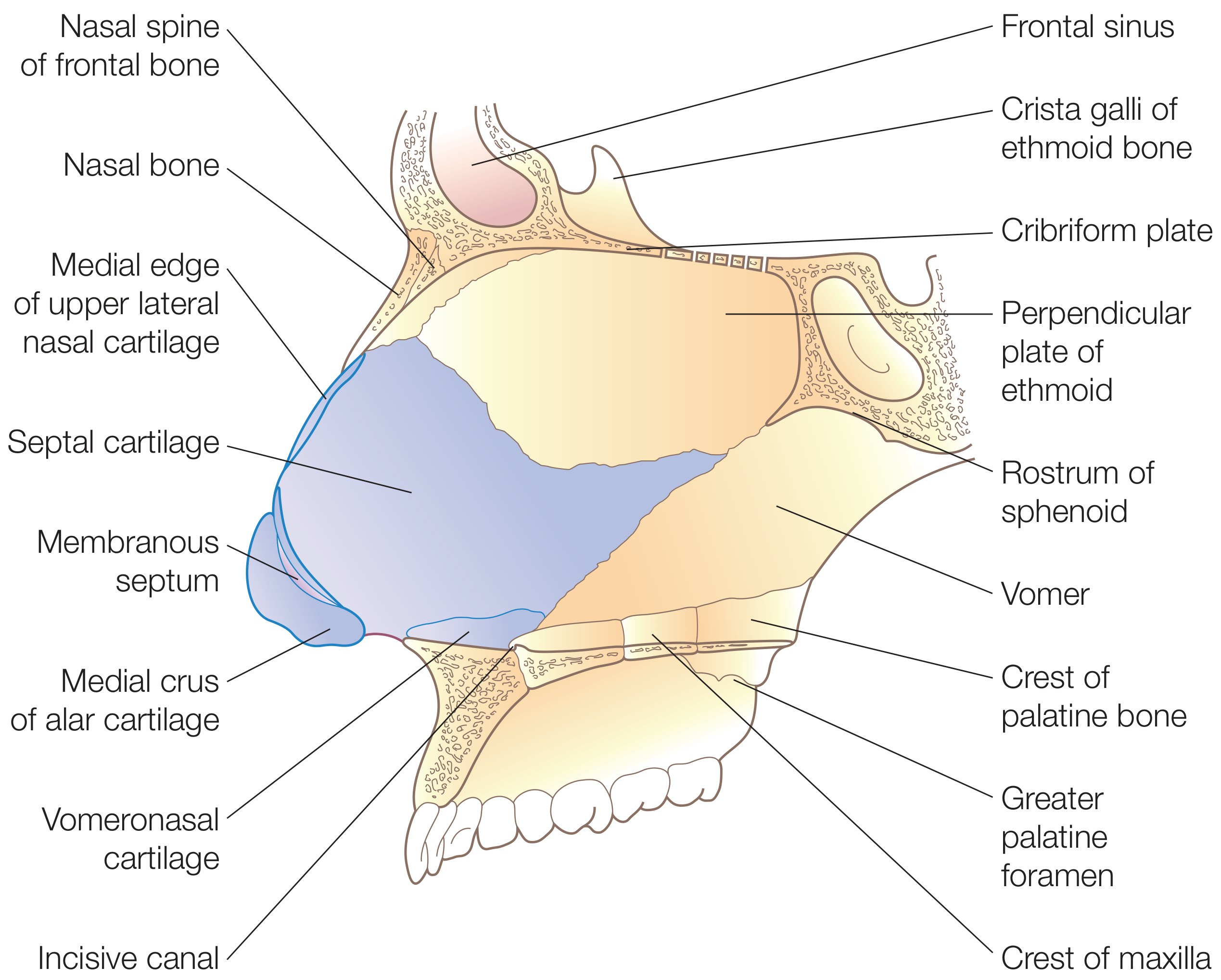
1. Bony Portion
| Bone | Location on Septum | Key Relations |
|---|---|---|
| Perpendicular plate of ethmoid | Upper one-third | Continuous superiorly with cribriform plate and crista galli; articulates posteriorly with sphenoid crest, posteroinferiorly with vomer, anteroinferiorly with septal cartilage |
| Vomer | Posterior and inferior | Articulates by its two alae with the sphenoid rostrum forming vomerovaginal canals (through which pharyngeal branches of maxillary artery travel); inferior border articulates with nasal crest of maxilla and palatine bones |
| Maxillary crest | Inferior floor | Forms nasal crest with palatine bone |
| Palatine bone (nasal crest) | Posteroinferior | Completes the inferior nasal crest |
Key Point - Perpendicular Plate of Ethmoid:
- Forms upper 1/3 of the septum
- Superiorly continuous with the cribriform plate and crista galli
- Articulates anteroinferiorly with the septal cartilage
Key Point - Vomer:
- Forms posterior free edge of the septum
- The posterior edge of the vomer is the posterior free edge of the nasal septum
- Articulates anteriorly with the septal cartilage
2. Cartilaginous Portion - The Quadrilateral (Septal) Cartilage
- Bound firmly by collagenous fibers to the nasal bones, perpendicular plate of ethmoid, and vomer
- Continuous with the upper lateral cartilages (medial edge of upper lateral nasal cartilages) toward the nasal bridge - this forms the keystone region
- A posterior projection called the sphenoidal process (septal tail) extends posteriorly between the vomer and perpendicular plate of ethmoid - a useful source of graft cartilage especially in revision rhinoplasty
- The inferior attachment sits within the nasal crest of the maxilla, bound by loose connective tissue forming a pseudoarthrosis - this allows mobility during nasal flexion, reducing fracture risk with trauma
The L-Strut:
3. Membranous Portion
- A segment of connective tissue between the caudal (anterior) edge of the septal cartilage and the columella
- Also called the columellar septum
- Contains no cartilage or bone
Septal Swell Body (Clinically Important)
- A widened region of the anterior nasal septum located anterior to the middle turbinate at the internal nasal valve
- Histology shows an increased number of venous sinusoids and fewer glandular elements compared to adjacent septal mucosa
- Functions to alter nasal airflow, similar to the inferior turbinate
- Can contribute to nasal obstruction when engorged
Blood Supply
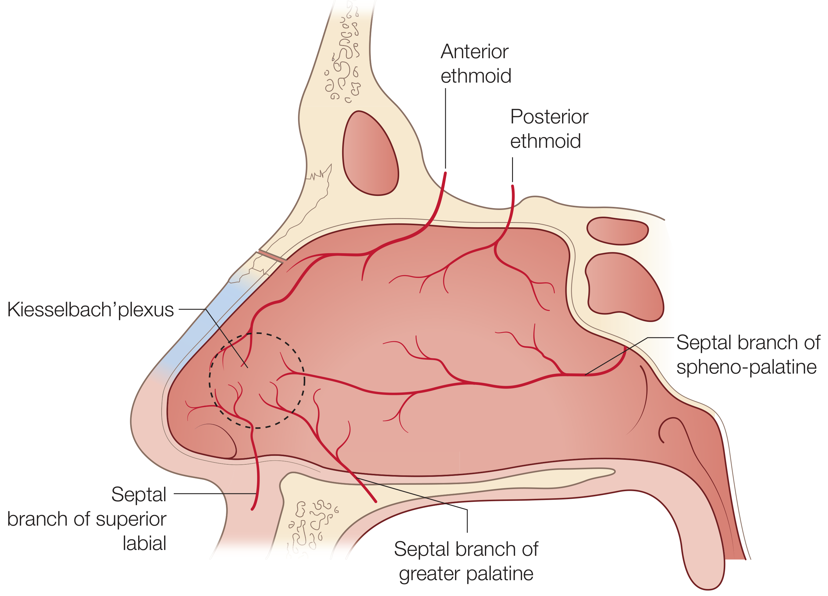
From External Carotid Artery (via Maxillary Artery):
| Artery | Supply to Septum |
|---|---|
| Sphenopalatine artery (posterior septal branch) | Posteroinferior septum; basis of the nasoseptal mucosal flap used in endoscopic skull base reconstruction |
| Greater palatine artery | Enters through the incisive canal; supplies anteroinferior septum |
| Septal branch of superior labial artery (branch of facial artery) | Caudal septum and columella |
From Internal Carotid Artery (via Ophthalmic Artery - Ethmoid Arteries):
| Artery | Supply to Septum |
|---|---|
| Anterior ethmoid artery | Anterosuperior septum |
| Posterior ethmoid artery | Posterosuperior septum |
Kiesselbach's Plexus (Little's Area) - HIGH YIELD
- Located at the anteroinferior nasal septum (approximately 1 cm posterior to the columella)
- Formed by anastomosis of:
- Anterior ethmoid artery (ICA branch)
- Posterior septal artery / sphenopalatine branch (ECA branch)
- Septal branch of superior labial artery (ECA branch)
- Greater palatine artery (via incisive canal)
- Rich vascular bed of long capillary loops
- Most common site of epistaxis (~80% of all nosebleeds are anterior/from this site) due to rich supply and susceptibility to turbulent airflow and digital trauma
Venous Drainage:
- Posteriorly via sphenopalatine vessels into the pterygoid plexus
- Anteriorly into the facial veins
- Superiorly via ethmoidal veins into the superior ophthalmic system - important as there may be direct intracranial connections through the foramen caecum into the superior sagittal sinus (route for intracranial spread of infection - a clinically important danger area)
Nerve Supply
Sensory Innervation (from Trigeminal Nerve - CN V):
| Nerve | Origin | Area Supplied |
|---|---|---|
| Nasopalatine nerve (long sphenopalatine nerve) | V2 (maxillary) via pterygopalatine ganglion | Anteroinferior septum; passes through incisive canal to hard palate |
| Anterior ethmoid nerve (internal nasal branch) | V1 (ophthalmic) via nasociliary nerve | Anterosuperior septum |
| Posterior superior nasal nerves | V2 via pterygopalatine ganglion | Posterosuperior septum |
- The first (V1) and second (V2) divisions of the trigeminal nerve supply sensory innervation to the nasal mucosa
- Trigeminal fibers pass through the sphenopalatine (pterygopalatine) ganglion transmitting pain, temperature, and touch
Autonomic Innervation:
- Presynaptic fibers travel via the facial nerve (CN VII) → become the greater superficial petrosal nerve at the geniculate ganglion → join the deep petrosal nerve → form the Vidian nerve (nerve of pterygoid canal) → synapse in the sphenopalatine (pterygopalatine) ganglion → postganglionic fibers innervate nasal mucosa
- Effect: increased secretion, vasodilation
- Postsynaptic sympathetic fibers pass through (without synapsing) the sphenopalatine ganglion and terminate in nasal mucosa
- Effect: vasoconstriction, reduced secretion
Lymphatic Drainage
- Anterior nasal cavity lymphatics drain to submandibular lymph nodes
- Posterior nasal cavity lymphatics drain to retropharyngeal lymph nodes and upper deep cervical chain
Mucosa of the Nasal Septum
- Covered by pseudostratified ciliated columnar epithelium (respiratory epithelium) over most of its surface
- The superior part (olfactory area / roof) is covered by olfactory epithelium (specialized sensory epithelium)
- The mucosa contains arteriovenous anastomoses and venous sinusoids (erectile tissue) - especially prominent on the anterior septum and inferior turbinates
- The mucoperichondrium and mucoperiosteum provide the blood supply to the underlying cartilage and bone respectively; loss of these linings (as during septal surgery) risks avascular necrosis
Embryology (Brief)
- The septum develops from the posterior midline growth of the frontonasal process in the root of the oral cavity, extending posteriorly to the opening of Rathke's pouch
- As the nasal cavities enlarge, the palatal processes (derived from lateral maxillary mesoderm) grow medially to meet the nasal septum
- Failure of fusion results in cleft palate
Clinical Correlates (HIGH YIELD for Exam)
| Condition | Anatomical Basis |
|---|---|
| Deviated nasal septum (DNS) | Trauma, birth compression, or asymmetric growth causes deviation; leads to unilateral/bilateral nasal obstruction, chronic sinusitis |
| Epistaxis (anterior) | Kiesselbach's plexus (Little's area) - most common; treated by anterior packing or cautery |
| Epistaxis (posterior) | Woodruff's plexus on the posterior lateral nasal wall near inferior meatus; or from sphenopalatine artery; managed by posterior packing or sphenopalatine artery ligation |
| Nasal septal hematoma | Blood collects between mucoperichondrium and cartilage; must be drained urgently as pressure cuts off cartilage nutrition → septal abscess → saddle nose |
| Saddle nose deformity | Loss of septal cartilage support (untreated hematoma, perichondritis, syphilis, relapsing polychondritis, cocaine abuse) |
| Septal perforation | Causes: trauma, cocaine, Wegener's granulomatosis, syphilis, iatrogenic (bilateral tears during septoplasty) |
| Nasal septal abscess | Complication of untreated hematoma; organisms: S. aureus; risks: cavernous sinus thrombosis (via ethmoid veins → superior ophthalmic vein) |
| L-strut preservation | During septoplasty, minimum 1 cm dorsal + 1 cm caudal strut must be preserved to prevent structural collapse |
Summary Table - Bones of the Nasal Septum
| Component | Bone/Structure | Position |
|---|---|---|
| Upper 1/3 | Perpendicular plate of ethmoid | Superior |
| Lower posterior | Vomer | Posteroinferior |
| Anteroinferior floor | Nasal crest of maxilla | Inferior-anterior |
| Posteroinferior floor | Nasal crest of palatine bone | Inferior-posterior |
| Anterior 2/3 (cartilage) | Septal (quadrilateral) cartilage | Anterior |
| Anterior tip | Membranous septum | Between cartilage and columella |