Describe the posterior triangle of the neck
posterior triangle of neck anatomy boundaries contents
Posterior Triangle of the Neck
Location
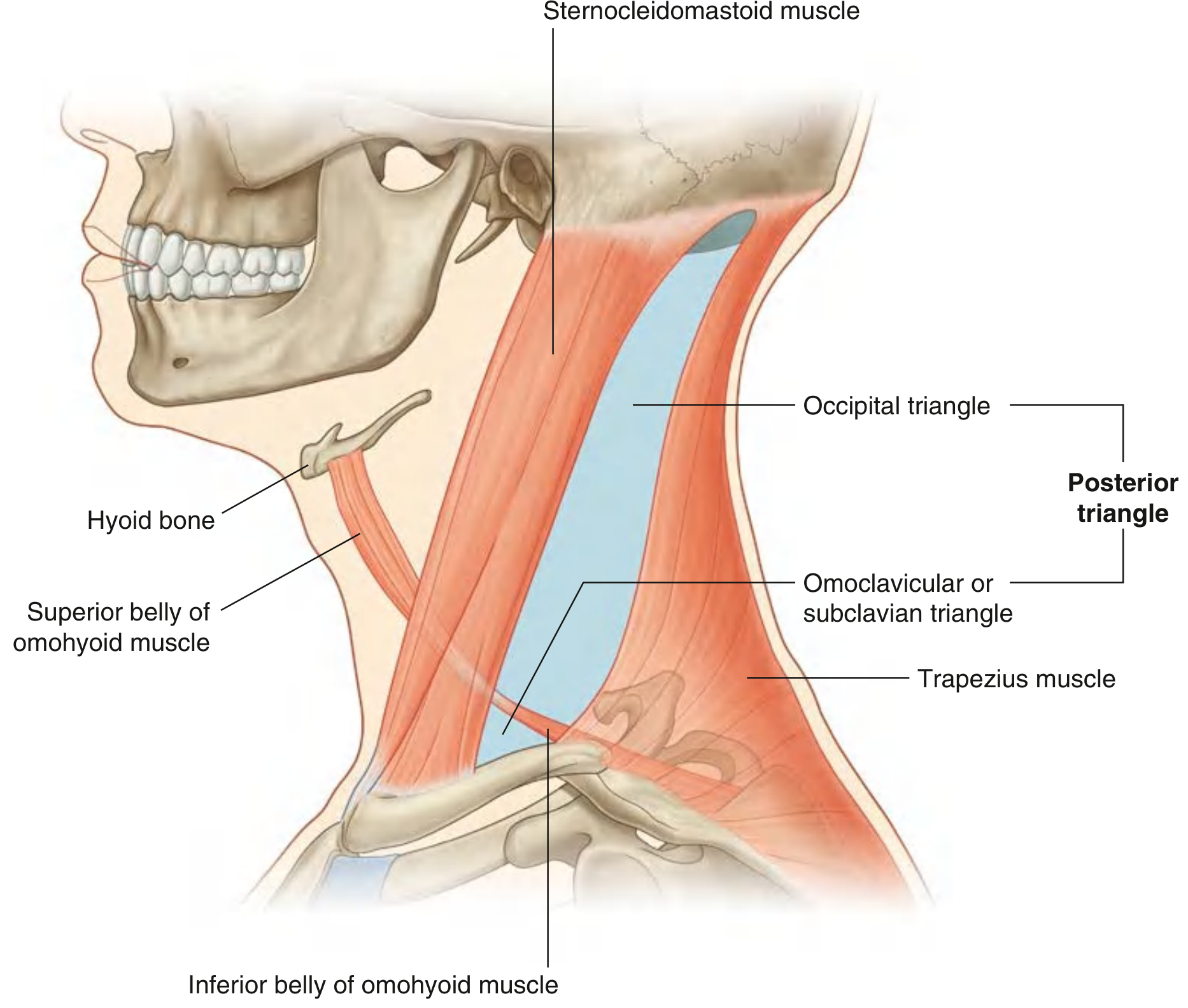
Boundaries
| Boundary | Structure |
|---|---|
| Anterior | Posterior border of the sternocleidomastoid muscle |
| Posterior | Anterior border of the trapezius muscle |
| Base (inferior) | Middle one-third of the clavicle |
| Apex (superior) | Occipital bone, just posterior to the mastoid process, where SCM and trapezius meet along the superior nuchal line |
| Roof | Investing layer of cervical fascia (with overlying skin, superficial fascia, and platysma) |
| Floor | Prevertebral layer of cervical fascia, covering (superior to inferior): splenius capitis, levator scapulae, posterior scalene, middle scalene, and anterior scalene muscles |
Subdivisions
- Occipital triangle (larger, superior): the main portion of the posterior triangle above the omohyoid
- Omoclavicular (subclavian) triangle (smaller, inferior): below the omohyoid, at the base of the posterior triangle
Contents
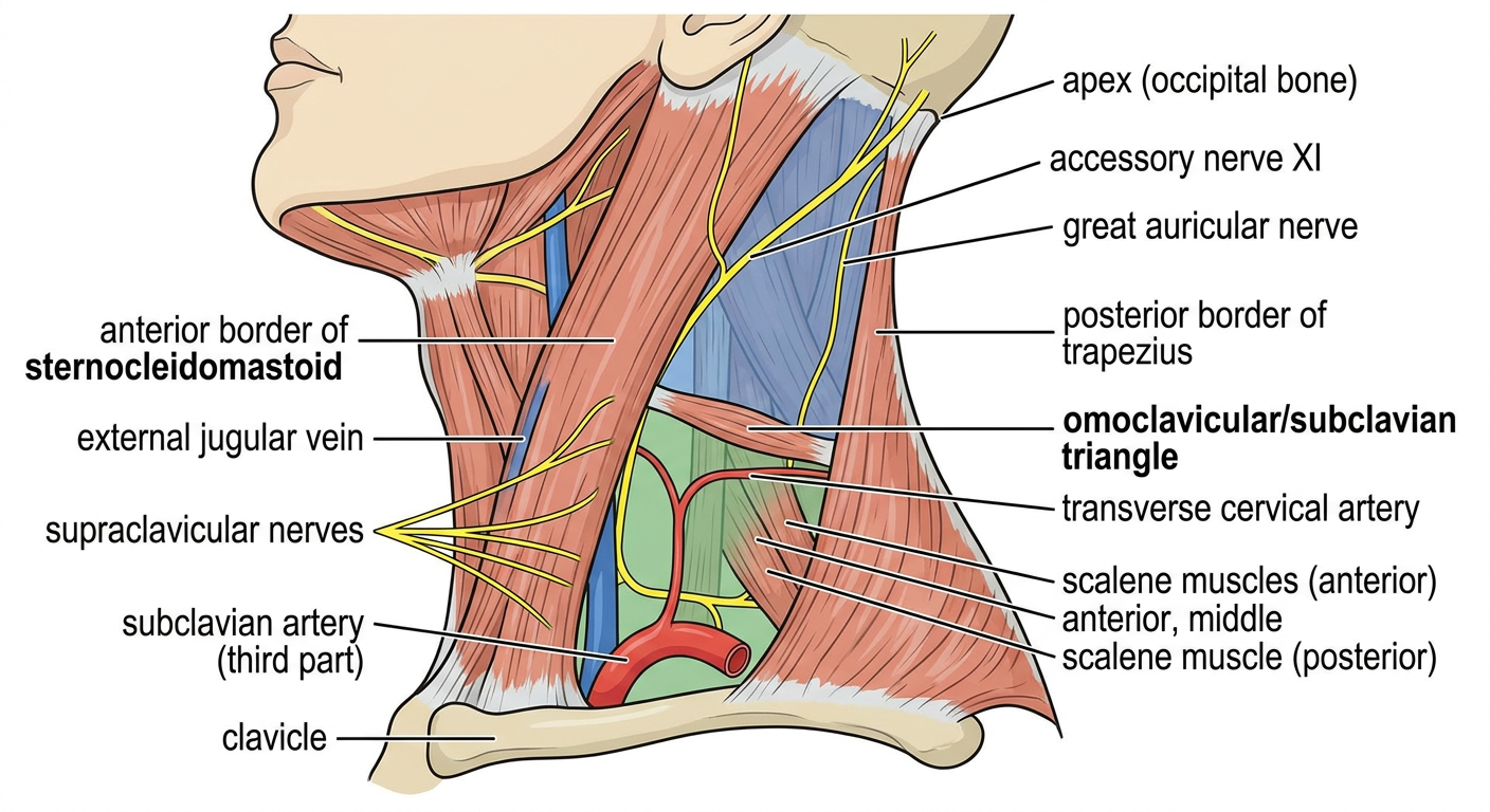
Nerves
![Accessory nerve [XI] and cutaneous branches of the cervical plexus in the posterior triangle](https://cdn.orris.care/cdss_images/e68a4a95724c26b485236b2ff367c8cfa7f23a2ccfb5226722ad6b95ee534c5b.png)
- Exits the skull via the jugular foramen and descends through the neck
- Pierces (or passes deep to) the SCM, which it innervates, then enters the posterior triangle within the investing layer of cervical fascia
- Crosses the triangle obliquely in a downward direction to reach the deep surface of the trapezius, which it also innervates
- Its superficial position within the posterior triangle makes it highly vulnerable to injury during lymph node biopsy or surgery
-
Cutaneous (superficial) branches emerge beneath the posterior border of SCM at the midpoint ("nerve point"):
- Lesser occipital nerve (C2) - skin of the lateral scalp and back of ear
- Great auricular nerve (C2, C3) - skin over the parotid and mastoid regions, and lower ear
- Transverse cervical nerve (C2, C3) - anterior neck skin
- Supraclavicular nerves (C3, C4) - skin over the clavicle and shoulder
-
Muscular (deep) branches include:
- Phrenic nerve (C3-C5) - descends across the anterior scalene muscle within the prevertebral fascia to enter the thorax
- Branches to prevertebral muscles and the ansa cervicalis (C1-C3, for infrahyoid muscles)
Arteries
- Transverse cervical artery (branch of thyrocervical trunk) - crosses the base of the triangle anterior to the anterior scalene and brachial plexus; divides into superficial and deep branches at the trapezius
- Suprascapular artery (also from the thyrocervical trunk) - passes across the lowest part of the posterior triangle, heading toward the superior border of the scapula
- Subclavian artery (third part) - crosses the base in the omoclavicular triangle, lateral to the anterior scalene
- Occipital artery may occasionally appear in the superior triangle
Veins
- External jugular vein - most superficial structure in the triangle; forms near the angle of the mandible, crosses the SCM, then descends vertically through the posterior triangle in the superficial fascia before draining into the subclavian vein just above the clavicle
- Subclavian vein - crosses the base of the triangle, lies anterior to and slightly below the subclavian artery; receives the external jugular vein here and then joins the internal jugular vein to form the brachiocephalic vein
- Transverse cervical and suprascapular veins accompany their respective arteries and drain into either the external jugular or subclavian veins
Lymph Nodes
Clinical Relevance
- Accessory nerve injury during posterior triangle surgery (e.g., lymph node biopsy) causes weakness of the trapezius, resulting in shoulder drop, pain, and lateral winging of the scapula
- Central venous access: the subclavian vein is accessed at the base of the triangle for central line placement
- Nerve blocks: the "nerve point" along the posterior SCM border is used for cervical plexus blocks; brachial plexus blocks can be performed in the supraclavicular region
- Cervical lymphadenopathy: the triangle is a common site for enlarged nodes in head/neck infections, lymphoma, and metastatic disease
- The phrenic nerve (C3-C5) crosses the anterior scalene muscle within the prevertebral fascia - deep procedures in this area risk phrenic nerve injury and consequent diaphragm paralysis
Write a notes on posterior triangle of neck with diagram
Detailed anatomical diagram of the posterior triangle of the neck, lateral view. Show and label: sternocleidomastoid muscle (anterior border), trapezius muscle (posterior border), clavicle (base), occipital bone at apex. Inside the triangle show: occipital triangle (upper, shaded blue), omoclavicular/subclavian triangle (lower, shaded green), inferior belly of omohyoid muscle crossing the triangle. Label nerves in yellow: accessory nerve XI crossing obliquely, lesser occipital nerve, great auricular nerve, transverse cervical nerve, supraclavicular nerves emerging at posterior border of SCM. Label vessels in red/blue: external jugular vein, subclavian artery (third part), transverse cervical artery. Floor muscles labeled: splenius capitis, levator scapulae, scalene muscles. Clear medical illustration style on white background.
mkdir -p /home/daytona/workspace/posterior-triangle && curl -s -o /home/daytona/workspace/posterior-triangle/diagram.png "https://cdn.orris.care/image-gen/794cc3e260684c299eeceaf4dc4056ce.png" && echo "Downloaded: $(du -sh /home/daytona/workspace/posterior-triangle/diagram.png)"
~/posterior-triangle/build.js
const {
Document, Packer, Paragraph, TextRun, Table, TableRow, TableCell,
ImageRun, AlignmentType, HeadingLevel, BorderStyle, WidthType,
ShadingType, VerticalAlign, PageNumber, Header, Footer, UnderlineType
} = require('docx');
const fs = require('fs');
const imgBuffer = fs.readFileSync('/home/daytona/workspace/posterior-triangle/diagram.png');
// ── helpers ──────────────────────────────────────────────────────────────────
const heading1 = (text) => new Paragraph({
text,
heading: HeadingLevel.HEADING_1,
spacing: { before: 320, after: 160 },
border: { bottom: { color: '1F4E79', size: 6, space: 4, style: BorderStyle.SINGLE } }
});
const heading2 = (text) => new Paragraph({
text,
heading: HeadingLevel.HEADING_2,
spacing: { before: 240, after: 120 },
});
const heading3 = (text) => new Paragraph({
text,
heading: HeadingLevel.HEADING_3,
spacing: { before: 200, after: 80 },
});
const body = (text) => new Paragraph({
children: [new TextRun({ text, size: 22, font: 'Calibri' })],
spacing: { after: 100 },
});
const bullet = (text, bold_prefix) => new Paragraph({
children: bold_prefix
? [
new TextRun({ text: bold_prefix, bold: true, size: 22, font: 'Calibri' }),
new TextRun({ text, size: 22, font: 'Calibri' })
]
: [new TextRun({ text, size: 22, font: 'Calibri' })],
bullet: { level: 0 },
spacing: { after: 80 },
});
const subbullet = (text, bold_prefix) => new Paragraph({
children: bold_prefix
? [
new TextRun({ text: bold_prefix, bold: true, size: 22, font: 'Calibri' }),
new TextRun({ text, size: 22, font: 'Calibri' })
]
: [new TextRun({ text, size: 22, font: 'Calibri' })],
bullet: { level: 1 },
spacing: { after: 60 },
});
const emptyLine = () => new Paragraph({ text: '', spacing: { after: 80 } });
// ── shaded info box ───────────────────────────────────────────────────────────
const infoBox = (rows) => new Table({
width: { size: 100, type: WidthType.PERCENTAGE },
rows: [
new TableRow({
children: [
new TableCell({
shading: { fill: 'DEEAF1', type: ShadingType.CLEAR },
borders: {
top: { style: BorderStyle.SINGLE, size: 4, color: '1F4E79' },
bottom: { style: BorderStyle.SINGLE, size: 4, color: '1F4E79' },
left: { style: BorderStyle.THICK, size: 12, color: '1F4E79' },
right: { style: BorderStyle.NONE }
},
children: rows,
margins: { top: 80, bottom: 80, left: 120, right: 120 }
})
]
})
]
});
// ── boundary table ────────────────────────────────────────────────────────────
const headerCell = (text) => new TableCell({
shading: { fill: '1F4E79', type: ShadingType.CLEAR },
children: [new Paragraph({
children: [new TextRun({ text, bold: true, color: 'FFFFFF', size: 22, font: 'Calibri' })],
alignment: AlignmentType.CENTER
})],
verticalAlign: VerticalAlign.CENTER,
margins: { top: 60, bottom: 60, left: 80, right: 80 }
});
const dataCell = (text, bold) => new TableCell({
children: [new Paragraph({
children: [new TextRun({ text, bold: !!bold, size: 22, font: 'Calibri' })]
})],
verticalAlign: VerticalAlign.CENTER,
margins: { top: 60, bottom: 60, left: 80, right: 80 }
});
const altCell = (text, fill, bold) => new TableCell({
shading: { fill: fill || 'FFFFFF', type: ShadingType.CLEAR },
children: [new Paragraph({
children: [new TextRun({ text, bold: !!bold, size: 22, font: 'Calibri' })]
})],
verticalAlign: VerticalAlign.CENTER,
margins: { top: 60, bottom: 60, left: 80, right: 80 }
});
const boundariesTable = new Table({
width: { size: 100, type: WidthType.PERCENTAGE },
rows: [
new TableRow({ children: [headerCell('Boundary'), headerCell('Structure')] }),
new TableRow({ children: [altCell('Anterior', 'EBF3FB', true), altCell('Posterior border of sternocleidomastoid (SCM)', 'EBF3FB')] }),
new TableRow({ children: [dataCell('Posterior', false), dataCell('Anterior border of trapezius muscle')] }),
new TableRow({ children: [altCell('Base (inferior)', 'EBF3FB', false), altCell('Middle one-third of the clavicle', 'EBF3FB')] }),
new TableRow({ children: [dataCell('Apex (superior)'), dataCell('Occipital bone posterior to mastoid process (where SCM & trapezius meet at the superior nuchal line)')] }),
new TableRow({ children: [altCell('Roof', 'EBF3FB'), altCell('Investing layer of deep cervical fascia (+ skin, superficial fascia, platysma)', 'EBF3FB')] }),
new TableRow({ children: [dataCell('Floor'), dataCell('Prevertebral fascia covering: splenius capitis → levator scapulae → posterior, middle, and anterior scalene muscles (sup → inf)')] }),
]
});
// ── subdivisions table ────────────────────────────────────────────────────────
const subdivisionsTable = new Table({
width: { size: 100, type: WidthType.PERCENTAGE },
rows: [
new TableRow({ children: [headerCell('Subdivision'), headerCell('Location'), headerCell('Key Contents')] }),
new TableRow({ children: [
altCell('Occipital Triangle', 'EBF3FB', true),
altCell('Superior (larger)', 'EBF3FB'),
altCell('Accessory nerve XI, cervical plexus cutaneous branches, upper brachial plexus, occipital artery', 'EBF3FB')
]}),
new TableRow({ children: [
dataCell('Omoclavicular (Subclavian) Triangle', true),
dataCell('Inferior (smaller)'),
dataCell('Subclavian artery (3rd part), subclavian vein, brachial plexus trunks, supraclavicular lymph nodes')
]}),
]
});
// ── contents table ────────────────────────────────────────────────────────────
const contentsTable = new Table({
width: { size: 100, type: WidthType.PERCENTAGE },
rows: [
new TableRow({ children: [headerCell('Category'), headerCell('Structure'), headerCell('Notes')] }),
new TableRow({ children: [altCell('Nerves', 'EBF3FB', true), altCell('Accessory nerve [XI]', 'EBF3FB', true), altCell('Crosses triangle within investing fascia; innervates SCM and trapezius; vulnerable to injury', 'EBF3FB')] }),
new TableRow({ children: [dataCell(''), dataCell('Lesser occipital nerve (C2)'), dataCell('Skin of lateral scalp and back of ear')] }),
new TableRow({ children: [altCell('', 'EBF3FB'), altCell('Great auricular nerve (C2, C3)', 'EBF3FB'), altCell('Skin over parotid, mastoid, and lower ear', 'EBF3FB')] }),
new TableRow({ children: [dataCell(''), dataCell('Transverse cervical nerve (C2, C3)'), dataCell('Anterior neck skin')] }),
new TableRow({ children: [altCell('', 'EBF3FB'), altCell('Supraclavicular nerves (C3, C4)', 'EBF3FB'), altCell('Skin over clavicle and shoulder', 'EBF3FB')] }),
new TableRow({ children: [dataCell(''), dataCell('Phrenic nerve (C3–C5)'), dataCell('Descends on anterior scalene within prevertebral fascia → thorax')] }),
new TableRow({ children: [altCell('', 'EBF3FB'), altCell('Brachial plexus (roots & trunks)', 'EBF3FB'), altCell('Between anterior and middle scalene; emerge into lower triangle', 'EBF3FB')] }),
new TableRow({ children: [dataCell('Arteries', true), dataCell('Transverse cervical artery'), dataCell('Branch of thyrocervical trunk; crosses base of triangle')] }),
new TableRow({ children: [altCell('', 'EBF3FB'), altCell('Suprascapular artery', 'EBF3FB'), altCell('Branch of thyrocervical trunk; crosses lowest part of triangle', 'EBF3FB')] }),
new TableRow({ children: [dataCell(''), dataCell('Subclavian artery (3rd part)'), dataCell('Crosses base of triangle, lateral to anterior scalene')] }),
new TableRow({ children: [altCell('Veins', 'EBF3FB', true), altCell('External jugular vein', 'EBF3FB', true), altCell('Most superficial structure; descends vertically in superficial fascia → drains into subclavian vein', 'EBF3FB')] }),
new TableRow({ children: [dataCell(''), dataCell('Subclavian vein'), dataCell('Crosses base; receives EJV; joins IJV → brachiocephalic vein')] }),
new TableRow({ children: [altCell('Lymph nodes', 'EBF3FB', true), altCell('Deep cervical lymph nodes', 'EBF3FB', true), altCell('Along accessory nerve and within triangle floor', 'EBF3FB')] }),
]
});
// ── document ──────────────────────────────────────────────────────────────────
const doc = new Document({
styles: {
paragraphStyles: [
{
id: 'Heading1',
name: 'Heading 1',
run: { size: 32, bold: true, color: '1F4E79', font: 'Calibri' },
paragraph: { spacing: { before: 320, after: 160 } }
},
{
id: 'Heading2',
name: 'Heading 2',
run: { size: 26, bold: true, color: '2E74B5', font: 'Calibri' },
paragraph: { spacing: { before: 240, after: 120 } }
},
{
id: 'Heading3',
name: 'Heading 3',
run: { size: 24, bold: true, color: '2E74B5', font: 'Calibri' },
paragraph: { spacing: { before: 200, after: 80 } }
}
]
},
sections: [{
properties: { page: { margin: { top: 720, bottom: 720, left: 900, right: 900 } } },
headers: {
default: new Header({
children: [new Paragraph({
children: [
new TextRun({ text: 'POSTERIOR TRIANGLE OF THE NECK', bold: true, size: 18, color: '1F4E79', font: 'Calibri' }),
new TextRun({ text: '\t\t', size: 18 }),
new TextRun({ text: 'Anatomy Notes', size: 18, color: '595959', font: 'Calibri' })
],
alignment: AlignmentType.LEFT,
border: { bottom: { style: BorderStyle.SINGLE, size: 4, color: '1F4E79', space: 4 } }
})]
})
},
footers: {
default: new Footer({
children: [new Paragraph({
children: [
new TextRun({ text: 'Source: Gray\'s Anatomy for Students', size: 18, color: '595959', font: 'Calibri' }),
new TextRun({ text: '\t\t', size: 18 }),
new TextRun({ text: 'Page ', size: 18, color: '595959' }),
new TextRun({ children: [PageNumber.CURRENT], size: 18, color: '595959' })
],
alignment: AlignmentType.LEFT,
border: { top: { style: BorderStyle.SINGLE, size: 4, color: '1F4E79', space: 4 } }
})]
})
},
children: [
// ── Title ──
new Paragraph({
children: [new TextRun({ text: 'POSTERIOR TRIANGLE OF THE NECK', bold: true, size: 48, color: '1F4E79', font: 'Calibri' })],
alignment: AlignmentType.CENTER,
spacing: { before: 0, after: 120 },
border: { bottom: { style: BorderStyle.THICK, size: 8, color: '1F4E79', space: 6 } }
}),
new Paragraph({
children: [new TextRun({ text: 'Anatomy Notes | Head & Neck', size: 22, color: '595959', italics: true, font: 'Calibri' })],
alignment: AlignmentType.CENTER,
spacing: { after: 320 }
}),
// ── 1. Introduction ──
heading1('1. Introduction'),
body('The posterior triangle of the neck is a clinically important anatomical region on the lateral aspect of the neck. It lies in direct continuity with the upper limb and contains major nerves, vessels, and lymph nodes that are frequently encountered in surgical procedures and physical examination.'),
emptyLine(),
// ── 2. Boundaries ──
heading1('2. Boundaries'),
body('The triangle is defined by the following borders:'),
emptyLine(),
boundariesTable,
emptyLine(),
// ── 3. Diagram ──
heading1('3. Diagram'),
new Paragraph({
children: [
new ImageRun({
data: imgBuffer,
transformation: { width: 560, height: 420 },
floating: undefined
})
],
alignment: AlignmentType.CENTER,
spacing: { before: 120, after: 80 }
}),
new Paragraph({
children: [new TextRun({ text: 'Fig. 1 — Posterior triangle of the neck: boundaries, subdivisions, and key contents', italics: true, size: 19, color: '595959', font: 'Calibri' })],
alignment: AlignmentType.CENTER,
spacing: { after: 240 }
}),
// ── 4. Subdivisions ──
heading1('4. Subdivisions'),
body('The inferior belly of the omohyoid muscle crosses the posterior triangle obliquely (lateral to medial, ~2.5 cm above the clavicle). Its central tendon is anchored to the clavicle by a fascial sling, dividing the triangle into two sub-triangles:'),
emptyLine(),
subdivisionsTable,
emptyLine(),
infoBox([
new Paragraph({ children: [new TextRun({ text: 'Omohyoid muscle:', bold: true, size: 22, font: 'Calibri' }), new TextRun({ text: ' Innervated by the ansa cervicalis (C1–C3). Function: depresses the hyoid bone. Superior belly lies in the anterior triangle; inferior belly crosses the posterior triangle.', size: 22, font: 'Calibri' })] })
]),
emptyLine(),
// ── 5. Floor muscles ──
heading1('5. Muscles of the Floor'),
body('The floor of the posterior triangle is formed (from superior to inferior) by:'),
bullet('Splenius capitis', ''),
bullet('Levator scapulae', ''),
bullet('Posterior scalene', ''),
bullet('Middle scalene', ''),
bullet('Anterior scalene', ''),
body('All covered by the prevertebral layer of deep cervical fascia.'),
emptyLine(),
// ── 6. Contents ──
heading1('6. Contents'),
emptyLine(),
contentsTable,
emptyLine(),
// ── 6a. Nerves detail ──
heading2('6a. Nerves — Detailed Notes'),
heading3('Accessory Nerve [CN XI]'),
bullet('Exits skull via the jugular foramen', ''),
bullet('Descends through the neck, pierces (or passes deep to) the SCM which it innervates', ''),
bullet('Enters posterior triangle within the investing fascia and crosses it obliquely downward', ''),
bullet('Reaches the deep surface of trapezius and innervates it', ''),
bullet('Superficial position makes it', ' Clinical note: '),
subbullet('Vulnerable to injury during lymph node biopsy or neck dissection', ''),
subbullet('Injury causes: trapezius weakness, shoulder drop, lateral scapular winging, chronic shoulder pain', ''),
emptyLine(),
heading3('Cervical Plexus (C1–C4)'),
bullet('Forms within the floor muscles, deep to prevertebral fascia', ''),
bullet('Cutaneous branches emerge at the "nerve point" (midpoint of posterior SCM border):', ''),
subbullet('Lesser occipital nerve (C2) — lateral scalp, back of ear', ''),
subbullet('Great auricular nerve (C2, C3) — parotid region, mastoid, lower ear', ''),
subbullet('Transverse cervical nerve (C2, C3) — anterior neck skin', ''),
subbullet('Supraclavicular nerves (C3, C4) — skin over clavicle and shoulder', ''),
bullet('Deep/muscular branches:', ''),
subbullet('Phrenic nerve (C3–C5) — descends on anterior scalene → supplies diaphragm', ''),
subbullet('Ansa cervicalis (C1–C3) — innervates infrahyoid (strap) muscles', ''),
emptyLine(),
heading3('Brachial Plexus'),
bullet('Roots (C5–T1) and trunks form between anterior and middle scalene muscles', ''),
bullet('Emerge into the lower posterior triangle (mainly the omoclavicular triangle)', ''),
bullet('Subclavian brachial plexus block is performed here in clinical practice', ''),
emptyLine(),
// ── 6b. Vessels detail ──
heading2('6b. Vessels — Detailed Notes'),
heading3('External Jugular Vein'),
bullet('Most superficial structure in the triangle', ''),
bullet('Forms near the angle of the mandible (posterior branch of retromandibular vein + posterior auricular vein)', ''),
bullet('Crosses SCM → enters posterior triangle → descends vertically in superficial fascia', ''),
bullet('Drains into the subclavian vein above the clavicle', ''),
emptyLine(),
heading3('Subclavian Artery (3rd Part)'),
bullet('Lateral to the anterior scalene muscle, crossing rib I', ''),
bullet('Becomes the axillary artery at the lateral border of rib I', ''),
emptyLine(),
heading3('Subclavian Vein'),
bullet('Continuation of axillary vein from lateral border of rib I', ''),
bullet('Receives external jugular, suprascapular, and transverse cervical veins', ''),
bullet('Joins internal jugular vein → brachiocephalic vein near sternoclavicular joint', ''),
bullet('Lies anterior to and slightly below the subclavian artery', ''),
emptyLine(),
heading3('Transverse Cervical & Suprascapular Arteries'),
bullet('Both arise from the thyrocervical trunk of the subclavian artery', ''),
bullet('Cross the base of the triangle, "pinning" the phrenic nerve to the anterior scalene', ''),
emptyLine(),
// ── 7. Lymph nodes ──
heading1('7. Lymph Nodes'),
bullet('Deep cervical lymph nodes lie along the accessory nerve and floor of the triangle', ''),
bullet('Enlarged nodes may signify local infection, lymphoma, or metastatic disease', ''),
bullet('Supraclavicular lymphadenopathy (especially left-sided "Virchow\'s node") can indicate abdominal malignancy', ''),
emptyLine(),
// ── 8. Clinical relevance ──
heading1('8. Clinical Relevance'),
emptyLine(),
infoBox([
new Paragraph({ children: [new TextRun({ text: 'Accessory Nerve Injury:', bold: true, size: 22, font: 'Calibri' }), new TextRun({ text: ' Most common iatrogenic injury in the posterior triangle. Results in trapezius palsy: shoulder drop, restricted abduction, lateral winging of scapula, and chronic pain. Occurs during lymph node biopsy or neck dissection.', size: 22, font: 'Calibri' })] }),
new Paragraph({ text: '', spacing: { after: 60 } }),
new Paragraph({ children: [new TextRun({ text: 'Cervical Plexus Block:', bold: true, size: 22, font: 'Calibri' }), new TextRun({ text: ' Performed at the nerve point on the posterior SCM border. Used for carotid endarterectomy and neck surgery.', size: 22, font: 'Calibri' })] }),
new Paragraph({ text: '', spacing: { after: 60 } }),
new Paragraph({ children: [new TextRun({ text: 'Subclavian Vein Cannulation:', bold: true, size: 22, font: 'Calibri' }), new TextRun({ text: ' Central venous access via the subclavian vein at the base of the triangle. Risk of pneumothorax (apex of lung nearby), haemothorax, and subclavian artery puncture.', size: 22, font: 'Calibri' })] }),
new Paragraph({ text: '', spacing: { after: 60 } }),
new Paragraph({ children: [new TextRun({ text: 'Phrenic Nerve Damage:', bold: true, size: 22, font: 'Calibri' }), new TextRun({ text: ' Deep procedures on the anterior scalene risk the phrenic nerve (C3–C5), causing ipsilateral diaphragm paralysis.', size: 22, font: 'Calibri' })] }),
new Paragraph({ text: '', spacing: { after: 60 } }),
new Paragraph({ children: [new TextRun({ text: 'Supraclavicular Brachial Plexus Block:', bold: true, size: 22, font: 'Calibri' }), new TextRun({ text: ' Trunks of the brachial plexus in the omoclavicular triangle are targeted for upper limb anaesthesia.', size: 22, font: 'Calibri' })] }),
]),
emptyLine(),
// ── 9. Summary ──
heading1('9. Quick Summary'),
body('The posterior triangle of the neck is bounded anteriorly by the SCM, posteriorly by trapezius, and inferiorly by the middle third of the clavicle. Its apex lies at the superior nuchal line. The omohyoid divides it into the occipital (larger, superior) and omoclavicular (smaller, inferior) triangles. Key contents include the accessory nerve (CN XI), cervical plexus cutaneous branches, phrenic nerve, brachial plexus trunks, external jugular vein, subclavian vessels, transverse cervical and suprascapular arteries, and deep cervical lymph nodes. The accessory nerve is the most vulnerable structure and the most clinically tested.'),
emptyLine(),
new Paragraph({
children: [new TextRun({ text: 'Reference: Gray\'s Anatomy for Students, pp. 1163–1169', italics: true, size: 19, color: '595959', font: 'Calibri' })],
alignment: AlignmentType.RIGHT
})
]
}]
});
Packer.toBuffer(doc).then(buf => {
fs.writeFileSync('/home/daytona/workspace/posterior-triangle/Posterior_Triangle_Neck_Notes.docx', buf);
console.log('Done. File written.');
});
cd /home/daytona/workspace/posterior-triangle && node build.js 2>&1
python3 /home/daytona/skills/docx/scripts/office/validate.py /home/daytona/workspace/posterior-triangle/Posterior_Triangle_Neck_Notes.docx 2>&1
cd /home/daytona/workspace/posterior-triangle && python3 -c "import zipfile; z = zipfile.ZipFile('Posterior_Triangle_Neck_Notes.docx'); print('Valid ZIP. Files:', z.namelist()[:8])"
du -sh /home/daytona/workspace/posterior-triangle/Posterior_Triangle_Neck_Notes.docx
Posterior Triangle Neck Notes
Word Document · DOCX
| Section | Content |
|---|---|
| 1. Introduction | Overview of the region |
| 2. Boundaries | Full colour-coded table (anterior, posterior, base, apex, roof, floor) |
| 3. Diagram | Embedded labelled anatomy illustration |
| 4. Subdivisions | Occipital vs omoclavicular triangle table, omohyoid note |
| 5. Floor Muscles | Splenius capitis → anterior scalene (sup → inf) |
| 6. Contents | Complete nerve, artery, vein, and lymph node table with detailed sub-sections |
| 7. Lymph Nodes | Deep cervical nodes + Virchow's node note |
| 8. Clinical Relevance | Accessory nerve injury, cervical plexus block, subclavian cannulation, phrenic nerve risk, brachial plexus block |
| 9. Quick Summary | High-yield exam paragraph |
Convert that word document into PDF please
python3 /home/daytona/skills/docx/scripts/office/soffice.py --headless --convert-to pdf /home/daytona/workspace/posterior-triangle/Posterior_Triangle_Neck_Notes.docx --outdir /home/daytona/workspace/posterior-triangle/ 2>&1
cp /home/daytona/workspace/posterior-triangle/Posterior_Triangle_Neck_Notes.docx /tmp/ptn.docx && python3 /home/daytona/skills/docx/scripts/office/soffice.py --headless --convert-to pdf /tmp/ptn.docx --outdir /tmp/ 2>&1 && ls -lh /tmp/ptn.pdf
cp /tmp/ptn.pdf /home/daytona/workspace/posterior-triangle/Posterior_Triangle_Neck_Notes.pdf && ls -lh /home/daytona/workspace/posterior-triangle/Posterior_Triangle_Neck_Notes.pdf
Posterior Triangle Neck Notes
PDF Document · PDF