
vitiligo
vitiligo
vitiligo overview causes diagnosis treatment American Academy of Dermatology
clinical photograph vitiligo sharply demarcated depigmented patches
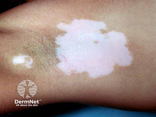
Clinical photography of axillary skin showing depigmented vitiligo patches. Modality: Clinical photograph, dermoscopy not used. The image depicts a large, well-demarcated, chalk-white macule/patch centered in the axillary fold with irregular but sharply defined borders. Adjacent skin exhibits relative hyperpigmentation surrounding the patch, consistent with repigmentation margins typical of vitiligo-affected areas. The surface appears smooth, non-scaly, and non-erythematous, with no crusting or ulceration. Hair within the depigmented areas may be reduced or absent, although follicular retention can occur. Lesions are unilateral and localized to the axilla in this view; a second smaller depigmented area is visible nearby. The clinical impression is consistent with localized vitiligo (leukoderma) presenting as hypopigmented to depigmented patches due to melanocyte destruction. The absence of scale distinguishes vitiligo from tinea versicolor or eczema; the uniform whiteness under standard lighting supports vitiligo rather than postinflammatory hypopigmentation. This image is relevant for educational descriptions of vitiligo morphology, differential diagnosis (hypopigmentation variants, pityriasis versicolor), and for demonstrating axillary involvement. Potential clinical utility includes patient counseling, Wood lamp correlation, and monitoring treatment response to topical corticosteroids, calcineurin inhibitors, or phototherapy over time. Image serves as a reference for clinicians evaluating depigmented lesions and educating patients about prognosis.
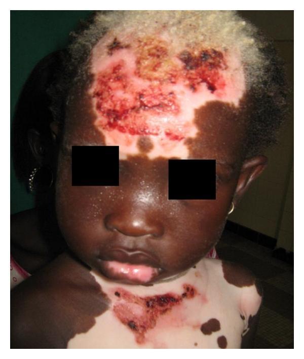
Clinical photograph of a pediatric patient with generalized vitiligo and secondary skin changes. The image displays extensive, sharply demarcated patches of depigmentation (achromia) on the forehead, chest, and lower lip, contrasting with the patient's natural dark skin tone. Notable leukotrichia (depigmented hair) is visible on the scalp. Within the larger depigmented macules on the forehead and upper chest, there are superimposed erythematous erosions, hemorrhagic crusting, and dark brown scabs, likely representing secondary cutaneous damage, such as actinic injury or superinfection in the unprotected amelanotic skin. The distribution is multi-focal, involving the face and trunk. This case illustrates the clinical presentation of early-onset vitiligo and the vulnerability of depigmented skin to environmental or secondary factors in a young child with a family history of the condition.
Vitiligo
Why it happens
Patterns
- Non-segmental vitiligo: patches on multiple body areas, often symmetrically, and may progress intermittently.
- Segmental vitiligo: typically limited to one side or region of the body and often stabilizes after an initial period.
Diagnosis
Treatment
- Sun protection with broad-spectrum SPF 30+ to prevent burns and reduce contrast
- Cosmetic camouflage or self-tanner
- Prescription topical treatments, such as corticosteroids, calcineurin inhibitors, or ruxolitinib cream in appropriate patients
- Narrowband UVB phototherapy for more extensive disease
- Excimer laser/light for selected localized patches
- Surgical grafting or cell-based procedures for stable, treatment-resistant localized disease
When to seek evaluation
Kidney stone
nephrolithiasis prevention
kidney stones symptoms when to seek emergency help diagnosis treatment prevention NIDDK
diagram kidney stone in ureter urinary tract obstruction
Kidney stone
Common symptoms
- Sudden, severe pain in the side, back, lower abdomen, or groin
- Pain that comes in waves
- Nausea or vomiting
- Blood in urine, or cloudy/foul-smelling urine
- Burning or urgency when urinating
Seek urgent medical care today if you have
- Fever, chills, or feel unwell with flank/back pain
- Severe or uncontrolled pain
- Vomiting or inability to keep fluids down
- Trouble passing urine or very little urine
- A single kidney, pregnancy, known kidney disease, or immunosuppression
How it is diagnosed
Treatment
- Small stones: pain control, anti-nausea medication, fluids as tolerated, and time to pass it. Some patients may be prescribed a medicine to relax the ureter.
- Large, obstructing, infected, or persistent stones: urology procedures such as shock-wave lithotripsy, ureteroscopy with laser fragmentation, or occasionally percutaneous removal.
Reducing future stones
??
- Where the pain is and when it started
- Whether you have fever/chills, vomiting, blood in urine, or trouble urinating
- Your age and whether you are pregnant or have one kidney/kidney disease

Read it
ureteral calculi hydronephrosis management
ureteral stone hydronephrosis emergency fever urine obstruction urology care foundation
Main findings
-
Right ureteric stone
- A 6.4 mm stone at the right UVJ, where the ureter joins the bladder.
- It is causing mild hydronephrosis and hydroureter, meaning urine is backing up and dilating the right kidney and ureter.
- There are also a few small 2-3 mm stones in the right kidney.
-
Left kidney stone
- A 7 mm stone in the mid calyx of the left kidney.
- It is causing minimal focal caliectasis, a small amount of dilation in part of the kidney.
- There are also a few small 2-3 mm stones in the left kidney.
-
Gallbladder
- A tiny 3 mm possible gallbladder polyp. It is not described as a mobile gallstone and has no shadowing.
- This is usually monitored with repeat ultrasound as advised by your clinician.
-
Otherwise
- Liver, spleen, visible pancreas, prostate, and bladder appear broadly normal.
- No abdominal fluid or pleural fluid seen.
What needs to happen
- Urine routine and culture
- Kidney function tests: creatinine, urea, electrolytes
- CBC if infection is possible
- Stone analysis if you pass a stone
Go to emergency care now if you have
- Fever or chills
- Severe or worsening pain not controlled with medication
- Repeated vomiting or inability to drink fluids
- Very low urine output or inability to pass urine
- Weakness, confusion, or feeling very unwell