
glomerular filtration barrier and its clinical aspect with UTI
"glomerular filtration barrier" AND proteinuria
Glomerular Filtration Barrier - Structure, Function, and Clinical Aspects with UTI
1. Overview of the Glomerular Filtration Barrier (GFB)
- Glycocalyx overlying the endothelial cells
- Fenestrated endothelium
- Glomerular basement membrane (GBM)
- Podocyte epithelial layer with slit diaphragms
2. Layers in Detail
Layer 1 - Fenestrated Endothelium
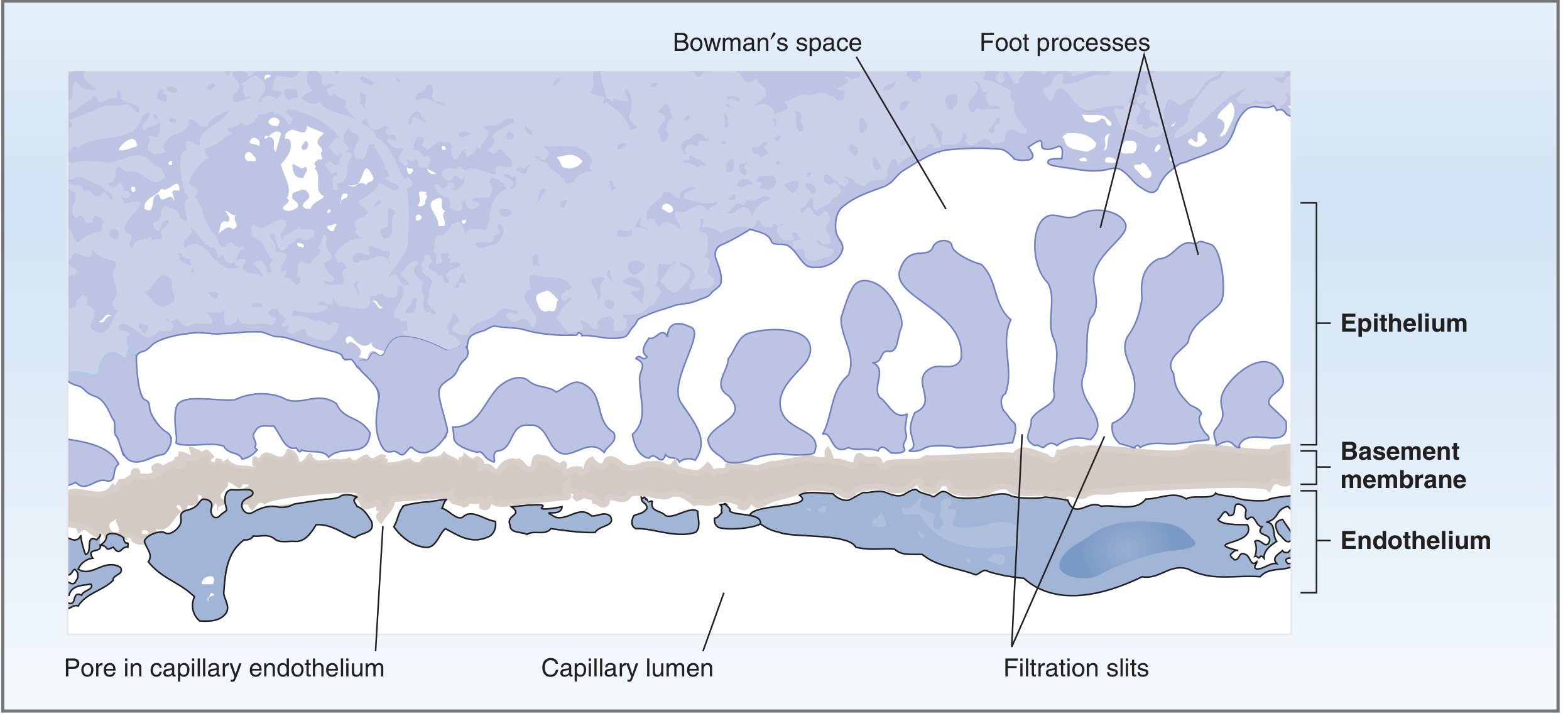
- Endothelial cells have fenestrations (pores) 70-100 nm in diameter
- These pores freely pass fluid, dissolved solutes, and plasma proteins
- They are not large enough to permit blood cells to cross
- The surface is coated by an endothelial glycocalyx (negatively charged), which acts as the first electrostatic barrier against anionic proteins like albumin
Layer 2 - Glomerular Basement Membrane (GBM)
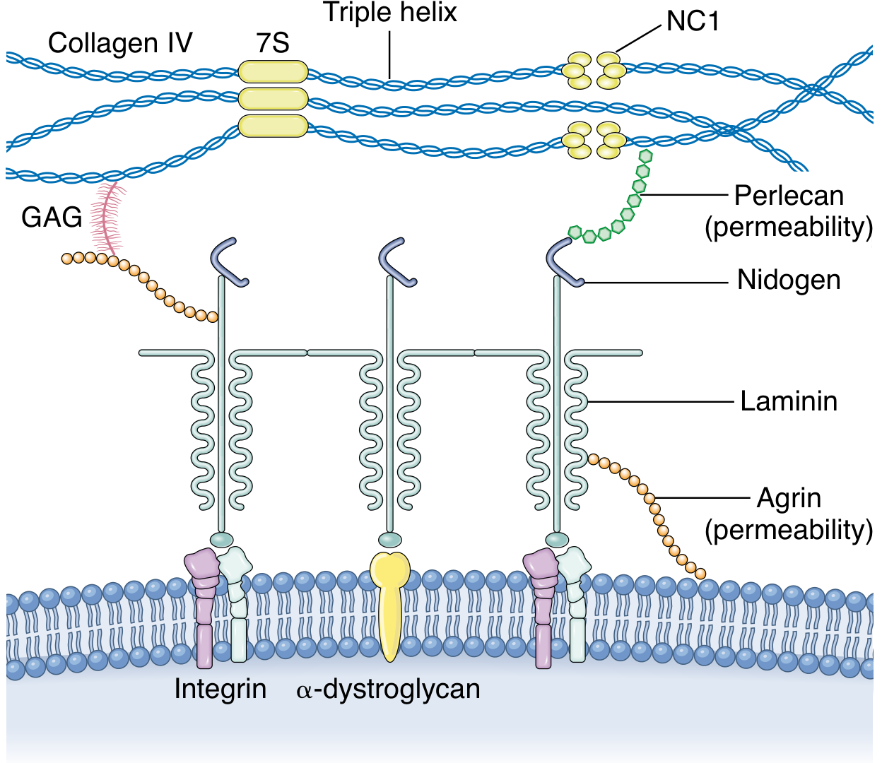
- Lamina rara interna - fused to the endothelium
- Lamina densa - central dense layer (primary structural barrier)
- Lamina rara externa - fused to the podocyte layer
- Type IV collagen (α3α4α5 chains in the mature GBM) - provides structural scaffold
- Laminin 521 - synthesized jointly by podocytes and endothelial cells
- Agrin and perlecan - heparan sulfate proteoglycans (HSPGs) providing anionic charge barrier
- Nidogen - links collagen IV to laminin networks
Layer 3 - Podocytes and Filtration Slit Diaphragm
- Extend major processes from the cell body
- Major processes give rise to secondary foot processes that interdigitate in a "zipper-like" pattern
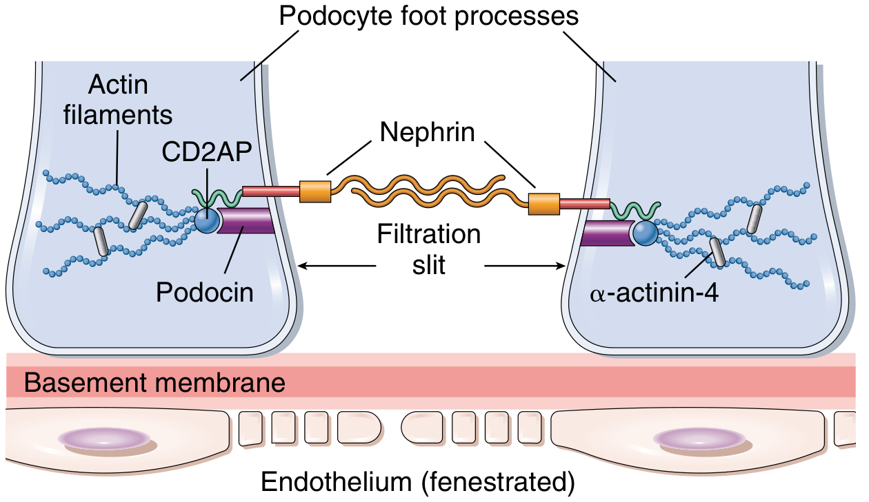
- Between adjacent foot processes are filtration slits (25-60 nm) bridged by the slit diaphragm
- Nephrin (NPHS1) - transmembrane immunoglobulin superfamily protein; forms the core structural scaffold of the slit with neph1; nephrin molecules are positioned on the apical side
- Podocin (NPHS2) - anchors nephrin to the plasma membrane; creates a signaling hub in lipid-rich membrane compartments; linked to TRPC6 channel
- CD2AP (CD2-associated protein) - links nephrin/podocin complex to the actin cytoskeleton
- α-actinin-4 - connects foot process actin filaments
- ZO-1, occludin - tight junction proteins associated with the slit diaphragm
- FAT1, P-cadherin - adherens junction proteins
- Podocalyxin - sialomucin on the apical surface; its highly negative charge keeps adjacent foot processes separated, maintaining the filtration barrier open
3. Filtration Selectivity - Size and Charge Barriers
Size Selectivity
| Component | Pore/Slit Size | Function |
|---|---|---|
| Endothelial fenestrae | 70-100 nm | Bars blood cells |
| GBM (lamina densa) | ~8 nm effective radius | Bars large proteins |
| Filtration slits | 25-60 nm | Additional barrier |
Charge Selectivity
- Fixed negative charges (from HSPGs agrin/perlecan, podocalyxin, endothelial glycocalyx) line all three layers
- These repel negatively charged macromolecules like albumin (which has net negative charge at physiologic pH)
- They attract positively charged solutes
- Small ions (Na⁺, K⁺, Cl⁻, HCO₃⁻) are freely filtered regardless of charge
- Classic experiment (Costanzo): Dextrans of the same molecular radius but different charges - cationic dextran filtered most, anionic dextran filtered least, neutral in between
4. Clinical Aspects - When the Filtration Barrier Fails
A. Nephrotic Syndrome - Loss of Charge/Structural Barrier
| Disease | Mechanism of GFB Disruption | Key Feature |
|---|---|---|
| Minimal Change Disease (MCD) | Loss of anionic charges (HSP loss), podocyte foot process effacement via CD80, c-mip, and angiopoietin-like 4 activation | Nephrotic range proteinuria; foot processes effaced on EM |
| Focal Segmental Glomerulosclerosis (FSGS) | Mutations in NPHS1 (nephrin), NPHS2 (podocin), ACTN4 (α-actinin-4), TRPC6 | Segmental scarring, massive proteinuria |
| Membranous Nephropathy | Anti-PLA2R antibodies attacking podocyte antigens; immune complex deposition in subepithelial space | "Spike and dome" on EM; complement activation |
| Diabetic Nephropathy | GBM thickening (increased synthesis), loss of HSPGs, mesangial expansion | Microalbuminuria → proteinuria |
| Alport Syndrome | Mutations in COL4A3/A4/A5 (type IV collagen α-chains) → "basket-weave" GBM appearance | Hematuria, sensorineural deafness, renal failure |
B. Nephritic Syndrome - Breach of Barrier by Inflammation
- Post-infectious GN (e.g., post-streptococcal): immune complex deposition activates complement and neutrophil infiltration
- IgA Nephropathy: galactose-deficient IgA1 forms mesangial deposits; characterized by synpharyngitic hematuria
- Crescent GN (RPGN): rupture of GBM; fibrin and plasma proteins leak into urinary space → crescent formation
5. UTI - Clinical Aspects and Renal Connection
Definitions (Comprehensive Clinical Nephrology 7th Ed.)
- Asymptomatic bacteriuria (ASB): >10⁵ organisms/mL without symptoms
- Cystitis: lower tract infection (frequency, dysuria, strangury); as few as 10² organisms/mL with pyuria and symptoms
- Acute pyelonephritis: >10⁵ organisms/mL + parenchymal infiltration + upper tract symptoms (fever, loin pain); may progress to systemic sepsis
Common Organisms
- E. coli (>70% of UTIs) - virulence factors include type 1 fimbriae (P-pili) for uroepithelial adhesion
- Klebsiella spp.
- Proteus spp. (especially in diabetics or urinary obstruction)
- Enterococci
- Staphylococcus saprophyticus
- Pseudomonas (especially in catheterized/immunocompromised patients)
Pathogenesis of Ascending Infection
- Perineal colonization (mostly E. coli)
- Ascent through urethra to bladder (cystitis)
- Ascent through ureters to renal pelvis and parenchyma (pyelonephritis)
- Bacterial fimbriae (pili) attach to urothelial receptors, facilitating ascent
UTI and Renal Damage - The GFB Connection
- Direct parenchymal bacterial infiltration causing inflammation that can extend to glomeruli
- Reflux nephropathy (VUR + recurrent UTI) leading to renal scarring, interstitial fibrosis, and reduced GFR
- Obstructive uropathy + UTI leading to hydronephrosis and permanent nephron loss
- Sepsis from pyelonephritis causing hemodynamic compromise → acute kidney injury (reduced GFR via decreased Kf and reduced glomerular capillary pressure)
UTI in Special Clinical Populations Affecting the Filtration Barrier
- UTI risk is increased post-transplant; ureteral stents and immunosuppression are risk factors
- Allograft pyelonephritis may cause graft dysfunction (reduced GFR) during acute episodes
- TMP-SMX prophylaxis reduces UTI incidence; VUR should be addressed to prevent recurrent UTI
- Progesterone-induced smooth muscle relaxation → ureteral dilation + stasis → increased risk of ascending UTI
- ASB affects 2-9% of pregnant women; 30% of untreated ASB progresses to pyelonephritis
- Pyelonephritis in pregnancy: sepsis, permanent renal injury, premature labor
- US Preventive Services Task Force: screen with urine culture at 12-16 weeks gestation (Grade A)
- Treatment of ASB reduces pyelonephritis incidence by >80%
Management of UTI (Rosen's Emergency Medicine)
| Condition | Treatment |
|---|---|
| ASB/Cystitis (pregnant) | Cephalexin 500 mg BID × 3-7 days, OR nitrofurantoin 100 mg BID × 3-7 days, OR amoxicillin 500 mg TID × 7 days (avoid nitrofurantoin near term) |
| Pyelonephritis (outpatient) | Oral fluoroquinolone × 7 days (if not pregnant); guided by local susceptibilities |
| Pyelonephritis (pregnant/inpatient) | IV ceftriaxone 1 g daily; adjust per culture; IV hydration + obstetric consultation |
| Recurrent UTI | Identify and treat anatomical abnormalities (VUR, obstruction); low-dose antibiotic prophylaxis |
6. Summary: GFB-UTI Clinical Integration
| GFB Impairment Mechanism | Resulting Condition | Clinical Link to UTI |
|---|---|---|
| Charge loss (HSP depletion) | Proteinuria (MCD-like) | Infections can trigger MCD relapse via TLR ligands (LPS from gram-negative bacteria, poly-IC from viruses) |
| GBM inflammation/breach | Post-infectious GN | Group A Streptococcal infections - both pharyngeal and skin infections; urinary infections less commonly implicated |
| Reflux nephropathy from recurrent UTI | Renal scarring → reduced GFR | Progressive loss of filtration surface area |
| Septic AKI from pyelonephritis | Reduced GFR (intrinsic + pre-renal) | Hemodynamic compromise + direct tubular toxicity |
| Allograft pyelonephritis | Acute graft dysfunction | Transient GFR reduction; long-term graft function may be preserved |
- Costanzo Physiology 7th Ed., Chapter 6 - Renal Physiology
- Brenner & Rector's The Kidney 2-Volume Set - Chapters on Podocyte Development and Structure
- Comprehensive Clinical Nephrology 7th Ed. - UTI in Pregnancy, Loss of Anionic Charges in GFB
- Robbins, Cotran & Kumar Pathologic Basis of Disease - Glomerular Filtration Barrier and Injury Patterns
- Rosen's Emergency Medicine - UTI Management in Pregnancy
- Medical Physiology (Boron & Boulpaep) - Filterability of Solutes
Recent evidence note: A 2025 systematic review (PMID: 41368354) confirms that genetic and environmental factors altering filtration slit proteins (nephrin, podocin) play a significant role in proteinuric kidney diseases - consistent with the above textbook content.
now make 4 slides content on this topic, 3 slide should focus on GFB only and last slide about corelation on uti.
**/SKILL.md in /tmp/skills
ls /tmp/workspace/ 2>/dev/null || mkdir -p /tmp/workspace && echo "ready"
mkdir -p /tmp/workspace/gfb-slides && python /tmp/skills/shared/scripts/fetch_images.py "https://cdn.orris.care/cdss_images/28c78c7afc9cffef0292ca68e59f2fc9dec02cf03a15db6f08038b3e316e1d57.png" "https://cdn.orris.care/cdss_images/6e8164abf32463bb1f42fcb5be4234153348d699743a3993d9420cc4fdf1d4b9.png" "https://cdn.orris.care/cdss_images/e49a35294b6460315647b8c10d91c4241e04a1cea7d9642169973d3456f696bb.png" > /tmp/workspace/gfb-slides/images.json && echo "done"
~/gfb-slides/make_slides.py
import json, base64
from io import BytesIO
from pptx import Presentation
from pptx.util import Inches, Pt, Emu
from pptx.dml.color import RGBColor
from pptx.enum.text import PP_ALIGN, MSO_ANCHOR
from pptx.util import Inches, Pt

# ── colour palette ──────────────────────────────────────────────────────────
NAVY      = RGBColor(0x0D, 0x2B, 0x55)   # deep navy - title BG
TEAL      = RGBColor(0x00, 0x7B, 0x83)   # accent bar / headings
LIGHT_BG  = RGBColor(0xF0, 0xF6, 0xF7)   # slide background
WHITE     = RGBColor(0xFF, 0xFF, 0xFF)
DARK_TXT  = RGBColor(0x1A, 0x1A, 0x2E)
ACCENT    = RGBColor(0xE8, 0x6A, 0x10)   # orange accent
LIGHT_TEAL= RGBColor(0xD6, 0xF0, 0xF2)   # light teal fill for boxes

# ── load images ──────────────────────────────────────────────────────────────
with open("/tmp/workspace/gfb-slides/images.json") as f:
    imgs = json.load(f)

def img_stream(idx):
    b64 = imgs[idx]["base64"]
    if b64 and "," in b64:
        b64 = b64.split(",", 1)[1]
    return BytesIO(base64.b64decode(b64))

# ── helpers ──────────────────────────────────────────────────────────────────
def new_prs():
    prs = Presentation()
    prs.slide_width  = Inches(13.333)
    prs.slide_height = Inches(7.5)
    return prs

def blank_slide(prs):
    return prs.slides.add_slide(prs.slide_layouts[6])

def fill_bg(slide, color):
    from pptx.oxml.ns import qn
    from lxml import etree
    bg = slide.background
    fill = bg.fill
    fill.solid()
    fill.fore_color.rgb = color

def add_rect(slide, l, t, w, h, fill_color, line_color=None, line_w=Pt(0)):
    shape = slide.shapes.add_shape(1, Inches(l), Inches(t), Inches(w), Inches(h))
    shape.fill.solid()
    shape.fill.fore_color.rgb = fill_color
    if line_color:
        shape.line.color.rgb = line_color
        shape.line.width = line_w
    else:
        shape.line.fill.background()
    return shape

def add_tb(slide, l, t, w, h, text, size, bold=False, color=DARK_TXT,
           align=PP_ALIGN.LEFT, wrap=True, italic=False):
    tb = slide.shapes.add_textbox(Inches(l), Inches(t), Inches(w), Inches(h))
    tf = tb.text_frame
    tf.word_wrap = wrap
    tf.margin_left = tf.margin_right = tf.margin_top = tf.margin_bottom = 0
    p = tf.paragraphs[0]
    p.alignment = align
    r = p.add_run()
    r.text = text
    r.font.name = "Calibri"
    r.font.size = Pt(size)
    r.font.bold = bold
    r.font.italic = italic
    r.font.color.rgb = color
    return tb

def add_para(tf, text, size, bold=False, color=DARK_TXT,
             align=PP_ALIGN.LEFT, italic=False, bullet=False, space_before=0):
    p = tf.add_paragraph()
    p.alignment = align
    p.space_before = Pt(space_before)
    if bullet:
        from pptx.oxml.ns import qn
        from lxml import etree
        pPr = p._p.get_or_add_pPr()
        buChar = etree.SubElement(pPr, qn('a:buChar'))
        buChar.set('char', '•')
    r = p.add_run()
    r.text = text
    r.font.name = "Calibri"
    r.font.size = Pt(size)
    r.font.bold = bold
    r.font.italic = italic
    r.font.color.rgb = color
    return p

def title_bar(slide, title, subtitle=None):
    """Navy top bar with white title."""
    add_rect(slide, 0, 0, 13.333, 1.25, NAVY)
    # teal left accent strip
    add_rect(slide, 0, 0, 0.18, 1.25, TEAL)
    add_tb(slide, 0.3, 0.1, 12.5, 0.8, title, 30, bold=True, color=WHITE, align=PP_ALIGN.LEFT)
    if subtitle:
        add_tb(slide, 0.3, 0.88, 12.5, 0.4, subtitle, 14, bold=False,
               color=RGBColor(0xC8, 0xE6, 0xF0), align=PP_ALIGN.LEFT)

# ════════════════════════════════════════════════════════════════════════════
prs = new_prs()

# ─────────────────────────────────────────────────────────────────────────────
# SLIDE 1 — Layers of the GFB
# ─────────────────────────────────────────────────────────────────────────────
s1 = blank_slide(prs)
fill_bg(s1, LIGHT_BG)
title_bar(s1, "Glomerular Filtration Barrier — Structural Layers",
          "Three-layered ultrafiltration unit separating capillary blood from Bowman's space")

# Main image (capillary wall diagram) — right column
s1.shapes.add_picture(img_stream(0), Inches(7.5), Inches(1.4), Inches(5.5), Inches(5.6))

# Left content column
# Layer boxes
layers = [
    ("1.  Fenestrated Endothelium",
     "Pores 70–100 nm in diameter  •  Allow fluid, solutes & plasma proteins\n"
     "Block blood cells  •  Coated by negatively charged glycocalyx",
     TEAL),
    ("2.  Glomerular Basement Membrane (GBM)",
     "Trilaminar: Lamina rara interna → Lamina densa → Lamina rara externa\n"
     "Most significant protein barrier  •  Contains Type IV collagen, Laminin,\n"
     "Agrin & Perlecan (heparan sulfate proteoglycans)",
     NAVY),
    ("3.  Podocytes & Slit Diaphragm",
     "Foot processes interdigitate in zipper pattern  •  Filtration slits 25–60 nm\n"
     "Slit diaphragm: Nephrin, Podocin, CD2AP, α-actinin-4\n"
     "Podocalyxin on apical surface — negative charge keeps slits open",
     TEAL),
]

y = 1.45
for title_l, body, col in layers:
    # colored header strip
    add_rect(s1, 0.25, y, 7.0, 0.35, col)
    add_tb(s1, 0.35, y+0.03, 6.8, 0.3, title_l, 12, bold=True, color=WHITE)
    # body box
    add_rect(s1, 0.25, y+0.35, 7.0, 0.9, WHITE, line_color=col, line_w=Pt(1))
    tb = s1.shapes.add_textbox(Inches(0.35), Inches(y+0.37), Inches(6.8), Inches(0.88))
    tf = tb.text_frame
    tf.word_wrap = True
    tf.margin_left = Inches(0.05)
    tf.margin_top = tf.margin_bottom = 0
    p = tf.paragraphs[0]
    r = p.add_run()
    r.text = body
    r.font.name = "Calibri"
    r.font.size = Pt(11)
    r.font.color.rgb = DARK_TXT
    y += 1.5

# Caption
add_tb(s1, 7.5, 7.05, 5.5, 0.4,
       "Fig. Structure of the glomerular capillary wall (Costanzo Physiology 7th Ed.)",
       9, italic=True, color=RGBColor(0x55,0x55,0x55), align=PP_ALIGN.CENTER)


# ─────────────────────────────────────────────────────────────────────────────
# SLIDE 2 — Filtration Selectivity (Size + Charge) & GBM Composition
# ─────────────────────────────────────────────────────────────────────────────
s2 = blank_slide(prs)
fill_bg(s2, LIGHT_BG)
title_bar(s2, "GFB — Filtration Selectivity: Size & Charge Barriers",
          "Dual mechanism restricts macromolecule passage across the filtration barrier")

# GBM composition image — right top
s2.shapes.add_picture(img_stream(2), Inches(7.4), Inches(1.35), Inches(5.6), Inches(3.5))
add_tb(s2, 7.4, 4.9, 5.6, 0.35,
       "Fig. GBM composition: Collagen IV, Laminin, Agrin, Perlecan, Nidogen\n(Robbins Pathologic Basis of Disease)",
       9, italic=True, color=RGBColor(0x55,0x55,0x55), align=PP_ALIGN.CENTER)

# Slit diaphragm image — right bottom
s2.shapes.add_picture(img_stream(1), Inches(7.4), Inches(5.3), Inches(5.6), Inches(2.0))
add_tb(s2, 7.4, 7.15, 5.6, 0.3,
       "Fig. Slit diaphragm proteins — Nephrin, Podocin, CD2AP (Robbins)",
       9, italic=True, color=RGBColor(0x55,0x55,0x55), align=PP_ALIGN.CENTER)

# LEFT: two side-by-side sub-boxes
# SIZE box
add_rect(s2, 0.25, 1.35, 3.4, 0.38, TEAL)
add_tb(s2, 0.35, 1.37, 3.2, 0.33, "SIZE SELECTIVITY", 13, bold=True, color=WHITE)
add_rect(s2, 0.25, 1.73, 3.4, 2.6, WHITE, line_color=TEAL, line_w=Pt(1.5))
tb = s2.shapes.add_textbox(Inches(0.35), Inches(1.76), Inches(3.2), Inches(2.5))
tf = tb.text_frame; tf.word_wrap = True
tf.margin_left = Inches(0.04); tf.margin_top = tf.margin_bottom = 0

size_rows = [
    ("Endothelial pores", "70–100 nm", "Bar blood cells"),
    ("GBM (lamina densa)", "~8 nm radius", "Primary protein barrier"),
    ("Filtration slits",   "25–60 nm",  "Additional barrier"),
]
header_p = tf.paragraphs[0]
header_p.alignment = PP_ALIGN.CENTER
hr = header_p.add_run()
hr.text = "Component           Pore/Slit Size   Role"
hr.font.name = "Calibri"; hr.font.size = Pt(9.5); hr.font.bold = True; hr.font.color.rgb = TEAL
for comp, sz, role in size_rows:
    add_para(tf, f"{comp:<22} {sz:<16} {role}", 9.5, color=DARK_TXT, space_before=4)

add_para(tf, "\nSmall ions (Na⁺, K⁺, Cl⁻) freely filtered regardless of size.",
         9.5, italic=True, color=TEAL)

# CHARGE box
add_rect(s2, 3.85, 1.35, 3.35, 0.38, NAVY)
add_tb(s2, 3.95, 1.37, 3.15, 0.33, "CHARGE SELECTIVITY", 13, bold=True, color=WHITE)
add_rect(s2, 3.85, 1.73, 3.35, 2.6, WHITE, line_color=NAVY, line_w=Pt(1.5))
tb2 = s2.shapes.add_textbox(Inches(3.95), Inches(1.76), Inches(3.15), Inches(2.5))
tf2 = tb2.text_frame; tf2.word_wrap = True
tf2.margin_left = Inches(0.04); tf2.margin_top = tf2.margin_bottom = 0

charge_lines = [
    ("Fixed –ve charges on all 3 layers", False),
    ("Sources: HSPGs (Agrin, Perlecan),", False),
    ("  Podocalyxin, Glycocalyx", False),
    ("", False),
    ("• Repel albumin (net –ve at pH 7.4)", True),
    ("• Attract +ve solutes", True),
    ("• Cationic dextran > neutral > anionic", True),
    ("  dextran filtration (classic experiment)", False),
    ("", False),
    ("Disease: Loss of anionic charges →", False),
    ("  proteinuria (MCD, Diabetic nephropathy)", False),
]
p0 = tf2.paragraphs[0]
r0 = p0.add_run()
r0.text = charge_lines[0][0]
r0.font.name = "Calibri"; r0.font.size = Pt(9.5); r0.font.color.rgb = DARK_TXT
for txt, blt in charge_lines[1:]:
    add_para(tf2, txt, 9.5, color=DARK_TXT, space_before=2)

# Bottom note
add_rect(s2, 0.25, 4.55, 7.0, 0.55, LIGHT_TEAL)
add_tb(s2, 0.35, 4.6, 6.8, 0.45,
       "Key concept: For large solutes (proteins), BOTH size AND charge determine filterability. "
       "Charge selectivity is negligible for small ions but critical for albumin-sized molecules.",
       10, italic=True, color=NAVY, wrap=True)

# Starling forces note
add_rect(s2, 0.25, 5.3, 7.0, 1.9, WHITE, line_color=TEAL, line_w=Pt(1))
add_tb(s2, 0.35, 5.35, 6.8, 0.3, "Starling Forces Driving GFR", 12, bold=True, color=TEAL)
tb3 = s2.shapes.add_textbox(Inches(0.35), Inches(5.68), Inches(6.8), Inches(1.4))
tf3 = tb3.text_frame; tf3.word_wrap = True
tf3.margin_left = Inches(0.04); tf3.margin_top = 0
p3 = tf3.paragraphs[0]
r3 = p3.add_run()
r3.text = "GFR = Kf [(PGC − PBS) − (πGC − πBS)]"
r3.font.name = "Calibri"; r3.font.size = Pt(11); r3.font.bold = True; r3.font.color.rgb = NAVY
starling_lines = [
    "PGC = glomerular capillary hydrostatic pressure (favours filtration)",
    "PBS = Bowman's space hydrostatic pressure (opposes filtration)",
    "πGC = glomerular oncotic pressure (opposes filtration)  |  πBS ≈ 0",
    "Kf = ultrafiltration coefficient (permeability × surface area)",
]
for ln in starling_lines:
    add_para(tf3, ln, 9.5, color=DARK_TXT, space_before=3)


# ─────────────────────────────────────────────────────────────────────────────
# SLIDE 3 — Clinical Diseases of the GFB
# ─────────────────────────────────────────────────────────────────────────────
s3 = blank_slide(prs)
fill_bg(s3, LIGHT_BG)
title_bar(s3, "GFB — Clinical Diseases & Pathological Mechanisms",
          "Disruption of the filtration barrier leads to characteristic nephropathies")

diseases = [
    ("Minimal Change Disease (MCD)",
     TEAL,
     [
       "Loss of anionic charges (heparan sulfate proteoglycans)",
       "Podocyte CD80 activation → actin reorganisation → foot process effacement",
       "Circulating factors: IL-13, hemopexin, anti-nephrin antibodies",
       "EM: diffuse foot process effacement  |  LM: normal glomeruli",
       "Result: Nephrotic syndrome — massive proteinuria, hypoalbuminaemia",
     ]),
    ("Focal Segmental Glomerulosclerosis (FSGS)",
     NAVY,
     [
       "Mutations: NPHS1 (nephrin), NPHS2 (podocin), ACTN4 (α-actinin-4), TRPC6",
       "Podocin anchors nephrin to membrane; TRPC6 regulates Ca²⁺ entry",
       "Focal & segmental scarring of glomeruli",
       "Primary (circulating permeability factor) vs Secondary (obesity, HIV, drugs)",
       "Result: Nephrotic syndrome — steroid-resistant proteinuria",
     ]),
    ("Diabetic Nephropathy",
     RGBColor(0x1A, 0x6B, 0x3A),
     [
       "GBM thickening (increased Type IV collagen synthesis)",
       "Loss of heparan sulfate proteoglycans → reduced anionic charge",
       "Mesangial expansion → reduced filtration surface area (reduced Kf)",
       "Microalbuminuria → overt proteinuria → progressive GFR decline",
       "Advanced glycation end-products (AGEs) further injure podocytes & endothelium",
     ]),
    ("Alport Syndrome",
     ACCENT,
     [
       "Mutations in COL4A3 / COL4A4 / COL4A5 → abnormal Type IV collagen α-chains",
       "GBM shows 'basket-weave' appearance on EM",
       "Triad: haematuria + sensorineural deafness + ocular defects",
       "X-linked (COL4A5), autosomal recessive/dominant (COL4A3/A4)",
       "Progressive → ESRD in males by 3rd decade",
     ]),
]

# 2×2 grid
positions = [(0.25, 1.35), (6.85, 1.35), (0.25, 4.45), (6.85, 4.45)]
box_w, box_h = 6.3, 2.85

for i, (dis_name, col, bullets) in enumerate(diseases):
    lx, ty = positions[i]
    # header bar
    add_rect(s3, lx, ty, box_w, 0.38, col)
    add_tb(s3, lx+0.1, ty+0.04, box_w-0.2, 0.32, dis_name, 12, bold=True, color=WHITE)
    # body
    add_rect(s3, lx, ty+0.38, box_w, box_h-0.38, WHITE, line_color=col, line_w=Pt(1))
    tb = s3.shapes.add_textbox(Inches(lx+0.12), Inches(ty+0.42), Inches(box_w-0.25), Inches(box_h-0.5))
    tf = tb.text_frame; tf.word_wrap = True
    tf.margin_left = Inches(0.02); tf.margin_top = 0
    p_first = tf.paragraphs[0]
    r_first = p_first.add_run()
    r_first.text = "• " + bullets[0]
    r_first.font.name = "Calibri"; r_first.font.size = Pt(9.5); r_first.font.color.rgb = DARK_TXT
    for b in bullets[1:]:
        add_para(tf, "• " + b, 9.5, color=DARK_TXT, space_before=3)

# bottom footnote
add_rect(s3, 0.25, 7.2, 12.9, 0.27, LIGHT_TEAL)
add_tb(s3, 0.4, 7.22, 12.7, 0.23,
       "Nephrotic features = massive proteinuria, hypoalbuminaemia, oedema, hyperlipidaemia  |  "
       "Nephritic features = haematuria, RBC casts, oliguria, hypertension",
       9, italic=True, color=NAVY)


# ─────────────────────────────────────────────────────────────────────────────
# SLIDE 4 — GFB Correlation with UTI
# ─────────────────────────────────────────────────────────────────────────────
s4 = blank_slide(prs)
fill_bg(s4, LIGHT_BG)
title_bar(s4, "UTI & the Glomerular Filtration Barrier — Clinical Correlation",
          "How urinary tract infections impact glomerular filtration and renal function")

# ── UTI definitions strip ──
add_rect(s4, 0.25, 1.38, 12.85, 0.35, NAVY)
add_tb(s4, 0.35, 1.41, 12.6, 0.3, "UTI Spectrum: Definitions", 11, bold=True, color=WHITE)
add_rect(s4, 0.25, 1.73, 12.85, 0.45, WHITE, line_color=NAVY, line_w=Pt(1))
def_tb = s4.shapes.add_textbox(Inches(0.35), Inches(1.75), Inches(12.6), Inches(0.42))
def_tf = def_tb.text_frame; def_tf.word_wrap = True
def_tf.margin_left = Inches(0.04); def_tf.margin_top = 0
dp = def_tf.paragraphs[0]; dr = dp.add_run()
dr.text = ("ASB: >10⁵ org/mL without symptoms   |   "
           "Cystitis: >10² org/mL + lower tract symptoms (dysuria, frequency)   |   "
           "Pyelonephritis: >10⁵ org/mL + parenchymal infiltration + fever + loin pain → may → sepsis")
dr.font.name = "Calibri"; dr.font.size = Pt(9.5); dr.font.color.rgb = DARK_TXT

# ── 3-column correlation section ──
cols_data = [
    ("DIRECT GFB IMPACT",
     TEAL,
     [
       "Ascending pyelonephritis → bacterial infiltration of renal parenchyma",
       "Inflammatory mediators (IL-6, TNF-α) damage podocytes → transient proteinuria",
       "Gram-negative LPS binds TLR-4 on podocytes → foot process injury",
       "Viral-like products (poly-IC) bind TLR-3 → MCD-like proteinuria",
       "Septic AKI from severe pyelonephritis → ↓ GFR via haemodynamic compromise",
       "Direct tubular toxicity compounds filtration defects",
     ]),
    ("REFLUX & CHRONIC DAMAGE",
     NAVY,
     [
       "Vesicoureteral Reflux (VUR) + recurrent UTI → reflux nephropathy",
       "Repeated infection → renal cortical scarring → nephron loss",
       "Reduced filtration surface area (↓ Kf) → progressive CKD",
       "Post-transplant UTI (especially with stents/VUR) → allograft pyelonephritis",
       "Transient graft dysfunction during acute episode",
       "Recurrent UTI post-transplant: address VUR; TMP-SMX prophylaxis",
     ]),
    ("PREGNANCY: HIGH-RISK UTI",
     ACCENT,
     [
       "Progesterone → ureteral dilation & stasis → ↑ ascending infection risk",
       "ASB affects 2–9% pregnant women; 30% progress to pyelonephritis if untreated",
       "Pyelonephritis → sepsis → permanent renal injury → ↓ long-term GFR",
       "Screen: Urine culture at 12–16 weeks (USPSTF Grade A)",
       "Tx ASB: Cephalexin / Nitrofurantoin × 3–7 days",
       "Pyelonephritis: IV Ceftriaxone 1 g daily + hydration + obstetric review",
     ]),
]

col_w = 4.2
starts_x = [0.25, 4.65, 9.05]
for j, (col_title, col_color, bullets) in enumerate(cols_data):
    lx = starts_x[j]
    add_rect(s4, lx, 2.28, col_w, 0.38, col_color)
    add_tb(s4, lx+0.08, 2.3, col_w-0.16, 0.34, col_title, 10.5, bold=True, color=WHITE)
    add_rect(s4, lx, 2.66, col_w, 3.6, WHITE, line_color=col_color, line_w=Pt(1))
    tb = s4.shapes.add_textbox(Inches(lx+0.1), Inches(2.7), Inches(col_w-0.2), Inches(3.5))
    tf = tb.text_frame; tf.word_wrap = True
    tf.margin_left = Inches(0.03); tf.margin_top = 0
    p0 = tf.paragraphs[0]; r0 = p0.add_run()
    r0.text = "• " + bullets[0]
    r0.font.name = "Calibri"; r0.font.size = Pt(9.5); r0.font.color.rgb = DARK_TXT
    for b in bullets[1:]:
        add_para(tf, "• " + b, 9.5, color=DARK_TXT, space_before=4)

# ── Summary table ──
add_rect(s4, 0.25, 6.4, 12.85, 0.3, TEAL)
add_tb(s4, 0.35, 6.42, 12.6, 0.26, "GFB Damage Pathway Summary", 10, bold=True, color=WHITE)
add_rect(s4, 0.25, 6.7, 12.85, 0.75, WHITE, line_color=TEAL, line_w=Pt(1))
sum_tb = s4.shapes.add_textbox(Inches(0.35), Inches(6.73), Inches(12.6), Inches(0.7))
sum_tf = sum_tb.text_frame; sum_tf.word_wrap = True
sum_tf.margin_left = Inches(0.04); sum_tf.margin_top = 0
sp = sum_tf.paragraphs[0]; sr = sp.add_run()
sr.text = ("Gram-negative UTI (LPS)  →  TLR-4 activation on podocytes  →  foot process effacement  →  proteinuria   |   "
           "Recurrent UTI + VUR  →  reflux nephropathy  →  ↓ GFR   |   "
           "Severe pyelonephritis  →  sepsis  →  AKI (↓ PGC, ↓ Kf)  →  acute GFR fall")
sr.font.name = "Calibri"; sr.font.size = Pt(9.5); sr.font.bold = False; sr.font.color.rgb = DARK_TXT

# ── Save ──────────────────────────────────────────────────────────────────────
out = "/tmp/workspace/gfb-slides/GFB_and_UTI.pptx"
prs.save(out)
print(f"Saved → {out}")
cd /tmp/workspace/gfb-slides && python make_slides.py 2>&1
python3 -c " from pptx import Presentation prs = Presentation('/tmp/workspace/gfb-slides/GFB_and_UTI.pptx') print(f'Slides: {len(prs.slides)}') print(f'Dimensions: {prs.slide_width.inches:.2f}\" x {prs.slide_height.inches:.2f}\"') for i, slide in enumerate(prs.slides, 1): texts = [s.text_frame.text[:60] for s in slide.shapes if s.has_text_frame and s.text_frame.text.strip()] print(f'Slide {i}: {texts[0] if texts else \"(no text)\"}') import os print(f'File size: {os.path.getsize(\"/tmp/workspace/gfb-slides/GFB_and_UTI.pptx\")/1024:.1f} KB') "
GFB and UTI
Presentation · PPTX
- Three labeled sections covering Fenestrated Endothelium (70-100 nm pores, glycocalyx), GBM (trilaminar structure: lamina rara interna/densa/externa; Type IV collagen, Laminin, Agrin, Perlecan), and Podocytes/Slit Diaphragm (nephrin, podocin, CD2AP, α-actinin-4, podocalyxin)
- Embedded capillary wall diagram from Costanzo Physiology
- Side-by-side boxes: Size selectivity table (pore sizes for each layer) vs Charge selectivity (fixed negative charges, dextran experiment, albumin repulsion)
- Starling forces equation with all four variables explained
- Embedded GBM composition and slit diaphragm protein diagrams
- 2×2 grid covering: MCD (foot process effacement, TLR activation, anionic charge loss), FSGS (NPHS1/NPHS2/ACTN4/TRPC6 mutations), Diabetic Nephropathy (GBM thickening, HSPG loss, mesangial expansion), and Alport Syndrome (COL4A3/A4/A5 mutations, basket-weave GBM)
- Footer distinguishing nephrotic vs nephritic syndrome features
- UTI spectrum definitions bar (ASB, cystitis, pyelonephritis thresholds)
- Three columns: Direct GFB Impact (LPS → TLR-4 → podocyte injury; septic AKI), Reflux & Chronic Damage (VUR → reflux nephropathy → ↓ GFR; transplant UTI), Pregnancy/High-Risk UTI (ASB prevalence, USPSTF screening, treatment regimens)
- Summary pathway strip at the bottom: LPS → TLR-4 → foot process effacement → proteinuria