
Noprilam BD?
Noprilam Bangladesh medicine generic name
Quick answer
- Bangladesh, as in whether the brand is sold there.
- "Twice daily" (bis die), as in the dose frequency.
- Something else, such as a company name.
- The generic name or strength printed on the strip or box, for example "X mg tablet".
- The manufacturer's name.
- What you want to know: what it is, the dose, the indication, or whether it's available in Bangladesh.
Amoxicillin , clavulenic acid ones or twice a day?
amoxicillin clavulanate dosing twice daily vs three times daily adults 875/125 500/125 Bangladesh Noprilam
"amoxicillin-potassium clavulanate combination"[MeSH Terms] AND dosing
| Tablet strength | Usual adult schedule |
|---|---|
| 875/125 mg | 1 tablet every 12 hours (twice daily) |
| 500/125 mg | 1 tablet every 8 hours (three times daily) |
| 625 mg (500/125) tablets, common in Bangladesh | Usually every 8 hours, though some prescribers use every 12 hours |
- Goldman-Cecil Medicine gives a usual adult dose of 500 mg every 12 hours for respiratory infections. The Cecil snippet doesn't give the clavulanate content.
- Tintinalli's Emergency Medicine lists 875/125 mg every 12 hours for 10 days for a skin and soft tissue infection.
- Fishman's Pulmonary Diseases lists 500/125 mg three times daily, or 875/125 mg twice daily, for outpatient community-acquired pneumonia.
- The WHO submission cites a randomised trial in 557 adults. Clinical success was about 92 to 94% with either 875/125 mg twice daily or 500/125 mg three times daily, and the two were equivalent.
- The Dermatology textbook warns not to double the dose of the combined tablet to raise the amoxicillin dose, because that gives too much clavulanate. It is a cause of diarrhoea.
- Take it at the start of a meal. This improves absorption and reduces nausea and diarrhoea.
- Space the doses evenly and finish the course your prescriber set. Courses usually run 5 to 7 days, but the length depends on the infection.
- Children are dosed by weight, so don't use adult tablets for them without a prescriber's instructions.
- Dose adjustment is needed with significant kidney impairment. The 875 mg tablet is generally avoided if creatinine clearance is below 30 mL/min.
- Don't use it if you have a penicillin allergy or previous liver injury from this drug.
Chalasion treatment
"chalazion"[MeSH Terms] AND treatment
chalazion eyelid
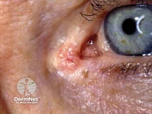
Clinical photograph of a solitary nodular lesion on the eyelid margin illustrating suspected eyelid basal cell carcinoma. The palpebral skin shows a flesh-colored to pink, slightly translucent nodule with a crusted surface and subtle surface telangiectasia. The lesion is localized to the eyelid/ocular adnexal region, near the margin, with mild surrounding dermal edema but no obvious pigmentary change. This presentation is characteristic of basal cell carcinoma in the periorbital area, a common eyelid malignancy. The image demonstrates features that raise suspicion for malignancy rather than a benign chalazion or inflammatory lesion: a persistent, slowly enlarging nodular lesion with surface crust and well-defined borders. While superior diagnostic confidence requires histopathology, clinically this pattern aligns with basal cell carcinoma of the eyelid, particularly the nodular subtype. Early recognition is important to minimize subcutaneous invasion and avoid functional impairment of the eyelid. Management implications include complete surgical excision with adequate margins, with Mohs micrographic surgery often preferred for periorbital tumors. Differential diagnoses include sebaceous carcinoma, squamous cell carcinoma, keratoacanthoma, chalazion, and benign nevi. This image is valuable for education on eyelid oncology, differential diagnosis of eyelid lesions, and contrast between malignant adnexal tumors and benign eyelid pathology. Correlate clinically with patient history and radiologic findings if available.
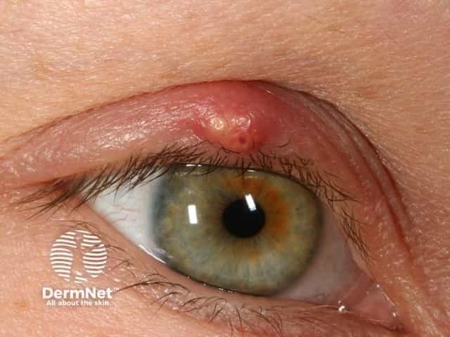
Clinical photograph of an acute hordeolum (stye) involving the upper eyelid margin. High‑resolution digital photography in a frontal close‑up view captures a small, erythematous, tender nodule with a central pustule along the lash line, accompanied by mild lid edema and slight crusting. The lesion most likely represents an infection of the eyelid gland (external Hordeolum involving Zeis or Moll glands) rather than a chronic, painless chalazion. The surrounding conjunctiva shows minimal injection; no orbital signs are evident. Key imaging features include focal erythema, localized induration, purulent content, and a localized surface lesion at the lid margin with preserved globe motility. Diagnostic significance: this appearance supports acute soft‑tissue infection of the eyelid, typically due to Staphylococcus aureus or other skin flora. Differential considerations include internal Hordeolum, chalazion, blepharitis with eyelid margin edema, and preseptal cellulitis if systemic symptoms develop. Clinical correlation should assess fever, spreading infection, pain severity, and response to warm compresses or topical antibiotics. Utility: this image is valuable for ophthalmology and dermatology education, telemedicine triage, clinical documentation, and imaging-based teaching on eyelid infections, eyelid anatomy, and differential diagnosis of eyelid lesions. Clinical teaching value includes recognizing edema patterns and treatment milestones.
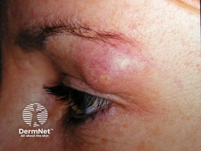
Imaging modality and technique: Clinical photography of the periorbital region, frontal close-up of the upper eyelid using diffuse illumination. Anatomical location: Upper eyelid (periorbital skin, lid margin). Visual features: a localized, dome-shaped erythematous nodule along the eyelid margin with mild surrounding edema and a possible central pustule, consistent with a stye (hordeolum) or blocked meibomian gland (chalazion) in early inflammation. Pathology and differential: The lesion reflects an inflammatory cyst-like lesion of eyelid glands; differential includes external hordeolum (stye), chalazion, sebaceous gland abscess, blepharitis, and preseptal cellulitis if spread occurs. Diagnostic significance: Distinguishes acute infectious eyelid inflammation from chronic eyelid granulomatous lesions; early recognition supports warm compress therapy, lid hygiene, topical antibiotics if indicated, and avoidance of squeezing. Clinical correlation: Correlate with symptoms such as tenderness, pain, eyelid swelling, fever, or discharge. Imaging context: Not applicable beyond clinical photography for dermatologic illustration. Utility: Educational reference for dermatology, ophthalmology, primary care; supports patient counseling, triage for antibiotic therapy, and monitoring for resolution or progression. Clinical outcome depends on timely management; signs of abscess formation, spreading cellulitis, or vision impairment require urgent evaluation. Documenting appearance aids clinicians in documenting severity, tracking response, and communicating prognosis to patients and trainees. Useful for teaching notes.
- At least a third resolve on their own, so watching is reasonable, especially if the lump is improving (Kanski's Clinical Ophthalmology, 10th ed.).
- Use a hot compress several times a day. It helps most in early lesions.
- Gentle lid massage and lid hygiene also help (Rosen's Emergency Medicine).
- Don't squeeze or pop it.
- Oral antibiotics are needed only if there is significant bacterial infection, not for sterile inflammation (Kanski).
- An acute chalazion can look the same as an internal hordeolum (stye), and the treatment is the same (Tintinalli's Emergency Medicine).
- Triamcinolone is injected into or around the lesion. Kanski reports resolution rates similar to incision and curettage.
- It is preferred for lesions at the lid margin or near the lacrimal punctum, where surgery risks damaging nearby structures.
- Reported regimens use 0.2 to 2 ml of triamcinolone diluted with lidocaine to 5 mg/ml, or 0.1 to 0.2 ml of 40 mg/ml, through a 27 or 30 gauge needle.
- About 80% resolve after one injection. A second can be given 1 to 2 weeks later.
- Uncommon complications are skin depigmentation and fat atrophy. Retinal vascular occlusion is rare. A conjunctival approach makes the first two less likely.
- This is considered if the chalazion lasts more than 4 to 6 weeks (Textbook of Family Medicine, 9th ed.).
- Under local anaesthesia, the lid is clamped and everted, the cyst is incised vertically through the tarsal plate, and the contents are curetted. No suture is used.
- Solid material can be excised and sent for histology.
- A topical antibiotic is used three times daily for 5 days afterwards (Kanski).
- Tintinalli advises referral to an ophthalmologist within 1 to 2 weeks for chronic or recurrent chalazia.
- A recurrent chalazion in the same spot should be biopsied, because it can be a sebaceous carcinoma (Textbook of Family Medicine).
- Warning signs for a tumour are loss of eyelashes, distorted lid margin, and a lump that keeps growing.
- See a doctor urgently for spreading redness or swelling of the whole lid, fever, pain on eye movement, or any change in vision. These suggest preseptal or orbital cellulitis.
Stye treatment
"hordeolum"[MeSH Terms]
- Apply a warm compress several times a day. Goodman & Gilman's lists warm compresses as the usual treatment.
- Most styes drain on their own and clear in about a week (Pfenninger and Fowler's Procedures for Primary Care).
- Don't squeeze or pop it.
- Avoid eye makeup and contact lenses until it heals.
- Tintinalli's Emergency Medicine says warm compresses plus erythromycin ophthalmic ointment twice daily for 7 to 10 days are usually enough. Removing the offending eyelash can be considered.
- The Wills Eye Manual suggests a short course of topical antibiotic ointment (bacitracin, tobramycin or erythromycin twice daily for 1 to 2 weeks).
- Oral antibiotics are needed only if there is significant cellulitis around the lid. Pfenninger and Fowler's recommend an anti-staphylococcal oral antibiotic when the stye is more severe but not yet "pointing".
- Topical antibiotics have limited proof of benefit, so ask your doctor whether you need one.
- This is done if the stye points (forms a visible head) or fails to settle with compresses.
- The doctor makes a small incision with a no. 11 blade under local anaesthesia until pus drains, often with a chalazion clamp for stability (Pfenninger and Fowler's).
- The redness and swelling spread across the lid or cheek.
- You have fever, severe pain, pain with eye movement, or changes in vision. These suggest preseptal or orbital cellulitis.
- The lump lasts more than a few weeks. It may have become a chalazion, and a recurrent lump in the same spot needs a biopsy to exclude a tumour.
- Styes keep coming back. Look for blepharitis, which needs lid hygiene.