
Artical on compound fracture of distal tibia
"distal tibia" AND "open fracture" AND management
compound open fracture distal tibia classification treatment
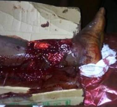
This clinical photograph displays a severe open fracture of the distal right lower leg (tibia and fibula), classified as a Gustilo-Anderson Type III injury. The image shows extensive soft tissue disruption with significant skin loss and exposed underlying musculature. Profuse active bleeding and dark clotted blood cover the wound site and surrounding areas. The anatomical region involved is the distal third of the leg extending toward the ankle joint. Visible in the immediate environment are improvised medical supports, including a blood-stained cardboard box and soiled white gauze, indicating high-energy trauma management in an initial presentation setting. Key educational concepts illustrated include the assessment of complex orthopedic trauma, classification of open fractures, and identification of severe soft tissue compromise associated with long bone fractures. This visual is relevant for surgical training in orthopedics and emergency medicine regarding the triage and initial stabilization of high-grade musculoskeletal injuries.
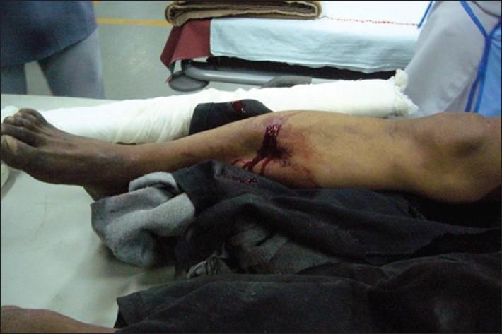
This clinical photograph displays a high-energy trauma injury to the right lower extremity of a patient in an emergency department setting. The image demonstrates a significant open (compound) fracture of the mid-shaft tibia and fibula. A large, transverse, irregular skin laceration is visible on the anterior-medial aspect of the leg, with active hematoma and fresh blood tracking down the skin. There is evident soft tissue maceration and substantial deformity of the limb's anatomical alignment, suggesting a complete fracture. The patient's leg is positioned on a medical table covered with dark clothing/fabric. Distal to the injury site, the lower leg and ankle are partially supported by white cotton padding/bandaging, serving as a temporary stabilization or splint. Proximally, near the knee, a black support material or brace is visible. This image serves as a clinical example of severe orthopedic trauma requiring immediate irrigation, stabilization, and surgical intervention.
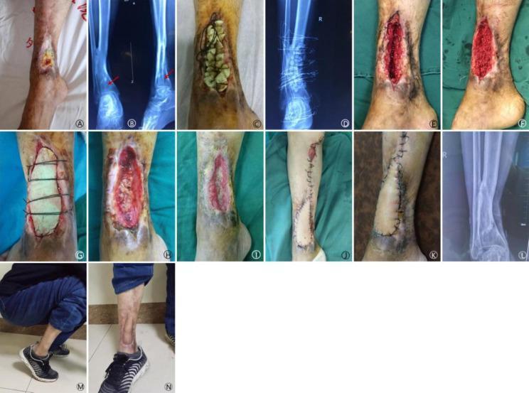
A multi-panel clinical photograph and radiographic sequence documenting the 15-month treatment progression of a 52-year-old male with a chronic open fracture and sinus tract of the right distal tibia. The series begins with (A-B) initial presentation showing a distal tibial wound and X-ray evidence of a metaphyseal bone defect. Subsequent panels (C-G) illustrate Masquelet-like technique stages, including the implantation of antibiotic-impregnated cement beads, followed by autologous bone grafting (iliac bone) and application of a bone cement sheet (BCS). Intermediate stages (H-I) demonstrate the debridement of necrotic graft particles and the formation of granulation tissue. Reconstructive surgery is shown in (J-K) via a medial leg flap for soft tissue coverage. The final panels (L-N) provide a 15-month follow-up, showing radiographic bone union, successful wound closure, and restored functional mobility (ankle dorsiflexion). This educational sequence highlights orthopedic management of complex bone defects, staged reconstruction using spacers/grafts, and soft tissue flap coverage.
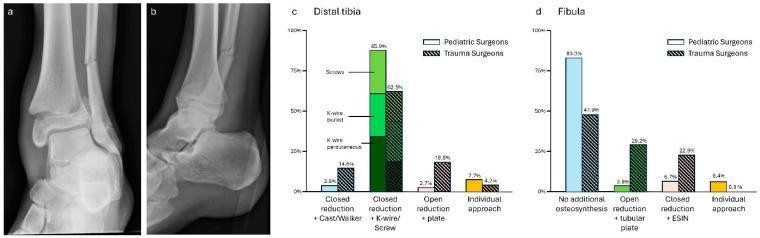
Educational material regarding pediatric trauma management, featuring diagnostic imaging and treatment preference data. (a) Anterior-posterior and (b) lateral X-ray views of a 15-year-old male demonstrate a displaced Salter-Harris Type 2 fracture of the distal tibia and a concomitant distal fibula shaft fracture. The growth plate is clearly visible, signifying skeletal immaturity. The distal tibia fracture shows a characteristic metaphyseal 'Thurston Holland' fragment and transverse extension through the physis. Graphs (c) and (d) provide a comparison of surgical preferences between pediatric surgeons and trauma surgeons for managing these specific injuries. For the distal tibia (c), pediatric surgeons favored closed reduction with percutaneous or buried K-wires, while trauma surgeons showed a higher preference for open reduction and internal fixation with plates. For the fibula (d), pediatric surgeons predominantly opted for no additional osteosynthesis, whereas trauma surgeons more frequently recommended open reduction with tubular plates or closed reduction with Elastic Stable Intramedullary Nailing (ESIN). The content illustrates the clinical intersection of pediatric orthopedics and general trauma surgery.
"distal tibia" AND "open fracture"
PMID: 36917286
Compound Fracture of the Distal Tibia: A Comprehensive Review
1. Definition and Terminology
- Distal tibial shaft fractures (metaphyseal-diaphyseal junction, lower third)
- Pilon (tibial plafond) fractures - intra-articular fractures of the distal tibial weight-bearing surface
- Distal tibial metaphyseal fractures without articular involvement
2. Anatomy and Why This Region Is Vulnerable
- Thin medial skin envelope: The anteromedial cortex is nearly subcutaneous, so even low-energy injuries can breach the skin
- Watershed vascular territory: The distal tibia is the most poorly vascularized zone of the tibia, predisposing to nonunion and avascular bone loss
- Adjacent ankle joint: Intra-articular extension threatens articular cartilage and long-term joint function
- Compartments: Four compartments of the leg run to the ankle; swelling can produce compartment syndrome even in open injuries
3. Mechanisms of Injury
| Mechanism | Fracture Type | Energy |
|---|---|---|
| Road traffic accident (direct blow) | Transverse/oblique shaft | High |
| Falls from height (axial load) | Pilon fracture | High |
| Sports/twisting injury | Spiral shaft | Low-medium |
| Gunshot / blast | Comminuted, segmental | Extreme |
| Industrial crush injury | Comminuted + skin loss | High |
4. Classification
4.1 Gustilo-Anderson Classification (the primary system for open fractures)
| Grade | Description |
|---|---|
| Type I | Low-energy, wound < 1 cm, clean |
| Type II | Laceration > 1 cm, no extensive soft-tissue damage, flaps, or avulsion |
| Type III | High-energy, extensive damage to soft tissue including muscle, skin and neurovascular structures; high contamination; unstable fracture |
| Type IIIA | Adequate soft-tissue cover after stabilisation |
| Type IIIB | Inadequate soft-tissue cover; flap coverage required |
| Type IIIC | Open fracture with an arterial injury requiring repair |
- Bailey and Love's Short Practice of Surgery 28th Ed, Table 32.2
4.2 OTA/AO Open Fracture Classification (OTA-OFC)
4.3 AO/OTA Fracture Classification (Bone morphology)
- 42- = tibial shaft (lower third when metaphyseal extension)
- 43- = distal tibia (metaphyseal)
- 43-C = intra-articular (pilon) - most severe, subdivided C1 (simple), C2 (metaphyseal comminution), C3 (complete articular comminution)
4.4 Ruedi-Allgower Classification (Pilon fractures)
| Type | Description |
|---|---|
| I | Cleavage fracture, no displacement |
| II | Displacement with minimal comminution |
| III | Comminuted articular surface with impaction |
5. Clinical Presentation
History
- Mechanism: RTA, fall, crush, sports
- Time since injury (critical for antibiotic timing)
- Contamination (agricultural, urban, water)
- Tetanus immunisation status
- Comorbidities: diabetes, vascular disease, smoking, alcohol (all significantly increase infection risk)
Physical Examination
- Wound assessment: Size, depth, contamination, skin loss
- Limb deformity: Shortening, angulation, rotation
- Neurovascular exam: Posterior tibial and dorsalis pedis pulses, capillary refill, sensation in the foot (posterior tibial, superficial peroneal, deep peroneal, sural nerve territories)
- Compartment syndrome signs: Pain out of proportion, pain on passive stretch, firmness of compartments, paraesthesiae
- Skin blistering: Fracture blisters are common and affect surgical planning
6. Investigations
Radiographs
- AP and lateral of the ankle (2 views minimum)
- Full-length tibia/fibula views
- Rule of "2s": two views, two joints (above and below), two occasions (before and after manipulation)
CT Scan
- Essential for pilon fractures to define articular comminution, impaction, and fragment geometry
- Guides surgical approach and fixation strategy
- CT angiography if vascular injury suspected
Doppler/Angiography
- If ABI < 0.9 or asymmetric pulses - formal vascular assessment required
- Type IIIC fractures need urgent vascular surgery input
MRI
- Not routinely indicated acutely; may be used for soft-tissue mapping or occult injuries
7. Emergency Department Management
- Haemorrhage control - pressure dressings; tourniquet only as a last resort due to potential for further nerve and limb damage
- Wound photography and coverage - photograph wound once, then cover with saline-soaked gauze; avoid repeated re-examination to limit contamination
- Splinting - protect the extremity with sterile dressings and appropriate splinting until surgery
- Early antibiotic prophylaxis - begin as soon as possible (see Section 8)
- Tetanus prophylaxis
- Compartment syndrome recognition - watch for the 5 Ps; may require emergent fasciotomy
The limited anteromedial soft tissue envelope of the tibia makes open fractures more common here than in other locations. Infection and nonunion are more common with open tibia fractures. - Rockwood and Green's Fractures in Adults 10th ed
8. Antibiotic Prophylaxis
| Gustilo Type | Antibiotic Regimen |
|---|---|
| I and II | First-generation cephalosporin (e.g., cefazolin 2g IV) |
| III | Add gram-negative cover: aminoglycoside (gentamicin) or piperacillin-tazobactam |
| Farm/soil contamination | Add penicillin for Clostridium (gas gangrene risk) |
- Duration: 24-72 hours post-debridement for most types
- Iodine skin prep has been shown more effective than chlorhexidine in preventing surgical site infections in open fractures - Campbell's Operative Orthopaedics 15th ed
9. Surgical Management
9.1 Irrigation and Debridement (I&D)
- Thorough removal of devitalized tissue, foreign material, and contamination
- Copious irrigation (normal saline; low-pressure pulsed lavage preferred; high-pressure lavage may drive bacteria deeper)
- Serial debridements may be necessary for heavily contaminated wounds (every 48-72 hours until clean)
- Preserve all potentially viable bone fragments when possible, especially in the metaphysis
9.2 Fixation Strategies
Distal Shaft / Metaphyseal Fractures (non-articular)
- Most common treatment for open tibia fractures
- Infection rate ~3% and union rate ~89% for lower-grade open fractures with primary nailing after I&D
- The SPRINT trial showed reaming is safe in open tibia fractures
- Controversy exists for high-grade IIIB/IIIC injuries where some prefer external fixation initially
- For severe cases, infection rates of 16% and complication rates 33-57% (LEAP study)
- Preferred for: Gustilo IIIB/IIIC, severe contamination, significant bone loss, haemodynamically unstable patients, damage-control orthopaedics
- A delta frame or circular (Ilizarov) fixator spans the ankle
- Can be used as definitive treatment or as a bridge to plate fixation once soft tissues settle
- A recent multicenter RCT comparing ringed fixators to IM nailing for high-grade open tibia fractures found no difference in deep infection rate, but higher overall complications with ring fixators
- MIPO (Minimally Invasive Plate Osteosynthesis) - used for distal metaphyseal patterns
- Locking plates through small percutaneous incisions reduce soft-tissue stripping
Pilon Fractures (articular, AO 43-C)
- Emergency I&D
- Temporary spanning external fixation across the ankle joint (restores length, alignment, and stability while protecting soft tissues)
- Allows swelling to resolve before definitive fixation (typically 7-21 days)
- CT-guided ORIF: reconstruction of the articular surface using lag screws and buttress/locking plates
- Classic approach: fibula fixation first (restores lateral column length) then medial/anterior tibial plate
- Multiple approach options: anterolateral, anteromedial, posteromedial
- Indicated for: severe soft-tissue injury (IIIB/IIIC), elderly patients with osteoporosis, poor surgical candidates
- Recent systematic review (Necula et al., 2024) supports its role in high-energy distal tibia fractures with soft-tissue compromise
10. Soft-Tissue Reconstruction
| Soft-Tissue Defect | Reconstruction Option |
|---|---|
| Primary closure possible | Direct closure at initial or 48-72 h debridement |
| Small-medium defect | Split-thickness skin graft (STSG) |
| Exposed tendon/bone (no periosteum) | Local flap: soleus or flexor hallucis longus (FHL) muscle flap |
| Large proximal defect | Gastrocnemius flap |
| Large distal or complex defect | Free flap (anterolateral thigh, gracilis, or latissimus dorsi) |
- Widely used as a temporary wound management tool in high-energy open tibial shaft fractures between debridements
- Reduces oedema, promotes granulation, and protects against further contamination
11. Clinical Images
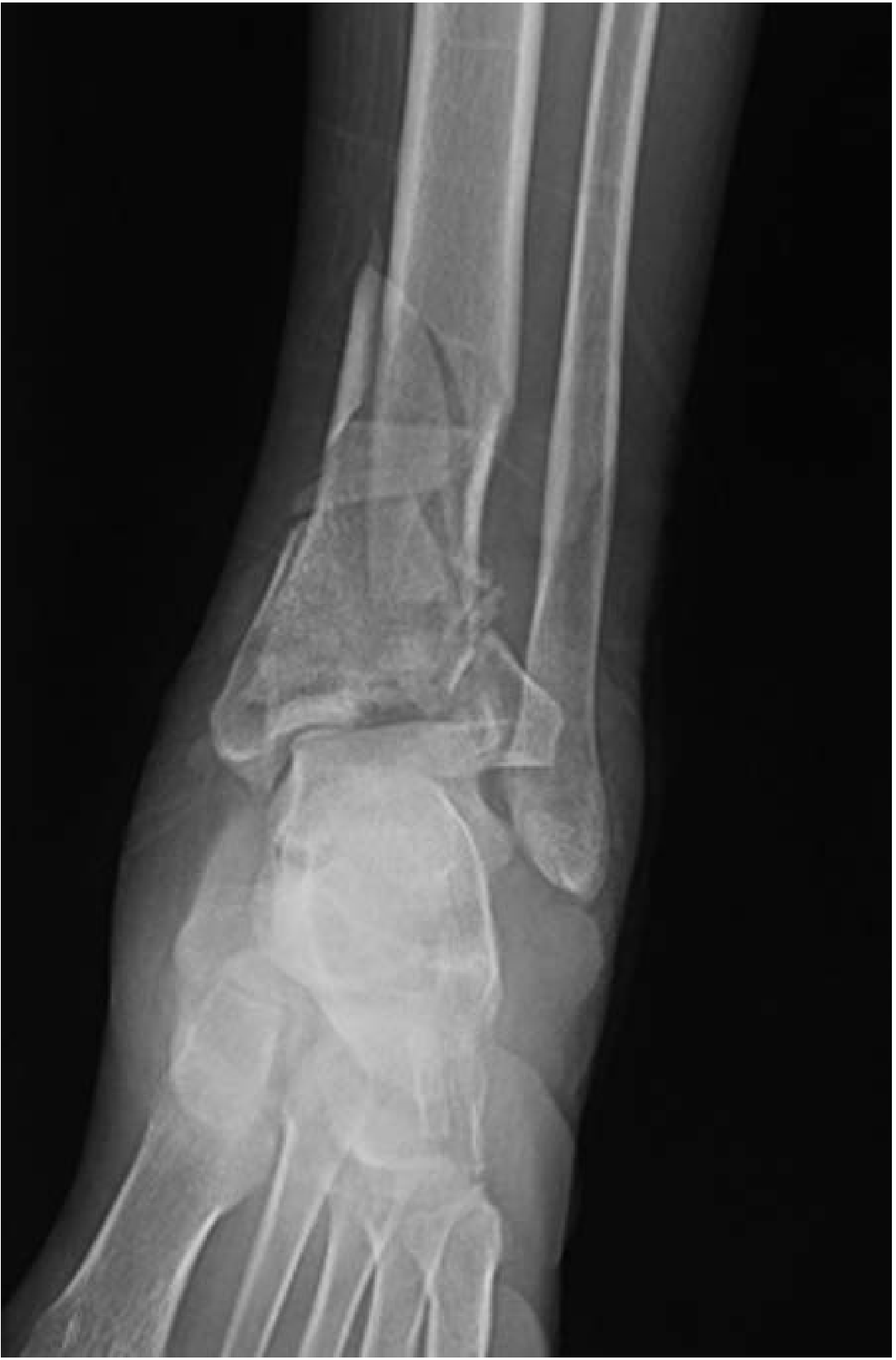
12. Complications
| Complication | Incidence | Notes |
|---|---|---|
| Infection / Osteomyelitis | 3-16% (grade dependent) | Most feared; often requires prolonged IV antibiotics, re-debridement, bone grafting |
| Nonunion | 10-15% | Higher in open, contaminated, and high-grade fractures |
| Malunion | Variable | Varus/valgus, shortening, rotational deformity |
| Compartment Syndrome | ~10% with open tibial fractures | Paradoxically can occur even with an open wound; requires fasciotomy |
| Post-traumatic Arthritis | Common with pilon fractures | Related to degree of articular comminution and quality of reduction |
| Wound dehiscence | Significant in distal tibia | Related to the poor soft-tissue envelope |
| Amputation | ~5-15% in IIIC fractures | Decision guided by mangled extremity severity score (MESS) |
| Bone loss / Defect | Requires staged reconstruction | Masquelet technique, bone transport (Ilizarov), or vascularised fibula graft |
13. The DANGER Score (Infection Risk Prediction)
| Letter | Factor |
|---|---|
| D | Diabetes mellitus |
| A | Antibiotic timing / adequacy |
| N | Nature of trauma (high vs. low energy) |
| G | Grade of fracture (AO/OTA classification) |
| E | Exposure severity (Gustilo-Anderson grade) |
| R | Relative risk (smoking, alcoholism, psychiatric disorders) |
- Total score 1-14; higher score = higher SSI risk
- Threshold of 7.5 distinguishes high-risk from low-risk patients (AUC on ROC analysis)
- Mean DANGER score was 8.2 in patients who developed SSI vs. 4.8 in those who did not (p<0.001)
14. Prognosis and Outcomes
- Closed distal tibial fractures generally do well with appropriate management
- Low-grade open fractures (I-II): Infection ~3%, union ~89%
- High-grade open fractures (IIIA-IIIC): Infection 9-16%, complication rates 33-57%
- Pilon fractures: Even with anatomic reduction, post-traumatic arthritis of the ankle is common; many eventually require ankle arthrodesis
- Outcome is heavily influenced by: energy of injury, Gustilo grade, degree of articular involvement, soft-tissue reconstruction, time to definitive fixation, and patient factors (diabetes, smoking, vascular disease)
15. Summary Algorithm
Compound Distal Tibia Fracture
         |
         ├── Emergency: ABCDE, haemostasis, splint, photograph wound
         ├── IV antibiotics (within 1-3 hours of injury)
         ├── Tetanus prophylaxis
         |
         ├── IMAGING: X-ray AP + lateral ± CT (always for pilon)
         |
         ├── NON-ARTICULAR (shaft/metaphysis)
         |       └── I&D → IM nail (most cases)
         |               → Ex-fix if IIIB/IIIC or contaminated
         |
         └── ARTICULAR (pilon)
                 ├── STAGE 1: I&D + spanning external fixator
                 ├── Soft tissue optimisation (5-21 days)
                 └── STAGE 2: ORIF (plates + screws) or circular fixator
- Rockwood and Green's Fractures in Adults, 10th ed (2025) - Open fracture classification
- Campbell's Operative Orthopaedics, 15th ed (2026) - Tibial shaft and pilon management
- Bailey and Love's Short Practice of Surgery, 28th ed - Gustilo-Anderson classification, Table 32.2
- Tintinalli's Emergency Medicine - Pilon fracture mechanism
- Messori et al. (2023). The DANGER score for SSI prediction in open distal tibial fractures. Eur J Orthop Surg Traumatol
- Necula et al. (2024). Orthoplastic reconstruction using circular external fixator for distal tibia high-energy fractures - Systematic Review. J Clin Med