
what is sperm
sperm cell anatomy diagram structure
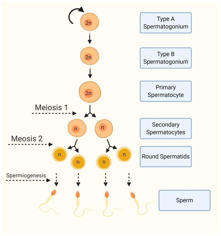
An educational pathophysiology diagram illustrating the stages of human spermatogenesis and the progression of germ cell differentiation. The flowchart follows a vertical linear-to-branched structure. The process begins with diploid (2n) germ cells: Type A Spermatogonium, which shows a self-renewal loop, followed by Type B Spermatogonium and Primary Spermatocyte. The diagram then depicts Meiosis I, transitioning from one primary spermatocyte to two haploid (n) Secondary Spermatocytes. Meiosis II follows, leading to the formation of four haploid (n) Round Spermatids. The final stage, labeled Spermiogenesis, demonstrates the morphological transformation from round cells into mature Sperm (spermatozoa), characterized by the development of a distinct head and flagellum. The diagram emphasizes key cellular events, including chromosomal reduction from diploid to haploid states and the structural evolution of the male gamete, making it a central resource for understanding male reproductive biology and gametogenesis.
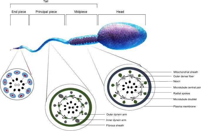
Anatomical diagram and comparison chart illustrating the structure of a mature human spermatozoon. The top section shows a longitudinal view of the sperm, segmented into the Head (containing the nucleus) and the Tail, which is further divided into the Midpiece, Principal piece, and End piece. Below the main illustration are three detailed cross-sectional diagrams of the flagellum (axoneme) at different tail levels. The Midpiece cross-section highlights the central '9+2' microtubule arrangement (nine microtubule doublets and a central pair) surrounded by outer dense fibers and a mitochondrial sheath. The Principal piece cross-section demonstrates the axoneme enclosed by a fibrous sheath. The End piece cross-section shows the simplified 9+2 microtubule structure within the plasma membrane. Labels identify critical ultra-structural components including nexin links, radial spokes, and inner and outer dynein arms. This diagram serves as an educational resource for reproductive biology and andrology, emphasizing the mechanical components necessary for sperm motility and male fertility.
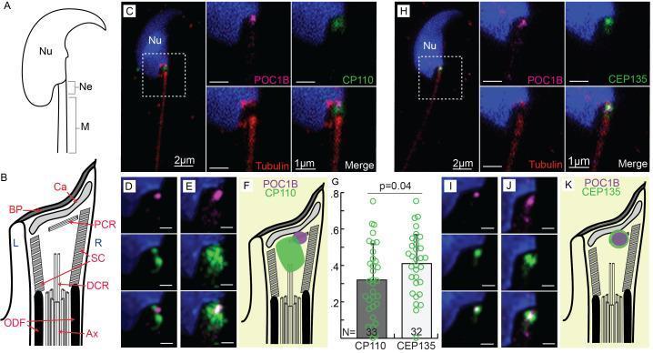
This composite educational graphic illustrates the ultrastructure and protein localization within the mouse sperm neck (centriolar adjunct). Panel A provides an anatomical diagram of a murine sperm, identifying the nucleus (Nu), neck (Ne), and midpiece (M). Panel B details the complex neck anatomy, including the basal plate (BP), capitulum (Ca), proximal and distal centriolar remnants (PCR, DCR), striated column (SC), and axoneme (Ax). Panels C-E and H-J utilize high-resolution immunofluorescence and HyVolution microscopy to demonstrate the spatial organization of centriolar proteins. The nucleus is stained blue (Hoechst), while red (anti-tubulin) marks the axoneme. Specific protein markers POC1B (magenta), CP110 (green, C-E), and CEP135 (green, H-J) are shown localized at the neck region. Panels F and K provide schematic interpretations of these protein signals relative to the neck structures. A bar graph (G) quantifies the colocalization coefficients of POC1B with CP110 and CEP135, indicating significant spatial proximity. This content is designed for advanced study of reproductive cell biology and centriole remodeling during spermiogenesis.
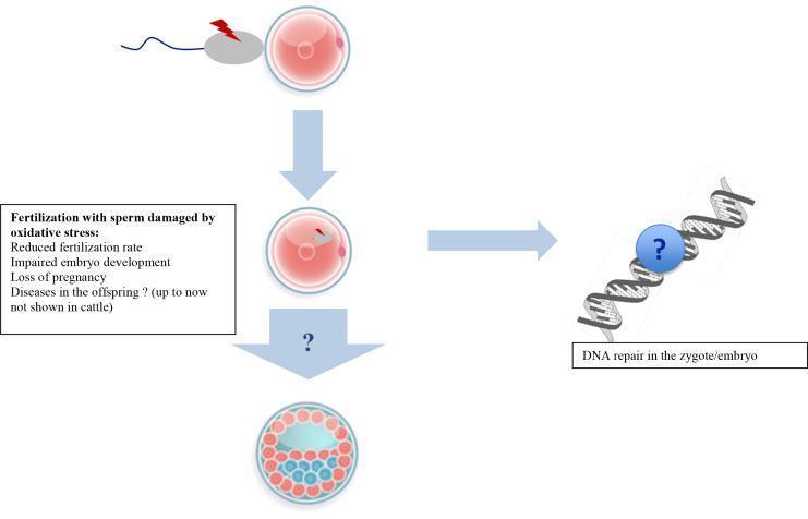
A pathophysiology diagram illustrating the effects of oxidative stress on sperm and subsequent embryo development. The vertical flow on the left depicts fertilization stages: 1) Initial contact between an oocyte and a sperm cell marked with a red lightning bolt symbol, indicating oxidative damage; 2) Incorporation of the damaged paternal genome into the oocyte (zygote stage); and 3) Progression to a blastocyst-like multicellular structure with differentiated cell clusters. A text box lists clinical outcomes of fertilization with oxidatively stressed sperm, including reduced fertilization rates, impaired embryo development, and pregnancy loss. A horizontal arrow from the zygote stage points to a DNA double helix illustration on the right, which is highlighted with a question mark symbol and labeled 'DNA repair in the zygote/embryo'. This visual highlights the uncertainty and critical role of maternal or embryonic DNA repair mechanisms in mitigating paternal oxidative damage. The diagram serves as an educational summary of male-factor infertility mechanisms and developmental biology.
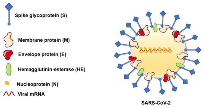
This medical illustration depicts the structural anatomy of a SARS-CoV-2 virion. The diagram shows a spherical viral particle with its major structural components identified by a color-coded legend. The external surface is dominated by blue, diamond-headed spike glycoproteins (S), which are distributed around the viral envelope and are responsible for host cell entry. Interspersed among the spikes are tan membrane proteins (M), which maintain viral shape; red envelope proteins (E), involved in assembly and release; and green hemagglutinin-esterase (HE) proteins. Internally, the yellow viral core contains a central line of yellow bead-like nucleoproteins (N) associated with a red wavy line representing the viral mRNA genome. This educational diagram is designed for virology and infectious disease studies, illustrating the key structural targets for antiviral therapies and vaccines, such as the S protein. It clearly differentiates between the superficial glycoproteins and the internal nucleocapsid structure.
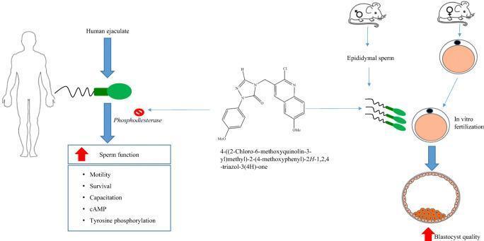
A graphical abstract summarizing a pharmacological study on sperm function and embryogenesis. The diagram illustrates a novel quinoline derivative, identified by the chemical name 4-((2-Chloro-6-methoxyquinolin-3-yl)methyl)-2-(4-methoxyphenyl)-2H-1,2,4-triazol-3(4H)-one, which acts as a phosphodiesterase (PDE) inhibitor. The pathway shows that the application of this compound to human ejaculate leads to enhanced sperm function, specifically increasing motility, survival, capacitation, intracellular cAMP levels, and tyrosine phosphorylation. In parallel, a mouse model demonstrates that the treatment of epididymal sperm facilitates in vitro fertilization (IVF), ultimately resulting in blastocyst formation with improved quality, as indicated by red up-arrows. Key visual elements include the chemical structure of the quinoline-triazole hybrid, silhouettes of human and murine figures, anatomical representations of spermatozoa and oocytes, and a schematic of a blastocyst with an inner cell mass. The content is relevant to reproductive medicine, andrology, and pharmacology, focusing on the development of synthetic PDE inhibitors to improve assisted reproductive outcomes.
What is Sperm?
Structure of a Sperm Cell
1. Head
- Contains the nucleus with tightly compacted DNA (condensed to about 10% of its original size)
- Capped by the acrosome - a membrane-bound vesicle derived from the Golgi apparatus that covers the tip of the head
- The acrosome contains enzymes needed to penetrate the egg's protective zona pellucida
2. Midpiece (Neck)
- Contains a spiral arrangement of mitochondria that provide the energy (ATP) to power the tail's movement
3. Tail (Flagellum)
- A long whip-like structure built around the "9+2" axoneme - nine outer microtubule doublets surrounding a central pair
- Outer dense fibers and dynein arms generate the beating motion for swimming
- Divided into: midpiece, principal piece, and end piece
How Sperm Are Made: Spermatogenesis
- Spermatogonia (diploid stem cells) divide by mitosis to produce progenitor cells
- Primary spermatocytes (diploid, 46 chromosomes) undergo Meiosis I to produce secondary spermatocytes
- Secondary spermatocytes (haploid) undergo Meiosis II to produce spermatids
- Spermiogenesis: Round spermatids transform into mature sperm cells through:
- Formation of the acrosome from the Golgi body
- Formation of the flagellum from the centriole
- Reorganization of mitochondria around the midpiece
- Extensive nuclear compaction
Sperm Transport and Capacitation
- A man typically deposits 150-600 million sperm per ejaculation into the vagina; only 50-100 reach the fallopian tube where fertilization occurs
- Sperm reach the fallopian tube within ~5 minutes, aided by uterine contractions (not just swimming alone)
- In the female tract, sperm undergo capacitation - a maturation process in which a protective protein coat is removed, allowing sperm to penetrate the zona pellucida of the egg - Medical Physiology
Fertilization
- Attach to the zona pellucida surrounding the egg
- Undergo the acrosomal reaction - releasing enzymes to digest through the zona
- Fuse with the egg cell membrane
- The sperm nucleus enters the egg, and the two haploid nuclei (pronuclei) fuse to form a diploid zygote with 46 chromosomes
Key Facts
| Feature | Value |
|---|---|
| Size | ~60 micrometers long |
| Chromosomes | 23 (haploid) |
| Production site | Seminiferous tubules of testes |
| Lifespan in female tract | Up to 5 days |
| Normal count | >15 million/mL (WHO threshold) |
| Motility needed for fertility | >40% motile |