
Congenital diaphragmatic hernia in detail
I'll start by reading the medical library skill, then search for comprehensive information on congenital diaphragmatic hernia.
Reading File
Now I'll run parallel searches across the library for CDH content:
Finding Sources
Finding Sources
Searching PubMed
"congenital diaphragmatic hernia"[MeSH Terms]
Excellent - I have strong hits from multiple textbooks. Now I'll read the most comprehensive sources in parallel:
Reading File
Reading File
Reading File
Excellent content. Now let me read the Sabiston block 29 for management, the Tintinalli emergency section, and the Creasy block4 for fetal interventions:
Reading File
Reading File
Reading File
Let me get the postnatal management section from Sabiston and Miller's anesthesia section, plus read the CDH chest X-ray image:
Reading File
Reading File
Loading Image
I now have comprehensive content from multiple authoritative textbooks. Let me fetch one more image and get the postnatal management details:
Reading File
Loading Image
I now have all the information needed for a comprehensive, well-cited answer. Let me compile it:
Congenital Diaphragmatic Hernia (CDH)
Definition and Epidemiology
Congenital diaphragmatic hernia (CDH) is a spectrum of developmental conditions marked by a defect in the diaphragm that allows abdominal contents to herniate into the thoracic cavity, disrupting lung and pulmonary vascular development. The reported incidence is approximately 1:2,000 to 5,000 live births, with most cases sporadic, isolated, and non-syndromic.
- Left-sided (Bochdalek): 85% of cases
- Right-sided: 13%
- Bilateral or complete agenesis: <2%
- Isolated CDH (no associated anomalies): ~60% of cases; if associated with other anomalies, mortality exceeds 85%
(Sabiston Textbook of Surgery, p. 2675; Creasy & Resnik's Maternal-Fetal Medicine, p. 776)
Embryology and Types
The diaphragm develops embryologically from four structures:
- Septum transversum
- Pleuroperitoneal folds
- Components of the abdominal wall
- Dorsal mesentery
These begin fusing at 3-4 weeks gestation, separating the pleural and peritoneal cavities. Muscular ingrowth is complete by ~9 weeks gestation. Incomplete fusion at different locations results in distinct hernia types:
| Type | Location | Frequency |
|---|---|---|
| Bochdalek | Posterolateral | 70-75% |
| Morgagni | Anterior/retrosternal | 23-28% |
| Central | Central tendon | 2-7% |
Bochdalek hernias are most common on the left side (85%) because the right side is protected by the early-developing liver. Morgagni hernias are often asymptomatic until childhood or adulthood.
(Sabiston Textbook of Surgery, p. 2675)
Pathophysiology
Once abdominal organs herniate through the defect (during the 2nd trimester), a cascade of consequences occurs:
Pulmonary Hypoplasia
- The ipsilateral lung is compressed and undergoes reduced bronchial branching and reduced alveolar surface area
- The contralateral lung is also affected by compression and intrinsic genetic factors (e.g., FOG2, GATA4 mutations)
- Lungs have smaller bronchi, fewer airway branches, and less alveolar area on both sides
Pulmonary Hypertension
- Pulmonary vascular smooth muscle is hypertrophied, with increased arteriolar wall thickness
- Arteriolar vasculature is hypersensitive to vasoactive stimuli
- This leads to right-to-left shunting (persistent fetal circulation) at the foramen ovale and ductus arteriosus
Cardiac Effects
- Mediastinal shift compresses the contralateral lung and distorts cardiac anatomy
- Significant congenital heart defects co-occur in 25-50% of CDH patients
- Left ventricular hypoplasia may occur from reduced preload
Genetic Associations
- Trisomies, microdeletions, and single gene defects are all possible
- Array CGH detects abnormalities in ~6.3% of CDH cases
- Associated syndromes: Turner syndrome, Fryns syndrome, VACTERL association
(Bailey & Love's Surgery, p. 1763; Creasy & Resnik's, p. 776; Sabiston, p. 2675)
Prenatal Diagnosis and Imaging
Ultrasound screening has led to prenatal diagnosis in roughly two-thirds of cases, sometimes as early as 15 weeks gestation.
Sonographic Features:
- Visualization of stomach, bowel, or liver in the thorax
- Rightward cardiac/mediastinal shift (in left CDH)
- Polyhydramnios
- Absence of the normal stomach bubble below the diaphragm
Right-sided CDH is harder to detect: the liver's echogenicity mimics fetal lung. Doppler of the umbilical vein, hepatic vessels, and gallbladder position are used as landmarks. Cardiac compression and polyhydramnios are indirect signs.
Differential Diagnoses (Prenatal):
- Congenital pulmonary airway malformation (CPAM)
- Bronchopulmonary sequestration
- Bronchogenic/enteric cysts
- Mediastinal teratoma
In all these conditions, intraabdominal organs are NOT displaced.
Upon diagnosis, referral to a tertiary center is mandatory for advanced imaging (MRI for fetal lung volumes), genetic testing, echocardiography, and multidisciplinary counseling.
(Creasy & Resnik's, p. 776)
Prognostic Assessment
Two key prenatal prognostic tools:
1. Lung-to-Head Ratio (LHR)
The contralateral lung area is divided by the head circumference at the level of the atria.
| LHR | Prognosis |
|---|---|
| <1.0 | Poor (severe pulmonary hypoplasia) |
| 1.0-1.4 | Moderate |
| >1.4 | Near 100% survival |
2. Observed-to-Expected LHR (O/E LHR)
Corrects LHR for gestational age using normal reference values:
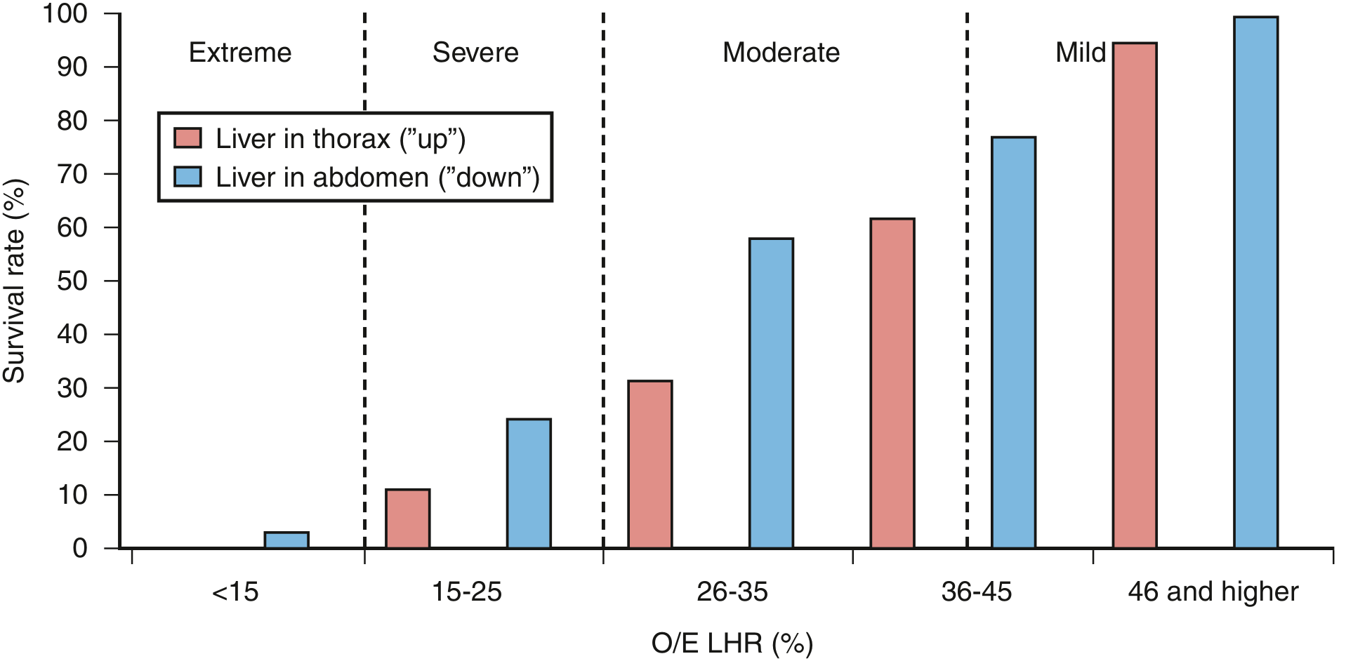
Figure: Relationship between O/E LHR, liver herniation, and survival. Liver "up" (in thorax) consistently worsens prognosis at every LHR category. (Creasy & Resnik's Maternal-Fetal Medicine)
| O/E LHR | Severity | Survival (expectant) |
|---|---|---|
| <15% | Extreme | ~0-3% |
| 15-25% | Severe | ~11-25% |
| 26-35% | Moderate | 31-58% |
| 36-45% | Moderate-mild | 62-77% |
| >46% | Mild | ~95-100% |
Liver herniation ("liver up") independently worsens prognosis at every severity level.
(Creasy & Resnik's, p. 776; Sabiston, p. 2716)
Clinical Presentation (Postnatal)
Classic Neonatal Presentation:
- Persistent respiratory distress at birth
- "Seesaw" or "rocking" respiratory pattern due to severely hypoplastic ipsilateral lung
- Persistent cyanosis and oxygen desaturation
- Scaphoid abdomen (hallmark - abdominal organs are displaced into chest)
- Bowel sounds audible in the chest
- Displacement of heart tones away from side of hernia
- Preductal and post-ductal SpO2 difference indicating right-to-left shunting
Delayed Presentation (10-20% of cases): CDH may present after the first 24 hours with feeding difficulties, recurrent respiratory infections, or pneumonia. Morgagni hernias are often asymptomatic until childhood.
(Sabiston, p. 2675; Tintinalli's Emergency Medicine)
Radiological Findings
Chest X-ray (confirmatory):
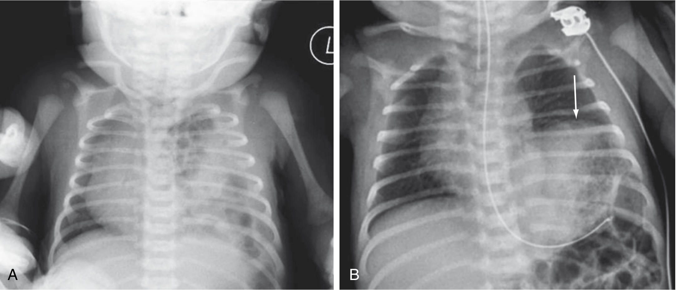
Figure: (A) Left CDH - multiple gas-filled bowel loops in the left hemithorax with mediastinal shift to the right. (B) Left diaphragmatic eventration for comparison (elevated hemidiaphragm, arrow). (Sabiston Textbook of Surgery)
Key X-ray findings:
- Gas-filled bowel loops within the hemithorax
- Mediastinal/cardiac shift to the contralateral side
- Absence or reduction of bowel gas in the abdomen
- Absence of visible diaphragmatic outline
(Sabiston, p. 2675; Grainger & Allison's Diagnostic Radiology)
Management
A. Emergency Stabilization (Birth/ER)
Critical "Do NOT" rule: Bag-mask ventilation is contraindicated - it inflates GI contents in the chest and further compromises ventilation.
Immediate steps:
- Immediate endotracheal intubation - avoid bag-mask ventilation
- Large-bore (10F) orogastric tube on continuous low suction - decompress herniated bowel
- Gentle ventilation: rate 40-50 bpm, lowest peak inspiratory pressure that allows normal chest rise (avoid barotrauma to hypoplastic lungs)
- Target PaCO2 30-35 mmHg (permissive hypercapnia is also acceptable) to reduce pulmonary vascular resistance
- Pulse oximetry monitoring pre- and post-ductal
(Tintinalli's Emergency Medicine)
B. ICU Stabilization - "Gentilation" Protocol
The mainstay is aggressive management of pulmonary hypertension before surgical repair. Surgery does NOT directly correct pulmonary hypertension - the child must be stabilized first.
Ventilation Strategy:
- High-frequency oscillatory ventilation (HFOV) - preferred in severe cases
- Permissive hypercapnia (limiting peak inspiratory pressures)
- Target pH 7.35-7.45, preductal SpO2 >85%
Pulmonary Hypertension Management:
- Inhaled nitric oxide (iNO) - selective pulmonary vasodilator
- Sildenafil (PDE-5 inhibitor) - oral/IV
- Prostacyclin (PGI2) and PGE1 (prostaglandins)
- Milrinone - inotrope + pulmonary vasodilator
- Inotropes for cardiac dysfunction
ECMO (Extracorporeal Membrane Oxygenation):
Reserved for the most severe cases not responding to maximal medical therapy. Used as a bridge to surgical repair or as rescue therapy.
(Sabiston, p. 2676; Miller's Anesthesia; Bailey & Love's)
C. Fetal Intervention - FETO
Fetoscopic Endoluminal Tracheal Occlusion (FETO) is offered at specialized centers for severe CDH:
- A balloon is inserted into the fetal trachea under fetoscopic guidance between the vocal cords and carina
- Typically performed at 27-29 weeks gestation
- Tracheal occlusion prevents lung fluid egress → lung fluid accumulation → lung growth
- Balloon is retrieved fetoscopically or percutaneously at ~34 weeks
- If removal fails, an EXIT procedure at delivery ensures airway patency
TOTAL Trials Results:
- Severe CDH (O/E LHR <25%): FETO improved survival to 40% vs. 15% with expectant management (stopped early for efficacy)
- Moderate CDH (O/E LHR 25-35%): FETO at 30-32 weeks showed no survival benefit over expectant management
Risks of FETO: Preterm labor, premature rupture of membranes (PROM), preterm birth, fetal demise.
(Sabiston, p. 2716; Creasy & Resnik's, p. 776)
D. Surgical Repair
Timing: Surgery is delayed until pulmonary hypertension is controlled and the infant is hemodynamically stable. Unlike traumatic hernias, urgent reduction of bowel in CDH does NOT improve gas exchange.
Approach Options:
| Approach | Route |
|---|---|
| Open subcostal (transabdominal) | Most common for complex cases |
| Thoracoscopic | Minimally invasive option |
| Thoracotomy | Less common |
Technique:
- Herniated viscera (stomach, bowel, occasionally liver/spleen) are reduced back into the abdomen
- The diaphragmatic defect is assessed:
- Small defect: Primary repair with interrupted non-absorbable sutures
- Large defect: Patch repair using Gore-Tex (PTFE) or Silastic/conical patch
- A hernia sac may be present - it can be excised or plicated
- Chest drain placed to expand lung
Anesthesia Considerations:
- Avoid bag-mask ventilation pre-intubation
- High risk of pneumothorax (ipsilateral or contralateral) from barotrauma to hypoplastic lungs
- Avoid hypoxemia and excessive hypercapnia to prevent pulmonary hypertension crisis
- Deep general anesthesia to blunt stress response
(Sabiston, p. 2676; Bailey & Love's, p. 1787; Miller's Anesthesia)
Outcomes and Survival
- Overall survival: 65-90% at specialized centers
- Prenatal diagnosis is paradoxically associated with worse prognosis (larger defects are more visible prenatally)
- Total lung volume >45% of normal is predictive of survival
Chronic Morbidities in Survivors:
| System | Complication |
|---|---|
| Pulmonary | Bronchopulmonary dysplasia (BPD), recurrent respiratory infections, obstructive/restrictive lung disease |
| Cardiovascular | Persistent pulmonary hypertension, left ventricular hypoplasia |
| GI | Gastroesophageal reflux disease (GERD), feeding difficulties |
| Nutritional | Failure to thrive |
| Structural | Thoracic deformity, scoliosis after repair |
| Neurodevelopmental | Hearing loss, developmental delay |
(Creasy & Resnik's, p. 776; Sabiston, p. 2676)
Summary
| Feature | Key Points |
|---|---|
| Incidence | 1:2,000-5,000 births |
| Most common type | Left-sided Bochdalek (85%) |
| Core pathology | Pulmonary hypoplasia + pulmonary hypertension |
| Prenatal diagnosis | Ultrasound (2/3 of cases); confirmed by MRI |
| Key prognostic tool | O/E LHR + liver position |
| Emergency rule | NO bag-mask ventilation; intubate immediately |
| Medical Rx | iNO, sildenafil, HFOV, ECMO for severe cases |
| Fetal intervention | FETO for severe CDH (O/E LHR <25%) |
| Surgery | After stabilization; primary suture or patch repair |
| Survival | 65-90% at tertiary centers |
Sources:
- Sabiston Textbook of Surgery, 21st Ed., pp. 2675-2676, 2716
- Creasy & Resnik's Maternal-Fetal Medicine, pp. 776-778
- Bailey & Love's Short Practice of Surgery, 28th Ed., p. 1763
- Tintinalli's Emergency Medicine
- Miller's Anesthesia, 10th Ed.
- Pye's Surgical Handicraft, 22nd Ed.
This is a shared conversation. Sign in to Orris to start your own chat.