Cavernous sinus

Reading File
Finding Sources
Searching Images

cavernous sinus anatomy contents relations diagram
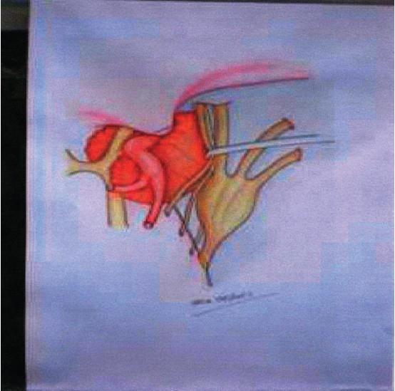
This medical anatomical diagram illustrates the surgical field during a pituitary adenoma resection, specifically highlighting the cavernous sinus (CS) anatomy and the fronto-orbito-zygomatic (FOZ) approach. The diagram depicts the intracavernous portion of a pituitary adenoma (rendered in reddish-orange) fully exposed. Centrally, a red vascular structure represents the internal carotid artery (ICA) and its proximal branches. To the right, a yellowish nerve structure representing the III cranial nerve (oculomotor nerve) is shown mobilized and laterally displaced using a surgical self-retaining microretractor. This maneuver widens the access through Hakuba’s triangle (anteromedial triangle), allowing direct visualization and removal of the tumor within the cavernous sinus. The illustration emphasizes the spatial relationship between the tumor mass, the protective pseudocapsule, the ICA, and the mobilized cranial nerves, which is critical for preventing neurovascular injury during complex skull base surgery.
This medical anatomical diagram illustrates the surgical field during a pituitary adenoma resection, specifically highlighting the cavernous sinus (CS) anatomy and the fronto-orbito-zygomatic (FOZ) approach. The diagram depicts the intracavernous portion of a pituitary adenoma (rendered in reddish-orange) fully exposed. Centrally, a red vascular structure represents the internal carotid artery (ICA) and its proximal branches. To the right, a yellowish nerve structure representing the III cranial nerve (oculomotor nerve) is shown mobilized and laterally displaced using a surgical self-retaining microretractor. This maneuver widens the access through Hakuba’s triangle (anteromedial triangle), allowing direct visualization and removal of the tumor within the cavernous sinus. The illustration emphasizes the spatial relationship between the tumor mass, the protective pseudocapsule, the ICA, and the mobilized cranial nerves, which is critical for preventing neurovascular injury during complex skull base surgery.

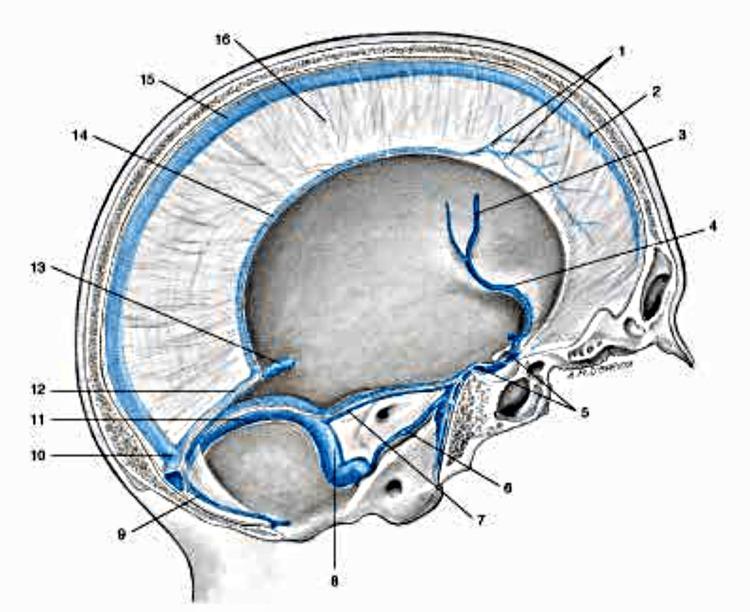
This medical anatomical diagram provides a sagittal perspective of the human cranium, illustrating the complex venous sinus system and dural membranes. The superior sagittal sinus is highlighted along the superior margin within the falx cerebri, receiving tributary veins from the dura mater. Inferiorly, the inferior sagittal sinus runs along the free margin of the falx, joining the great cerebral vein (of Galen) to form the straight sinus. Posteriorly, these structures converge at the confluence of sinuses, which connects to the transverse and occipital sinuses. The diagram further details the anterior and lateral drainage pathways, including the sphenoparietal sinus, cavernous sinus (with intercavernous connections), and the superior and inferior petrosal sinuses. These ultimately drain into the sigmoid sinus before exiting the skull as the internal jugular vein. This illustration is an essential educational tool for understanding intracranial venous anatomy, relevant to neurosurgery, radiology, and managing conditions like dural venous sinus thrombosis.
This medical anatomical diagram provides a sagittal perspective of the human cranium, illustrating the complex venous sinus system and dural membranes. The superior sagittal sinus is highlighted along the superior margin within the falx cerebri, receiving tributary veins from the dura mater. Inferiorly, the inferior sagittal sinus runs along the free margin of the falx, joining the great cerebral vein (of Galen) to form the straight sinus. Posteriorly, these structures converge at the confluence of sinuses, which connects to the transverse and occipital sinuses. The diagram further details the anterior and lateral drainage pathways, including the sphenoparietal sinus, cavernous sinus (with intercavernous connections), and the superior and inferior petrosal sinuses. These ultimately drain into the sigmoid sinus before exiting the skull as the internal jugular vein. This illustration is an essential educational tool for understanding intracranial venous anatomy, relevant to neurosurgery, radiology, and managing conditions like dural venous sinus thrombosis.

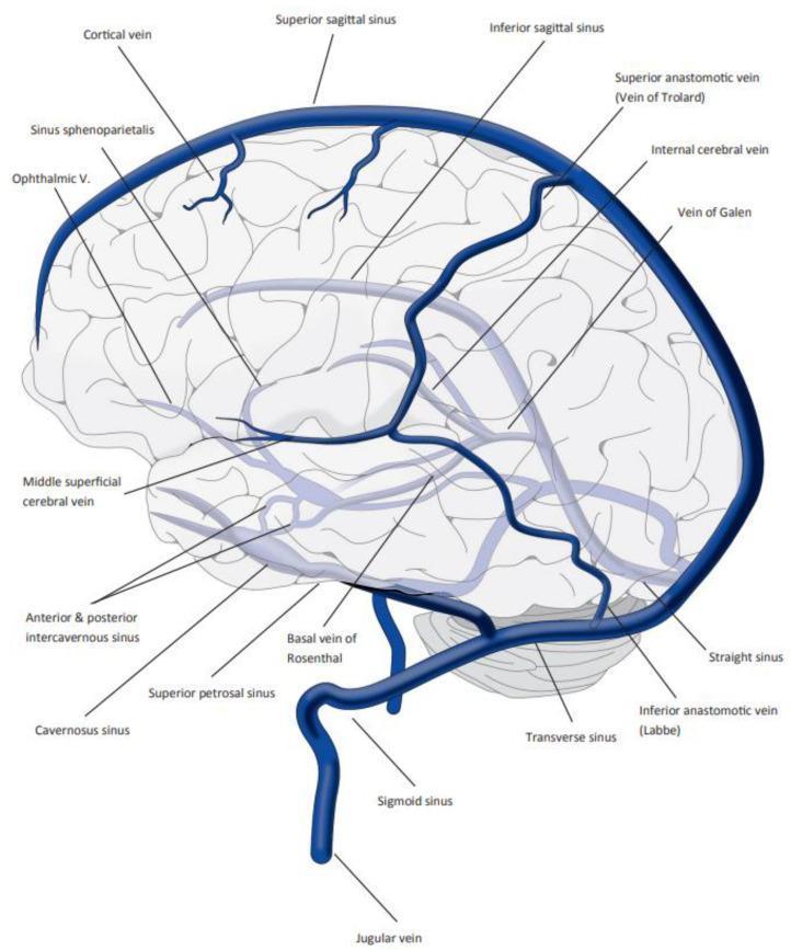
This medical anatomical diagram provides a lateral view of the human brain, illustrating the complex organization of the cerebral dural venous sinuses and deep cerebral veins. The illustration highlights the superficial and deep drainage systems through a network of blue-colored vessels overlaid on a stylized brain outline. Key structures labeled include the superior sagittal sinus along the longitudinal fissure, cortical veins, the inferior sagittal sinus, and the straight sinus leading to the vein of Galen. Anastomotic pathways are represented by the superior anastomotic vein (Vein of Trolard) and the inferior anastomotic vein (Vein of Labbé). The inferior drainage components shown include the cavernous sinus, anterior and posterior intercavernous sinuses, sphenoparietal sinus, ophthalmic vein, superior petrosal sinus, and the sigmoid sinus draining into the jugular vein. Deep structures like the basal vein of Rosenthal and internal cerebral veins are also depicted. This diagram serves as an educational reference for understanding cerebral venous anatomy, which is clinically significant for diagnosing conditions such as cerebral venous sinus thrombosis (CVST).
This medical anatomical diagram provides a lateral view of the human brain, illustrating the complex organization of the cerebral dural venous sinuses and deep cerebral veins. The illustration highlights the superficial and deep drainage systems through a network of blue-colored vessels overlaid on a stylized brain outline. Key structures labeled include the superior sagittal sinus along the longitudinal fissure, cortical veins, the inferior sagittal sinus, and the straight sinus leading to the vein of Galen. Anastomotic pathways are represented by the superior anastomotic vein (Vein of Trolard) and the inferior anastomotic vein (Vein of Labbé). The inferior drainage components shown include the cavernous sinus, anterior and posterior intercavernous sinuses, sphenoparietal sinus, ophthalmic vein, superior petrosal sinus, and the sigmoid sinus draining into the jugular vein. Deep structures like the basal vein of Rosenthal and internal cerebral veins are also depicted. This diagram serves as an educational reference for understanding cerebral venous anatomy, which is clinically significant for diagnosing conditions such as cerebral venous sinus thrombosis (CVST).

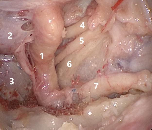
This endoscopic clinical photograph displays the microsurgical anatomy of the cavernous sinus and parasellar region, likely viewed via a transnasal transpterygoid approach. The central landmark is the cavernous segment of the internal carotid artery (1), shown as a prominent, vertically oriented vessel. To its medial side are the hypophysis (2) and the inferiorly positioned clivus (3). The lateral wall of the cavernous sinus is dissected to reveal several cranial nerves in their anatomical order. From superior to inferior, the oculomotor nerve (4), the abducens nerve (5), and the ophthalmic branch (V1) of the trigeminal nerve (6) are visible, following a longitudinal course. The maxillary branch (V2) of the trigeminal nerve (7) is seen most inferiorly and laterally, emerging toward the pterygopalatine fossa. The image serves as an educational reference for skull base surgery, illustrating the critical spatial relationships between vascular structures and cranial nerves during surgical decompression or tumor resection in Meckel's cave and the cavernous sinus.

This endoscopic clinical photograph displays the microsurgical anatomy of the cavernous sinus and parasellar region, likely viewed via a transnasal transpterygoid approach. The central landmark is the cavernous segment of the internal carotid artery (1), shown as a prominent, vertically oriented vessel. To its medial side are the hypophysis (2) and the inferiorly positioned clivus (3). The lateral wall of the cavernous sinus is dissected to reveal several cranial nerves in their anatomical order. From superior to inferior, the oculomotor nerve (4), the abducens nerve (5), and the ophthalmic branch (V1) of the trigeminal nerve (6) are visible, following a longitudinal course. The maxillary branch (V2) of the trigeminal nerve (7) is seen most inferiorly and laterally, emerging toward the pterygopalatine fossa. The image serves as an educational reference for skull base surgery, illustrating the critical spatial relationships between vascular structures and cranial nerves during surgical decompression or tumor resection in Meckel's cave and the cavernous sinus.

Reading File
Reading File
Reading File
Reading File
Finding Sources
Searching Images

cavernous sinus thrombosis carotid cavernous fistula clinical
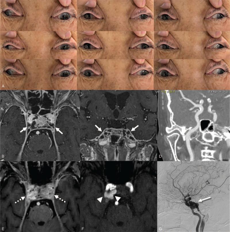
This composite figure illustrates the clinical and radiographic features of septic cavernous sinus thrombosis (SCST) progressing to a carotid-cavernous fistula (CCF). Panel A shows a series of nine cardinal gaze clinical photographs demonstrating bilateral eyelid swelling, incomplete ptosis, and restricted extraocular motility (ophthalmoplegia), notably bilateral exotropia with impaired medial and vertical gaze. Panels B and C are gadolinium-enhanced axial and coronal brain MRI images showing bilateral cavernous sinus expansion with prominent thrombotic filling defects (arrows). Panel D is a CT angiogram of the neck, used to screen for initial arterial involvement. Panel E (follow-up MRI) reveals persistent thrombosis and significant contrast enhancement/enlargement of the cavernous sinuses (dotted arrows). Panel F (Time-of-Flight MR Angiography) displays abnormal high-flow-related signal in the right cavernous sinus (arrowheads), diagnostic of a secondary CCF. Panel G (Digital Subtraction Angiography) confirms the fistula at the distal cavernous segment of the right internal carotid artery (shadowed arrow). The collection documents the evolution of infectious cavernous sinus disease into a vascular complication.
This composite figure illustrates the clinical and radiographic features of septic cavernous sinus thrombosis (SCST) progressing to a carotid-cavernous fistula (CCF). Panel A shows a series of nine cardinal gaze clinical photographs demonstrating bilateral eyelid swelling, incomplete ptosis, and restricted extraocular motility (ophthalmoplegia), notably bilateral exotropia with impaired medial and vertical gaze. Panels B and C are gadolinium-enhanced axial and coronal brain MRI images showing bilateral cavernous sinus expansion with prominent thrombotic filling defects (arrows). Panel D is a CT angiogram of the neck, used to screen for initial arterial involvement. Panel E (follow-up MRI) reveals persistent thrombosis and significant contrast enhancement/enlargement of the cavernous sinuses (dotted arrows). Panel F (Time-of-Flight MR Angiography) displays abnormal high-flow-related signal in the right cavernous sinus (arrowheads), diagnostic of a secondary CCF. Panel G (Digital Subtraction Angiography) confirms the fistula at the distal cavernous segment of the right internal carotid artery (shadowed arrow). The collection documents the evolution of infectious cavernous sinus disease into a vascular complication.

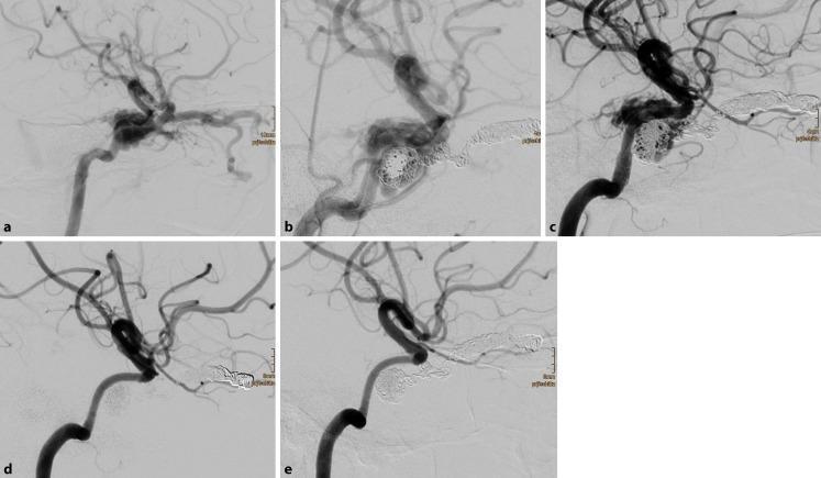
This series of digital subtraction angiograms (DSA) illustrates the endovascular management and longitudinal progression of a traumatic direct carotid-cavernous sinus fistula (CCSF). Image (a) demonstrates the baseline pathology: a massive arteriovenous shunt from the cavernous segment of the right internal carotid artery (ICA) into the adjacent cavernous sinus. Image (b) shows the initial intervention, featuring densely packed detachable coils within the superior ophthalmic vein and cavernous sinus, combined with the deployment of flow diverters within the ICA. Image (c) shows a follow-up DSA 6 days later, identifying a residual shunt that necessitated the placement of additional flow diverters to increase the construct's metal coverage and further promote thrombosis. Images (d) and (e) represent long-term follow-up at 2 and 8 months, respectively, showing complete occlusion of the fistula, absence of venous shunting, and restoration of normal arterial flow. The series highlights a combined endovascular approach using both coil embolization and flow-diverting stents to achieve definitive occlusion of high-flow traumatic arteriovenous communications.
This series of digital subtraction angiograms (DSA) illustrates the endovascular management and longitudinal progression of a traumatic direct carotid-cavernous sinus fistula (CCSF). Image (a) demonstrates the baseline pathology: a massive arteriovenous shunt from the cavernous segment of the right internal carotid artery (ICA) into the adjacent cavernous sinus. Image (b) shows the initial intervention, featuring densely packed detachable coils within the superior ophthalmic vein and cavernous sinus, combined with the deployment of flow diverters within the ICA. Image (c) shows a follow-up DSA 6 days later, identifying a residual shunt that necessitated the placement of additional flow diverters to increase the construct's metal coverage and further promote thrombosis. Images (d) and (e) represent long-term follow-up at 2 and 8 months, respectively, showing complete occlusion of the fistula, absence of venous shunting, and restoration of normal arterial flow. The series highlights a combined endovascular approach using both coil embolization and flow-diverting stents to achieve definitive occlusion of high-flow traumatic arteriovenous communications.

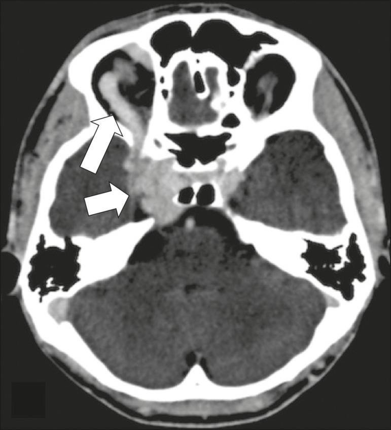
Axial CT angiography of the head demonstrating key diagnostic findings of a carotid-cavernous fistula (CCF). The image shows significant dilation and early contrast enhancement of the right cavernous sinus. This is accompanied by a markedly dilated and tortuous right superior ophthalmic vein (SOV), indicated by a white arrow, reflecting retrograde arterialized blood flow. Clinical manifestations secondary to these vascular changes are visible, including right-sided exophthalmos (proptosis) and enlargement of the extraocular muscles. The medial deviation of the right globe suggests convergent strabismus, clinically associated with abducens nerve (CN VI) compression within the cavernous sinus. This educational image serves to illustrate the classic radiological triad of cavernous sinus enlargement, superior ophthalmic vein dilation, and proptosis in the context of neurovascular pathology.

Axial CT angiography of the head demonstrating key diagnostic findings of a carotid-cavernous fistula (CCF). The image shows significant dilation and early contrast enhancement of the right cavernous sinus. This is accompanied by a markedly dilated and tortuous right superior ophthalmic vein (SOV), indicated by a white arrow, reflecting retrograde arterialized blood flow. Clinical manifestations secondary to these vascular changes are visible, including right-sided exophthalmos (proptosis) and enlargement of the extraocular muscles. The medial deviation of the right globe suggests convergent strabismus, clinically associated with abducens nerve (CN VI) compression within the cavernous sinus. This educational image serves to illustrate the classic radiological triad of cavernous sinus enlargement, superior ophthalmic vein dilation, and proptosis in the context of neurovascular pathology.

Reading File
Reading File
Loading Image
Loading Image
Finding Sources
Reading File
Reading File
I now have comprehensive material across anatomy, contents, venous drainage, pathology, and clinical syndromes. Let me compile the full answer.

Cavernous Sinus

Anatomy and Location

The cavernous sinus is a paired, trabeculated dural venous sinus situated on either side of the body of the sphenoid bone (sella turcica). It extends from the apex of the orbit anteriorly to the apex of the petrous temporal bone posteriorly. Its medial and inferior walls are formed by grooves in the body and base of the greater wing of the sphenoid. The medial, upper, and lateral walls are formed by a fold of the inner layer of dura, which is medially continuous with the diaphragma sellae. Posterolaterally, the wall is medial to the anterior part of Meckel's cave (which houses the trigeminal ganglion).
  • Scott-Brown's Otorhinolaryngology Head & Neck Surgery, Vol. 2, p. 1247
  • Gray's Anatomy for Students, p. 1026

Contents
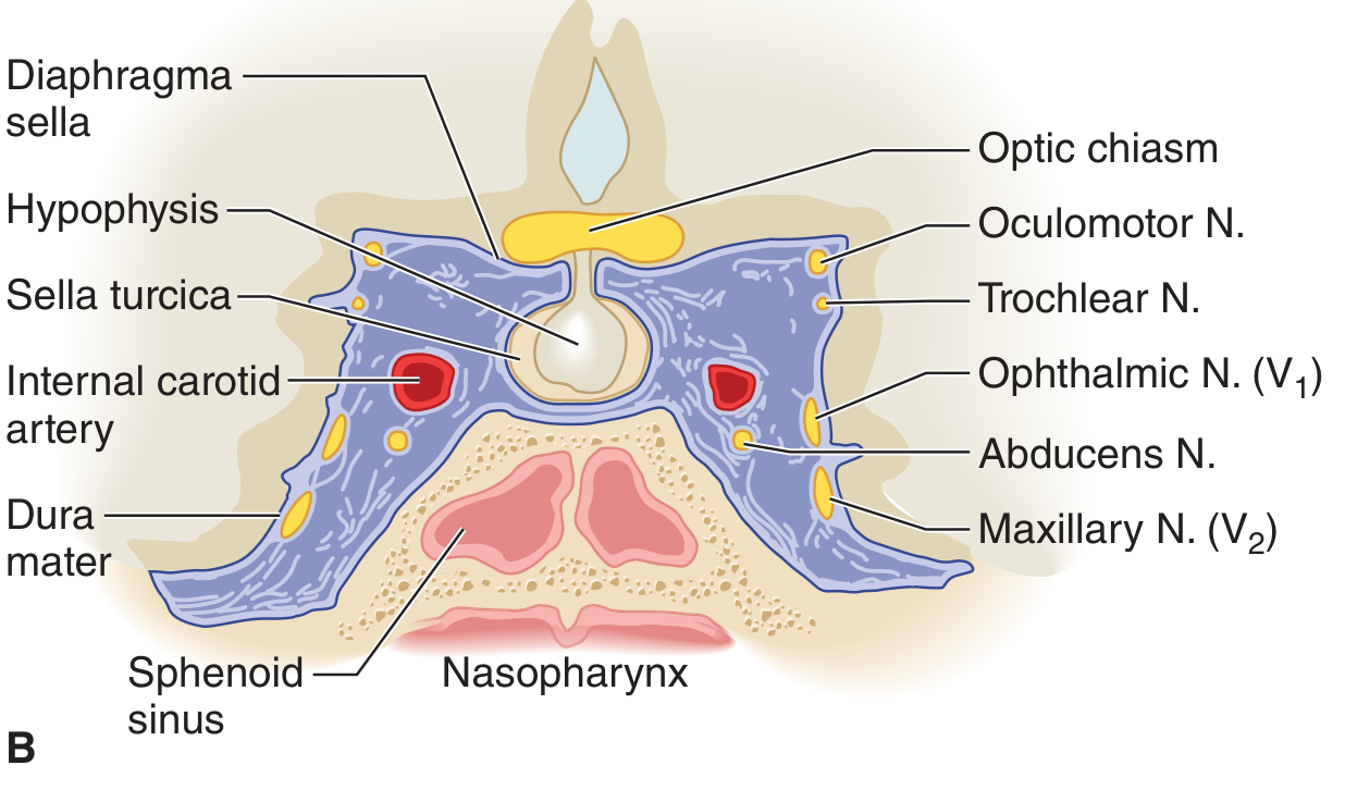
Coronal section view:
Coronal section through the cavernous sinus showing its contents and relations - from Adams and Victor's Principles of Neurology
Lateral view with meningeal layer removed:
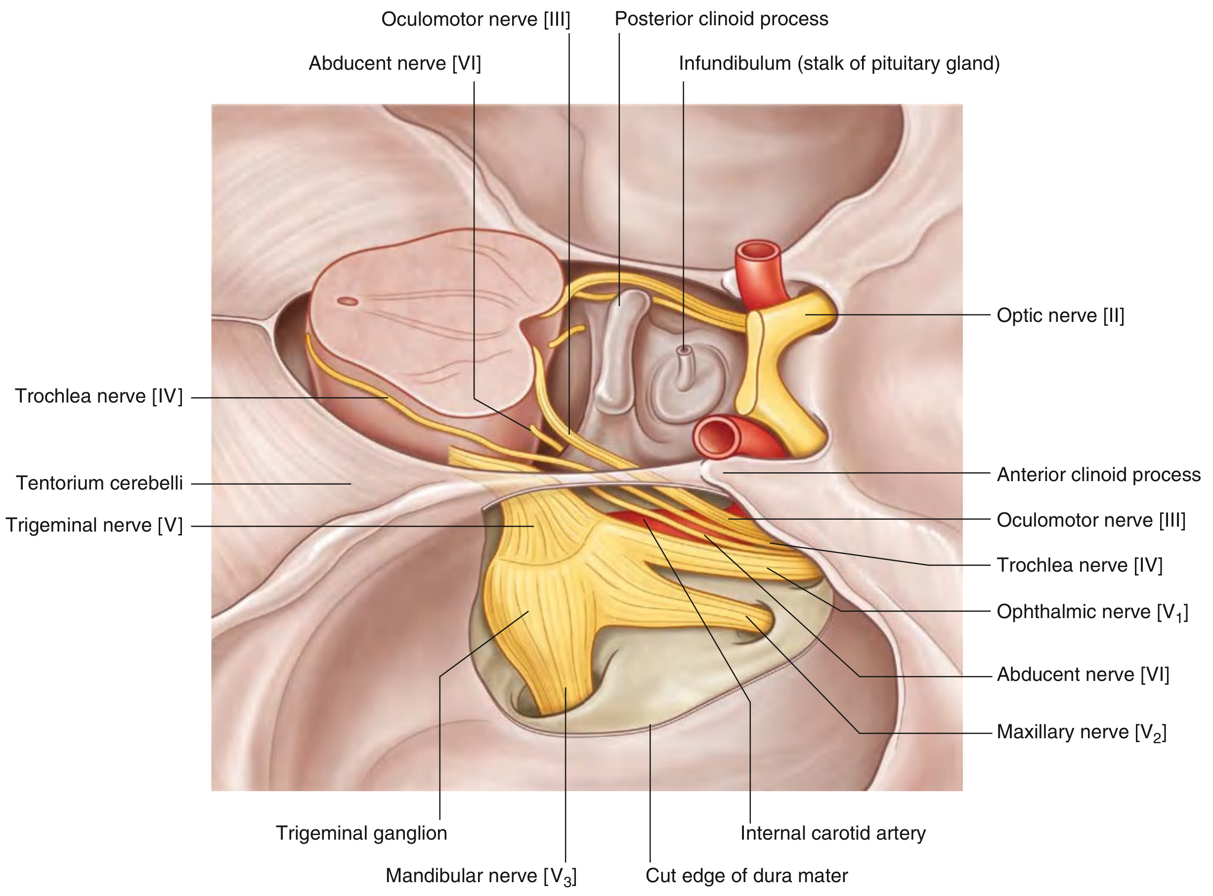
Lateral view of the right cavernous sinus with meningeal dura removed, showing cranial nerve contents - Gray's Anatomy for Students

Structures in the lateral wall (superior to inferior):

PositionStructure
1st (most superior)CN III - Oculomotor nerve
2ndCN IV - Trochlear nerve
3rdCN V1 - Ophthalmic nerve
4th (most inferior)CN V2 - Maxillary nerve

Structure running within the sinus (free in the venous blood):

  • CN VI - Abducens nerve (runs inferolaterally, within the sinus cavity itself - not embedded in the wall, making it uniquely vulnerable to raised intrasinus pressure)
  • Internal carotid artery (ICA) - the main content; it emerges from the petrous apex through the superior aspect of the foramen lacerum, courses upward, forward, and medially to the anterior clinoid process, where it perforates the dura to enter the middle cranial fossa
  • Sympathetic plexus - travels with the ICA
Key mnemonic: O TOM CAT - Oculomotor, Trochlear, Ophthalmic, Maxillary (wall), Carotid Artery, abducenT (free in sinus)
  • Gray's Anatomy for Students, p. 1026; Scott-Brown's Vol. 2, p. 1247

Relations

DirectionStructure
MedialSella turcica and pituitary gland (hypophysis), sphenoid sinus
SuperiorOptic chiasm, diaphragma sellae
InferiorForamen lacerum, body of sphenoid, nasopharynx
LateralTemporal lobe, trigeminal (Meckel's) cave
AnteriorSuperior orbital fissure (sinus communicates with orbit)
PosteriorPetrous apex, dorsum sellae
The two sinuses are connected by anterior and posterior intercavernous sinuses on either side of the pituitary stalk, forming a ring (circular sinus). This explains bilateral signs in many cavernous pathologies.

Venous Drainage and Connections

Each cavernous sinus:
  • Receives blood from the orbit (ophthalmic veins) and cerebral hemispheres
  • Receives from the sphenoparietal sinus (anteriorly)
  • Drains via the superior petrosal sinus → transverse sinus → sigmoid sinus → internal jugular vein
  • Drains via the inferior petrosal sinus → directly to internal jugular vein
  • Communicates with the pterygoid venous plexus via the foramen ovale and foramen of Vesalius
This rich communication with the facial and orbital veins - which lack valves - explains how infections from the "danger triangle of the face" can spread to cause cavernous sinus thrombosis.
  • Scott-Brown's Vol. 2, p. 1247; Gray's Anatomy for Students, p. 1027

Clinical Syndromes

1. Cavernous Sinus Syndrome

Involves multiple cranial nerves passing through the sinus. Presents with:
  • Painful ophthalmoplegia (CN III, IV, VI palsy)
  • Loss of corneal reflex and periorbital/facial numbness (CN V1, V2)
  • Sympathetic deficit (partial Horner syndrome - ptosis + miosis, without anhidrosis)
  • Proptosis, chemosis, congestion of conjunctival/retinal veins
An oculomotor palsy in the cavernous sinus may occur in isolation or with dysfunction of CN IV, VI, V1, V2 and sympathetic fibers simultaneously. - Bradley and Daroff's Neurology, p. 2516

2. Cavernous Sinus Thrombosis

Usually septic, resulting from spread of infection from:
  • Paranasal sinusitis (especially sphenoidal/ethmoidal)
  • Orbital/preseptal cellulitis
  • Facial skin infections spreading via the pterygoid plexus
Clinical features: rapid onset bilateral proptosis, chemosis, severe headache, fever, complete ophthalmoplegia, papilloedema, signs of meningeal irritation. Bilateral signs are a distinguishing feature (vs. orbital apex syndrome which is unilateral).
Mortality: ~20% treated; up to 100% untreated.
Imaging: MRI/MR venography (absence of flow in sinus); CT with contrast may show filling defects.
Treatment: IV antibiotics, anticoagulation (if intracranial hemorrhage excluded), surgical drainage of the source sinus. - Kanski's Clinical Ophthalmology, p. 131; Scott-Brown's Vol. 2

3. Carotid-Cavernous Fistula (CCF)

An arteriovenous fistula between the carotid artery and cavernous sinus causes raised venous pressure → orbital signs.
Direct (high-flow): ICA blood passes directly into the sinus through a defect in the intracavernous ICA wall, usually from trauma (75%) or spontaneous aneurysm rupture.
  • Classic triad: pulsatile proptosis + conjunctival chemosis + pulsatile bruit ("whooshing noise")
Indirect (low-flow / "dural shunt"): intracavernous ICA intact; meningeal branches of external or internal carotid feed the sinus. More subtle presentation; gradual-onset conjunctival redness. Caused by rupture of atherosclerotic artery or congenital malformation.
Imaging: CT angiography - dilated cavernous sinus, enlarged superior ophthalmic vein, proptosis. Digital subtraction angiography (DSA) is the gold standard.
Treatment: Endovascular embolization (coils ± flow diverters). - Kanski's Clinical Ophthalmology, p. 131

4. Tolosa-Hunt Syndrome

Idiopathic painful granulomatous inflammation of the cavernous sinus, typically:
  • Periorbital pain + ipsilateral oculomotor palsy (III, IV, VI)
  • Responsive to corticosteroids (hallmark)
  • Recurrence with steroid taper should raise suspicion for lymphoma (lymphoma is also steroid-responsive but recurs)
  • Differential includes cavernous sinus metastasis (especially in older patients), IgG4-related disease, and mucormycosis
  • Bradley and Daroff's Neurology, p. 2516; Localization in Clinical Neurology, 8e

5. Neoplastic Involvement

Common tumors involving the cavernous sinus:
  • Pituitary adenoma (direct extension)
  • Meningioma
  • Nasopharyngeal carcinoma (direct invasion)
  • Lymphoma, plasmacytoma
  • Skull base tumors, sphenoid wing tumors

Abducens Nerve - Special Vulnerability

CN VI is unique because it runs free within the venous blood of the sinus (not embedded in the lateral wall like CN III, IV, V1, V2). This means:
  • It is the first nerve affected by any increase in intra-sinus pressure
  • An isolated CN VI palsy may be an early or subtle sign of cavernous sinus pathology
  • Pituitary apoplexy and raised ICP (Gradenigo syndrome) can also stretch CN VI at this location

Summary Diagram

Surgical endoscopic view of cavernous sinus showing ICA (1), hypophysis (2), clivus (3), CN III (4), CN VI (5), V1 (6), V2 (7)
Endoscopic anatomy of the cavernous sinus via a transnasal transpterygoid approach: internal carotid artery (1), hypophysis (2), clivus (3), CN III (4), CN VI (5), V1 (6), V2 (7)

Sources: Gray's Anatomy for Students; Adams and Victor's Principles of Neurology, 12e; Bradley and Daroff's Neurology in Clinical Practice; Localization in Clinical Neurology, 8e; Kanski's Clinical Ophthalmology, 10e; Scott-Brown's Otorhinolaryngology Head & Neck Surgery, Vols. 1 & 2.
This is a shared conversation. Sign in to Orris to start your own chat.