
Uterine factors for infertility
uterine factors infertility fibroids polyps Asherman syndrome
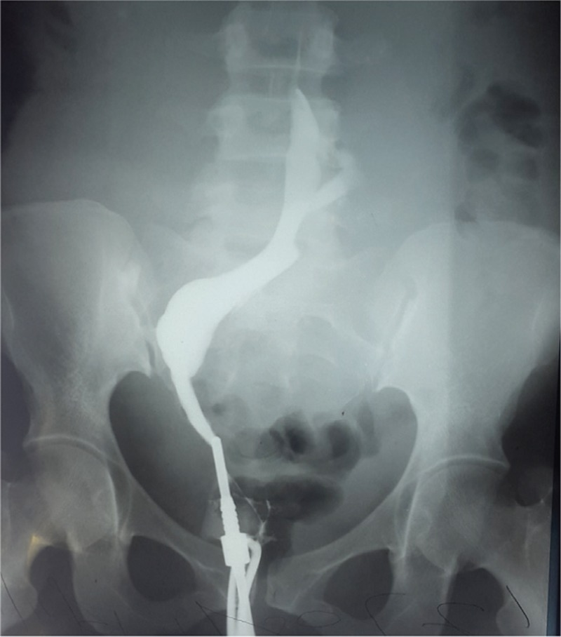
**Imaging Modality:** Hysterosalpingogram (HSG); fluoroscopic contrast study. **Anatomical Region:** Female pelvis, specifically the uterus and fallopian tubes. **Observed Pathology:** Intrauterine synechiae (Asherman syndrome). **Characteristic Visual Features:** The image demonstrates a hysterosalpingogram showing the uterine cavity opacified with radiopaque contrast media. The uterine contour is highly irregular and distorted. Key findings include multiple well-defined, linear, and irregular radiolucent filling defects within the contrast-filled uterine body. These filling defects represent intrauterine adhesions or synechiae that bridge the uterine walls, preventing uniform contrast distribution. **Differentiating Features:** The presence of sharp, angular, and non-mobile filling defects distinguishes synechiae from air bubbles (which are typically rounded and mobile) or endometrial polyps and fibroids (which usually present as smooth, rounded intraluminal masses). There is limited visualization of normal cornual anatomy, and no definitive bilateral tubal spillage is observed in this view, suggesting significant cavitary obliteration.
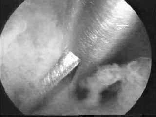
This monochrome diagnostic clinical image captures a hysteroscopic view of an intrauterine adhesiolysis procedure. The visual field demonstrates intrauterine adhesions, also known as Asherman Syndrome, appearing as cloudy, whitish, irregular fibrous bands and bridges of tissue within the uterine cavity. The surrounding endometrial tissue exhibits a granular, darker texture. A polished, reflective metallic hysteroscopic scissor is actively engaged in 'cold-knife' dissection of the adhesive structures. The instrument has a slim, sharp-edged profile designed for precise mechanical cutting without the use of electrosurgical energy, thereby minimizing thermal damage to the residual endometrium. This procedural image illustrates a key surgical intervention for restoring uterine cavity morphology to address symptoms such as infertility, amenorrhea, or cyclic pelvic pain.
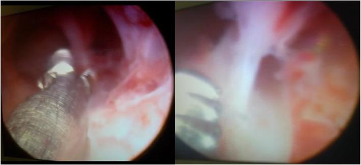
This diagnostic and interventional hysteroscopy image consists of two panels demonstrating intrauterine adhesions (Asherman syndrome) within the uterine cavity. The left panel shows the mid-uterine cavity with reddish-pink endometrial lining and whitish, fibrous tissue bands stretching across the lumen. A metallic grasping instrument with a textured surface is visible, engaging with the adhesions. The right panel provides a closer view of dense, opaque, avascular synechiae bridging the uterine walls. These adhesions partially obliterate the cavity, characteristic of moderate-to-severe intrauterine scarring. Surgical microscissors or grasping forceps are seen in the foreground, indicating the therapeutic lysis of these adhesions. The images serve to illustrate the visual appearance of fibrotic bands and the use of hysteroscopic instrumentation in the treatment of infertility related to endometrial pathology.
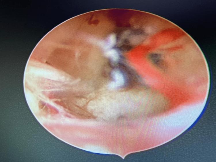
This clinical diagnostic image is a hysteroscopic view of the uterine cavity during a surgical adhesiolysis procedure. The image depicts intrauterine adhesions (Asherman syndrome) characterized by thick, whitish, fibrous bands and opaque web-like structures bridging the uterine walls. The background shows the healthy pinkish-red mucosal lining of the endometrium and visible branching blood vessels. A dark gray, thin, 5-Fr surgical scissor is positioned within the frame, actively engaging with the dense adhesions to restore the normal volume and architecture of the uterine cavity. The educational focus of this image is the visual identification of intrauterine synechiae and the procedural technique of hysteroscopic lysis in a patient with infertility or recurrent pregnancy loss.
uterine factors infertility treatment outcomes
uterine congenital anomalies Mullerian bicornuate septate arcuate uterus classification
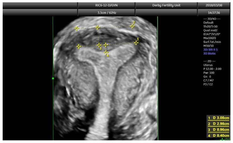
A 3D coronal plane ultrasound image of a human uterus, demonstrating the standard protocol for assessing congenital uterine anomalies (CUAs). The image features four key quantitative measurements used to differentiate between normal, arcuate, and septate morphologies. Line 1 (3.08 cm) represents the interostial line, connecting the interstitial portions of the fallopian tubes. Line 2 (2.98 cm) is a parallel reference along the serosal surface of the fundus. Measurement 3 (0.96 cm) indicates the uterine wall thickness, calculated as the vertical distance between the serosal surface and the interostial line. Measurement 4 (0.40 cm) quantifies the internal fundal indentation depth from the interostial line. The external fundal contour is smooth with no significant cleft, ruling out a bicornuate (U3) classification. The mild internal indentation of 0.40 cm is consistent with an arcuate uterus or a normal variant, as it does not meet the ESHRE/ESGE or ASRM criteria for a septate uterus. This diagnostic imaging is essential in reproductive medicine for evaluating Mullerian duct anomalies and fertility.
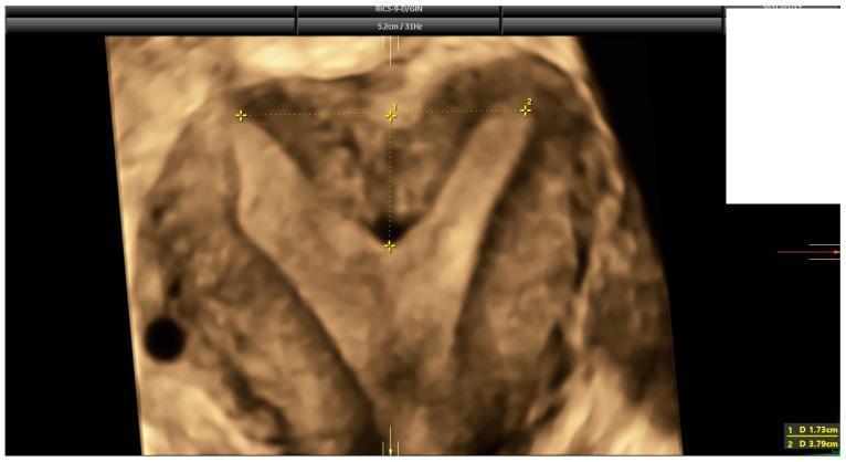
This diagnostic image is a 3D ultrasound of the uterus in the coronal plane, specifically reconstructed to evaluate congenital uterine anomalies. The image demonstrates a septate uterus, characterized by a significant internal midline indentation of the uterine fundus that divides the upper endometrial cavity into two horns while maintaining a relatively flat or minimally indented external serosal contour. Caliper measurements are visible for diagnostic classification: marker '1' measures the fundal indentation depth at 1.73 cm (17.3 mm), and marker '2' measures the inter-cornual distance at 3.79 cm. These measurements are critical for applying diagnostic criteria such as ESHRE-ESGE, ASRM, and CUME to differentiate between septate, bicornuate, and arcuate uterine morphologies. The educational focus is on identifying internal cavity architecture and utilizing specific measurement thresholds (e.g., indentation >10-15mm) to classify Mullerian duct anomalies in reproductive medicine.
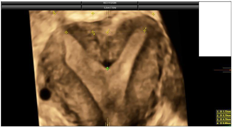
This diagnostic image is a 3D coronal ultrasound reconstruction of a human uterus, specifically highlighting a congenital uterine anomaly. The visual demonstrates a septate uterus, characterized by a prominent internal fundal indentation that divides the superior portion of the uterine cavity into two sections. Visible electronic calipers and numbered annotations (1–4) mark critical anatomical measurements used for classification according to ESHRE-ESGE and ASRM criteria. Measurement 1 (1.73 cm) indicates the depth of the fundal indentation, while Measurement 4 (0.90 cm) assesses the uterine wall thickness, resulting in a high indentation-to-wall-thickness (I:WT) ratio of 192%. The external fundal contour remains relatively flat or convex, a key feature distinguishing the septate uterus from a bicornuate uterus. This educational visual is used in reproductive medicine and radiology to teach the standardized assessment of Müllerian duct anomalies, focusing on the differentiation between septate, subseptate, and arcuate uterine morphologies through quantitative morphometric analysis.
Uterine Factors for Infertility
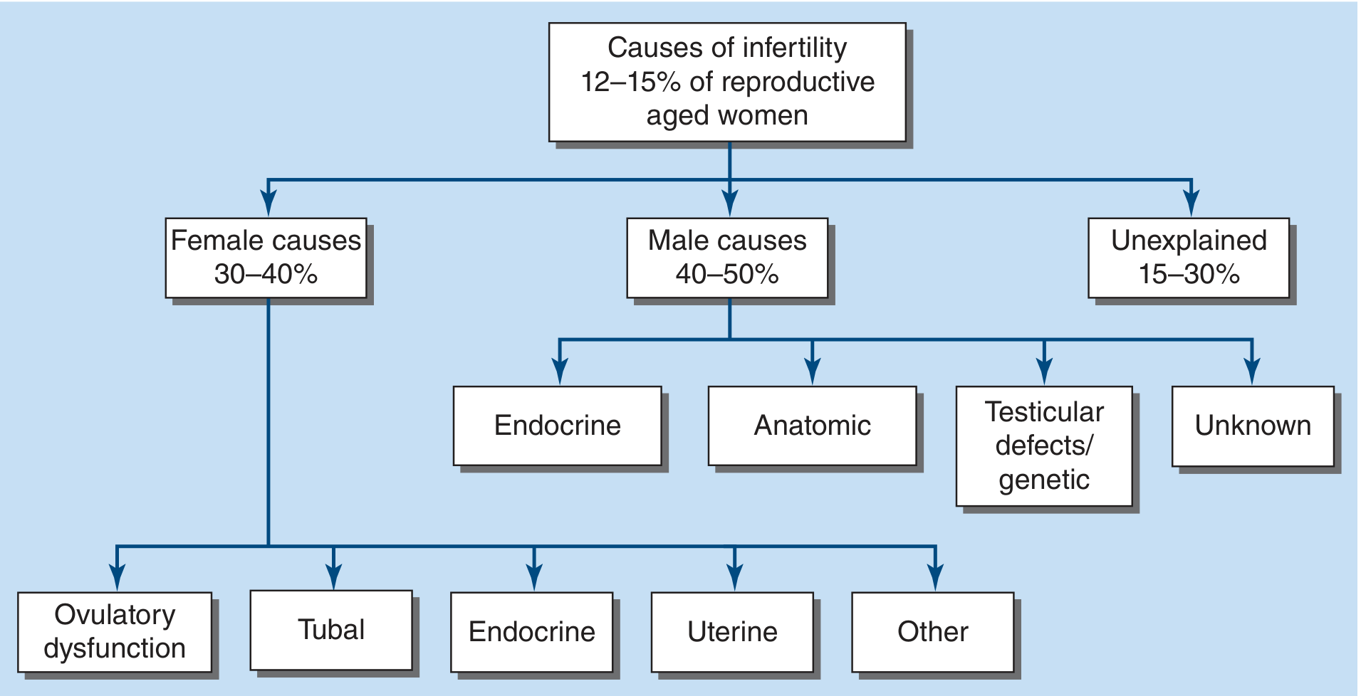
Classification of Uterine Factors
- Congenital (Mullerian) anomalies
- Acquired abnormalities
1. Congenital Uterine Anomalies (Mullerian Duct Anomalies)
| Anomaly | Description | Fertility Impact |
|---|---|---|
| Mullerian agenesis (MRKH) | Absent uterus and upper vagina | Cannot carry pregnancy; IVF + gestational carrier required |
| Unicornuate uterus | Only one Mullerian duct develops | Poor obstetric outcomes; rudimentary horn requires removal |
| Uterus didelphys | Failure of fusion - two separate uterine cavities | Primarily obstetric complications |
| Bicornuate uterus | Incomplete fusion - two uterine horns with one cervix | Poor obstetric outcomes; surgical repair controversial |
| Septate uterus | Failure of resorption of midline septum | Most common; hysteroscopic metroplasty significantly reduces pregnancy loss |
| Arcuate uterus | Mildest anomaly - minor fundal indentation | Live birth rates comparable to normal uterus |
| T-shaped uterus | DES-related in utero exposure (banned 1971) | Higher uterine malformation and obstetric complications |
Key point: Congenital anomalies are more closely associated with pregnancy wastage than infertility per se - the prevalence of uterine defects is generally similar in fertile and infertile women. The exception is Mullerian agenesis. (Berek & Novak's Gynecology)
2. Acquired Uterine Abnormalities
A. Leiomyomas (Fibroids)
- Most common benign tumor of the reproductive tract; occur in 50-70% of reproductive-age women
- The relationship between fibroids and infertility depends on location:
| Location | Effect on Fertility |
|---|---|
| Submucosal (distorts cavity) | Clearly reduces pregnancy rates and increases miscarriage risk |
| Intramural (distorts cavity) | May lower pregnancy rates |
| Subserosal (does not distort cavity) | Little to no impact on fertility |
- Treatment: Hysteroscopic removal of submucosal fibroids may improve pregnancy rates. Myomectomy is the fertility-preserving option for symptomatic intramural fibroids. (Harrison's 22E, p. 3194)
B. Endometrial Polyps
- Focal overgrowth of endometrial glands and stroma projecting into the uterine cavity
- Impair implantation by causing mechanical disruption
- Diagnosed by HSG, saline infusion sonography, or hysteroscopy
- Treatment: Hysteroscopic polypectomy significantly improves subsequent pregnancy rates
C. Intrauterine Adhesions (Asherman Syndrome)
- Intrauterine synechiae (adhesions) caused by destruction of the basal endometrium
- Common causes: Post-curettage (especially after dilation and curettage for missed/incomplete abortion), postpartum hemorrhage, uterine infection (tuberculous endometritis)
- Clinical features: Hypomenorrhea/amenorrhea, cyclic pelvic pain, infertility, recurrent pregnancy loss
- Severity: Ranges from thin filmy adhesions to dense fibrotic bands obliterating the cavity
- Treatment: Hysteroscopic adhesiolysis + postoperative estrogen therapy to promote endometrial regeneration
D. Adenomyosis
- Endometrial glands and stroma within the myometrium
- Impairs implantation and embryo development via altered uterine contractility, inflammatory milieu, and impaired endometrial receptivity
- Diagnosed by MRI or transvaginal ultrasound (globular uterus, myometrial cysts)
- Management is complex; GnRH agonist pre-treatment before IVF may improve outcomes
E. Endometrial Hyperplasia
- Abnormal proliferation of the endometrium, often secondary to unopposed estrogen
- Impairs normal implantation
- Treated with progestins; surgery reserved for atypical hyperplasia
F. Destroyed Endocervical Glands / Cervical Stenosis
- Occurs after cervical surgery (LEEP, cone biopsy) or cervical infection
- Impairs sperm transport; may require cervical dilation or IUI to bypass the cervix
3. Diagnostic Evaluation
| Investigation | What it Detects |
|---|---|
| Hysterosalpingogram (HSG) | Polyps, submucous myomas, adhesions, uterine anomalies, tubal patency |
| Transvaginal ultrasound (TVS) | Fibroids, adenomyosis, Mullerian anomalies, polyps |
| Saline infusion sonohysterography (SIS) | Better delineation of intracavitary lesions than TVS |
| Hysteroscopy | Gold standard for intracavitary pathology; also therapeutic |
| MRI | Best for adenomyosis; classifies complex Mullerian anomalies |
| 3D ultrasound | Non-invasive assessment of Mullerian anomalies |
4. Management Summary
| Condition | Treatment |
|---|---|
| Submucosal fibroid | Hysteroscopic myomectomy |
| Endometrial polyp | Hysteroscopic polypectomy |
| Asherman syndrome | Hysteroscopic adhesiolysis + postoperative estrogen |
| Septate uterus | Hysteroscopic metroplasty (reduces pregnancy loss) |
| Bicornuate/didelphys | Surgical repair controversial; mainly obstetric indication |
| Mullerian agenesis | IVF + gestational carrier |
| Adenomyosis | GnRH agonist pre-treatment + IVF |
5. Luteal Phase Defect (LPD)
- Berek & Novak's Gynecology (Uterine Factors chapter, pp. 2844-2890)
- Harrison's Principles of Internal Medicine 22E (Chapter 408, p. 3194)
- Goldman-Cecil Medicine, International Edition (Female Factors, p. 1956-1960)
- Textbook of Family Medicine, 9e (Female Infertility, p. 1378)