
What diagnosis and checks by 2d echo
2D echocardiography cardiac views parasternal apical
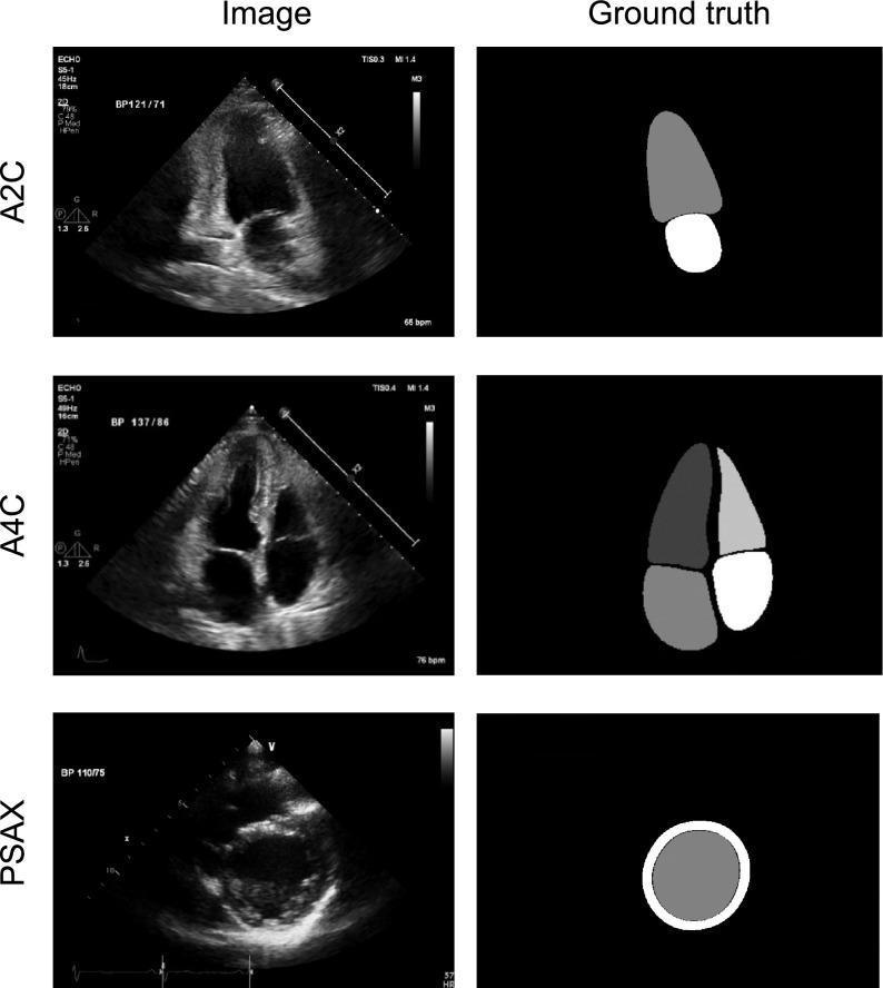
A multi-panel medical diagnostic graphic displaying standard 2D echocardiography views and their corresponding 'ground truth' segmentations for educational and machine-learning purposes. The images are organized in three rows representing primary echocardiographic orientations: Apical Two-Chamber (A2C), Apical Four-Chamber (A4C), and Parasternal Short Axis (PSAX) at the papillary muscle level. The left column shows the original grayscale B-mode ultrasound images, featuring anatomical landmarks such as the left ventricle (LV), right ventricle (RV), left atrium (LA), right atrium (RA), and interventricular septum. Overlaid metadata include heart rate (bpm) and blood pressure (BP) values. The right column displays expert-annotated segmentation masks where cardiac structures are color-coded as solid shapes: the LV cavity, LA cavity, RV cavity, and RA cavity in the A4C view; the LV and LA in the A2C view; and a circular cross-section of the LV myocardium and cavity in the PSAX view. This comparison highlights key anatomical boundaries used in clinical indices like ejection fraction (EF) and ventricular volume measurements.
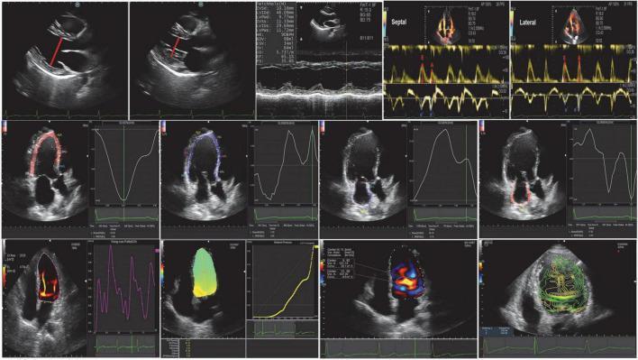
A multi-panel echocardiographic compilation demonstrating advanced cardiac imaging modalities. The top row illustrates conventional echocardiography, including 2D grayscale parasternal long-axis views with diameter measurements, M-mode tracings for ventricular wall motion assessment, and pulsed-wave Doppler for mitral inflow and tissue Doppler imaging for septal/lateral annular velocities. The middle row showcases Speckle-Tracking Echocardiography (STE), depicting apical four-chamber views with color-coded myocardial strain overlays and associated longitudinal strain/strain-rate curves, used to quantify regional and global myocardial deformation. The bottom row presents Vortex Flow Mapping (VFM) and intraventricular pressure gradient analysis. These panels visualize intracavitary hemodynamics using color-coded vector maps, vorticity contours, and energy loss distributions within the left ventricle. Quantitative graphs accompany these images, displaying flow velocity and pressure profiles across the cardiac cycle. The composite serves as an educational overview of integrated cardiac mechanics and hemodynamics, relevant for the advanced assessment of heart failure and ventricular function.
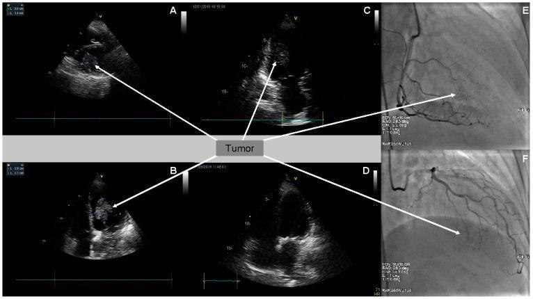
This composite figure illustrates the multi-modal diagnostic assessment and treatment response of a cardiac metastatic tumor, likely from melanoma. Panels A, B, and C display two-dimensional transthoracic echocardiography (2D TTE) in parasternal short-axis and apical four-chamber views. They reveal a large, well-defined, hyperechoic mass (arrows) situated within the left ventricle cavity, originating from the inferoseptal wall. Panel D shows a follow-up 2D TTE (4-chamber view) demonstrating complete tumor resolution following chemotherapy (pembrolizumab). Panels E and F present selective coronary angiography (CA) of the right coronary artery (RAO 28.3°, CAU 2.2°) and the left coronary artery circumflex branch (RAO 26.3°, CRA 23.1°). These images highlight sparse, poor tumor vascularity (arrows) during the late arterial phase, differentiating the mass from highly vascular primary cardiac tumors. This sequence serves as an educational tool for identifying metastatic cardiac lesions via echocardiography and angiography, as well as monitoring clinical response to immunotherapy.
2D Echocardiography - Diagnoses & Checks
What Is 2D Echo?
Diagnoses Made by 2D Echo
1. Valvular Heart Disease
| Condition | Key Echo Findings |
|---|---|
| Valve stenosis (aortic, mitral, etc.) | Valve anatomy and cause; transvalvular pressure gradient (AR); valve area (AVA by continuity equation); chamber hypertrophy; LV/RV systolic function |
| Valve regurgitation | Mechanism and cause; severity (vena contracta, EROA); chamber enlargement; pulmonary artery (PA) pressure estimate |
| Prosthetic valve function | Evidence of stenosis; detection of regurgitation; ventricular function |
| Endocarditis | Vegetations (TTE sensitivity 70-85%); degree of valve dysfunction; abscess detection; chamber function |
2. Coronary Artery Disease (CAD)
| Condition | Key Echo Findings |
|---|---|
| Acute coronary syndrome | Segmental wall motion abnormality (SWMA) reflecting myocardium at risk; global LV function (EF); complications: acute MR, VSD, LV thrombus/aneurysm/rupture, RV infarct |
| Stable angina | Global and segmental LV systolic function; exclude other causes (e.g., aortic stenosis, HCM) |
| Pre/post revascularization | Wall thickening and endocardial motion at baseline; improvement in segmental function post-procedure |
| End-stage ischemic disease | Overall LV EF; PA pressures; associated MR; LV thrombus; RV function |
3. Cardiomyopathies
| Type | Key Echo Findings |
|---|---|
| Dilated (DCM) | Chamber dilation (all 4 chambers); LV/RV systolic function and EF; coexisting AV regurgitation; LV thrombus |
| Restrictive (RCM) | LV wall thickness; LV systolic function; PA pressure; diastolic filling pattern |
| Hypertrophic (HCM) | Pattern and extent of LV hypertrophy; dynamic LVOT obstruction (imaging + Doppler); coexisting MR; diastolic LV dysfunction |
4. Hypertension
- LV hypertrophy (LVH)
- LV diastolic dysfunction (precedes systolic dysfunction)
- LV systolic function
- Aortic valve sclerosis, mitral annular calcification (MAC)
5. Pericardial Disease
| Condition | Key Echo Findings |
|---|---|
| Pericardial effusion | Echolucent space adjacent to heart; size grading: small (<0.5 cm), moderate (0.5-2 cm), large (>2 cm) |
| Cardiac tamponade | RA/RV diastolic collapse; reciprocal respiratory changes in RV and LV filling; IVC plethora |
| Constrictive pericarditis | Septal bounce; respiratory variation in mitral/tricuspid flow; annulus reversus on tissue Doppler |
6. Aortic Disease
| Condition | Key Echo Findings |
|---|---|
| Aortic dilation | Cause of dilation; accurate aortic diameter (aortic root, sinuses of Valsalva, ascending aorta); associated AR |
| Aortic dissection | 2D images of ascending aorta and arch; dissection "flap"; associated AR; ventricular function |
7. Cardiac Masses
| Type | Key Echo Findings |
|---|---|
| LV thrombus | High sensitivity/specificity; suspect with apical SWMA or diffuse LV dysfunction |
| LA thrombus | Lower sensitivity (TEE preferred); suspect with LA enlargement or MV disease |
| Cardiac tumors | Location, size, mobility, attachment; differentiate from thrombus |
8. Congenital Heart Disease
- Atrial septal defect (ASD), ventricular septal defect (VSD), patent ductus arteriosus (PDA)
- Great vessel anomalies, valvular anomalies
- Complex congenital defects
9. Heart Failure
- LV/RV systolic function (EF by Simpson's biplane method)
- Diastolic dysfunction grading (E/A ratio, deceleration time, E/e' ratio)
- PA pressures (estimated from TR jet velocity)
- HFrEF vs HFpEF differentiation
10. Pulmonary Hypertension
- RV size and function
- PA systolic pressure (PASP from tricuspid regurgitation jet)
- Paradoxical septal motion ("D-shaped" LV in severe cases)
Key Parameters Checked on 2D Echo
| Parameter | What It Tells You |
|---|---|
| Ejection Fraction (EF) | LV systolic function (normal >55%); calculated by Simpson's biplane method |
| LV dimensions | End-diastolic diameter (LVEDD), end-systolic diameter (LVESD) - detect dilation or hypertrophy |
| Wall thickness | Interventricular septum (IVS) and posterior wall (PW) thickness - assess hypertrophy |
| Wall motion | Regional SWMA (ischemia/infarction); graded as normal, hypokinetic, akinetic, dyskinetic |
| Valve morphology | Leaflet anatomy, calcification, mobility, prolapse, vegetation |
| Doppler gradients | Transvalvular pressure gradients in stenosis; severity of regurgitation |
| Valve area | AVA (aortic valve area) by continuity equation; MVA (mitral valve area) by pressure half-time |
| Chamber size | LA diameter/volume, RA size, RV size |
| PA systolic pressure | Estimated from TR jet velocity + RA pressure (4v² + RAP) |
| Diastolic function | E/A ratio, E-wave deceleration time, E/e' ratio, LA volume index |
| Pericardial effusion size | Location and quantification |
| IVC diameter and collapsibility | Estimate of RA pressure and volume status |
| Longitudinal strain (GLS) | More sensitive than EF for early LV dysfunction; assessed by speckle tracking |
Standard 2D Echo Views Used
- Parasternal Long Axis (PLAX) - Aortic valve, mitral valve, LV, aortic root
- Parasternal Short Axis (PSAX) - LV at multiple levels (mitral, papillary, apical); RV outflow
- Apical 4-Chamber (A4C) - All 4 chambers, mitral and tricuspid valves
- Apical 2-Chamber (A2C) - LV anterior and inferior walls
- Apical 3-Chamber (A3C/APLAX) - Aortic valve, LVOT, LV outflow
- Subcostal - IVC, RV, pericardial effusion (especially for pericardiocentesis guidance)
- Suprasternal - Aortic arch, great vessels
Limitations of 2D TTE
- Cannot visualize coronary artery anatomy directly (CT angiography or catheterization needed)
- LA thrombus detection requires TEE (transesophageal echo)
- Poor image quality in obese patients, COPD, or post-surgical chest anatomy
- Resting wall motion may be normal despite significant CAD (stress echo needed)
- Constrictive pericarditis may be difficult to differentiate from restrictive cardiomyopathy (cardiac catheterization sometimes required)
In 13 yrs old boy
congenital heart disease echocardiography ASD VSD child pediatric
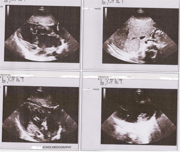
This diagnostic image displays a composite of four transthoracic echocardiography (TTE) frames obtained from a pediatric patient, utilized to evaluate complex congenital heart disease. The images are captured at a depth of 9 cm using a 7.5 MHz high-frequency transducer, appropriate for infant or small child imaging. Visually, the echocardiogram demonstrates a dextrocardia orientation with the heart situated in the right hemithorax. Key anatomical findings include significant chamber enlargement, specifically a dilated right ventricle and right atrium. The interventricular and interatrial septa show discontinuities consistent with a large perimembranous ventricular septal defect (VSD) and an ostium primum atrial septal defect (ASD). The parallel orientation of the great arteries suggests a Transposition of the Great Arteries (TGA). These views highlight the structural complexity associated with heterotaxy syndromes or situs inversus, illustrating the relationship between atrial and ventricular septal defects and conotruncal abnormalities in a single clinical case.
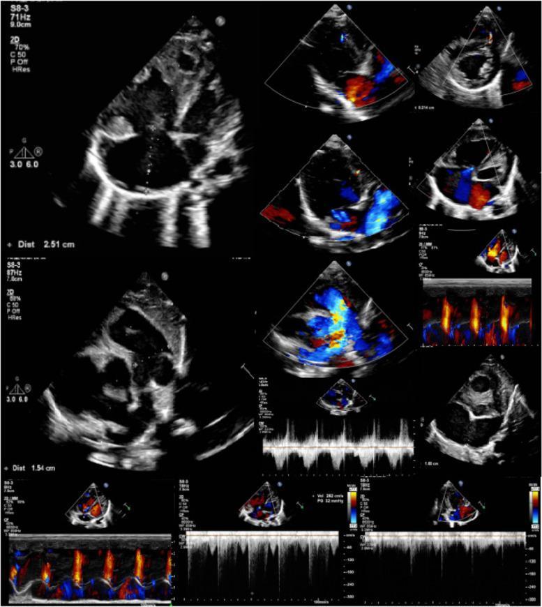
This diagnostic composite displays multiple echocardiography modalities, including 2D grayscale, color Doppler, and spectral Doppler (M-mode and pulse wave) views of a pediatric heart. The grayscale images show structural abnormalities characterized by biventricular hypertrophy and significant chamber enlargement. Visible septal discontinuities confirm a large atrial septal defect (ASD) measuring approximately 16mm and a muscular ventricular septal defect (VSD). Color Doppler imaging demonstrates complex, turbulent hemodynamic patterns with high-velocity mosaic signals indicating bidirectional (primarily left-to-right) shunting at the atrial and ventricular levels. Signs of mild-to-moderate tricuspid and mild mitral regurgitation are evident via reflux jets across the valvular planes. Spectral Doppler waveforms facilitate the assessment of pulmonary hypertension and abnormal flow velocities. The comprehensive imaging supports a diagnosis of multi-defect congenital heart disease often associated with syndromic presentations like OFCD. These views are essential for pediatric cardiology students to identify pathological shunting, valvular insufficiency, and secondary structural changes like hypertrophy.
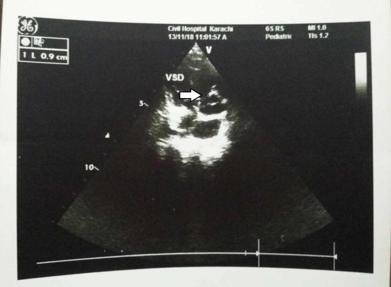
Diagnostic ultrasound: A black-and-white transthoracic echocardiogram in a pediatric patient. The image displays a cross-sectional view of the cardiac chambers and septa. A prominent white arrow points to a clear anatomical discontinuity in the ventricular septum, labeled 'VSD', which identifies a Ventricular Septal Defect measuring approximately 0.9 cm based on the on-screen caliper scale. The imaging demonstrates the structural interruption between the right and left ventricles. Metadata headers indicate the procedure was performed with a pediatric probe (65RS) at a mechanical index (MI) of 1.0. This visual serves to illustrate a core component of congenital heart disease, specifically associated with Tetralogy of Fallot in the clinical context of congenital CMV infection. The ultrasound highlights the use of echocardiography in identifying intracardiac shunts and septal abnormalities in neonatal and pediatric populations.
2D Echo in a 13-Year-Old Boy
- Congenital heart defects may present for the first time or be followed up
- Rheumatic heart disease is a major concern (especially in developing countries)
- Cardiomyopathies can manifest
- Pulmonary hypertension from long-standing shunts may be developing
- Normal reference ranges differ from adults (must use BSA-indexed values)
Priority Diagnoses to Evaluate
1. Congenital Heart Disease (CHD)
| Diagnosis | 2D Echo Findings |
|---|---|
| ASD (Atrial Septal Defect) | RV and RA volume overload; RV/RA enlargement; paradoxical septal motion; dropout in interatrial septum; color Doppler shows left-to-right shunt |
| VSD (Ventricular Septal Defect) | Septal discontinuity; color Doppler turbulent jet across septum; RV/LV volume load depending on size |
| Bicuspid Aortic Valve | Two leaflets in systole with raphe seen in diastole; aortic root/ascending aortic dilation; assess for stenosis or regurgitation |
| Aortic Coarctation | Descending thoracic aorta high-velocity systolic flow; holosystolic antegrade flow; ~50% have associated bicuspid aortic valve; pulsatile dilated aorta proximal to coarctation |
| Pulmonary Stenosis | Dysplastic/thickened pulmonic valve leaflets; systolic doming; high-velocity jet across RVOT; RV hypertrophy |
| Ebstein Anomaly | Septal tricuspid leaflet apically displaced; apparent RA enlargement; coexisting ASD/TR |
| Subaortic Membrane | Membrane from anterior MV leaflet to septum; high-velocity signal proximal to aortic valve |
| Sinus of Valsalva Aneurysm | Thin "wind-sock" projection from sinus; fistula into RA/RV/LV depending on cusp |
2. Rheumatic Heart Disease (RHD)
| Valve | Echo Findings |
|---|---|
| Mitral stenosis (MS) | Thickened, restricted leaflets; "hockey stick" doming of anterior leaflet; reduced MVA; elevated E-wave velocity; LA enlargement; Wilkins score for valve morphology |
| Mitral regurgitation (MR) | Leaflet thickening/prolapse/restriction; regurgitant jet into LA; LA and LV enlargement |
| Aortic regurgitation (AR) | Aortic leaflet thickening/prolapse; regurgitant jet into LV; LV volume overload (dilation) |
| Combined lesions | MS + MR is the most common RHD combination in children |
3. Cardiomyopathies
| Type | Echo Findings in a Child |
|---|---|
| Dilated CM (DCM) | LV/all 4 chambers dilated; EF reduced (<55%); global hypokinesia; mitral/tricuspid regurgitation secondary to annular dilation |
| Hypertrophic CM (HCM) | Asymmetric septal hypertrophy (IVS >15 mm or IVS/LVPW ratio >1.3); systolic anterior motion (SAM) of MV; dynamic LVOT obstruction; LA enlargement |
| Restrictive CM | Normal/small LV size; severely dilated atria (biatrial enlargement); diastolic dysfunction; elevated PA pressures |
4. Pulmonary Hypertension
- PASP estimated from TR jet velocity: PASP = 4v² + RAP
- RV hypertrophy and dilation
- "D-shaped" LV in short axis (flattened septum) - suggests severe RV pressure overload
- PA dilation
- Eisenmenger syndrome if shunt reversal occurs (bidirectional or right-to-left flow on color Doppler)
5. Kawasaki Disease Sequelae
- Coronary artery aneurysms (dilated left main, LAD, or RCA)
- Coronary artery stenosis or occlusion
- LV wall motion abnormalities from prior myocardial infarction
6. Myocarditis / Pericarditis
- Myocarditis: Dilated, poorly contracting LV; diffuse hypokinesia; reduced EF; pericardial effusion may coexist
- Pericarditis/effusion: Echolucent space surrounding heart; in tamponade - RA/RV diastolic collapse
Key Measurements & Parameters Specific to a 13-Year-Old
| Parameter | What to Check | Why Important |
|---|---|---|
| LV dimensions (LVEDD/LVESD) | Must be indexed to BSA (Body Surface Area) | Children have smaller hearts; raw values mislead |
| Ejection Fraction (EF) | Normal >55% (same as adults) | Global systolic function |
| IVS and LVPW thickness | Indexed to BSA | Detect HCM or hypertensive LVH |
| Aortic root / ascending aorta | Z-score indexed to BSA | Detect Marfan syndrome, bicuspid AV dilation |
| PA systolic pressure | From TR jet (4v² + RAP) | Screen for pulmonary hypertension from CHD |
| RV size and function | RV/LV ratio, TAPSE, FAC | Volume/pressure overload from shunts |
| Shunt quantification (Qp:Qs) | From PW Doppler at RVOT and LVOT | Assess hemodynamic significance of ASD/VSD |
| Mitral/Aortic valve morphology | Leaflet thickness, mobility, calcification | RHD screening |
| Coronary artery origins | Proximal LCA, RCA diameter and origin | Kawasaki, anomalous origin |
| Pericardial space | Effusion size and cardiac compression | Pericarditis, tamponade |
Z-Score Concept (Pediatric-Specific)
- Z-score 0 = exactly average for that BSA
- Z-score >2 = abnormally large (e.g., coronary aneurysm in Kawasaki if Z >2.5)
- Z-score <-2 = abnormally small
- Tools like Pediatric Heart Network nomograms are used
Clinical Scenarios to Flag in a 13-Year-Old
| Clinical Clue | Most Likely Diagnosis to Rule Out via Echo |
|---|---|
| Murmur since birth | ASD, VSD, PDA, pulmonary stenosis |
| Systolic murmur at LLSB + syncope on exertion | HCM with LVOT obstruction |
| Fever + recent sore throat + new murmur | Rheumatic carditis (MR, AR) |
| Easy fatigability + palpitations | DCM, arrhythmia-related cardiomyopathy |
| Chest pain on exertion | LVOTO (HCM), anomalous coronary, aortic coarctation |
| High BP in upper limbs, low BP in lower limbs | Aortic coarctation |
| Clubbing + cyanosis | Eisenmenger syndrome, cyanotic CHD |
| Prior history of prolonged fever in infancy | Kawasaki - check coronary arteries |
Grades of tonsillitis
Brodsky grading scale tonsil size grades 0 1 2 3 4 classification
tonsil grading Brodsky scale oropharynx enlargement grades
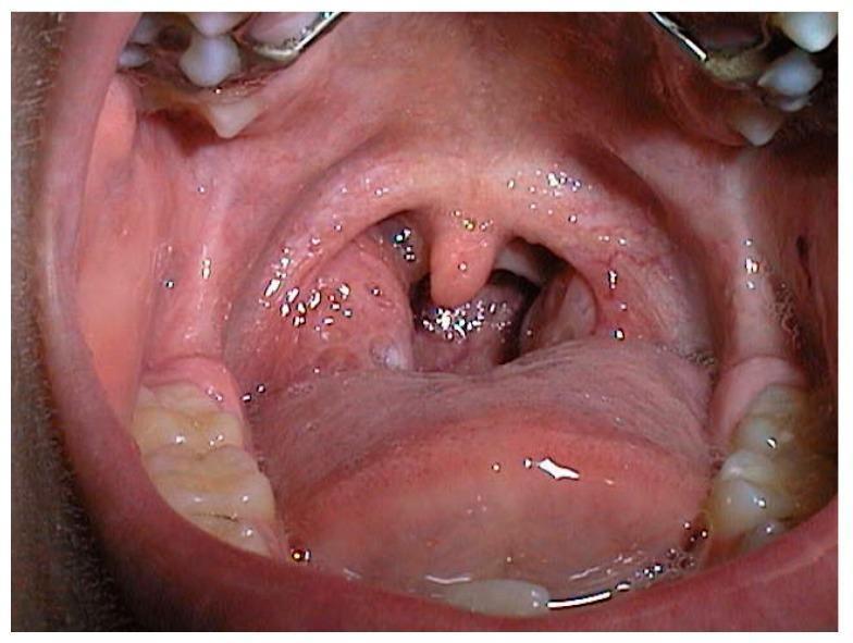
This clinical photograph displays an intraoral view of the oropharynx in a 6-year-old child, illustrating Grade III palatine tonsil hypertrophy. The tonsils are significantly enlarged and extend medially, occupying approximately 50% to 75% of the oropharyngeal airway width. The tonsillar tissue appears erythematous with visible surface crypts and an irregular morphology. This massive enlargement causes notable narrowing of the oropharyngeal space, with the central uvula appearing partially compressed between the two tonsillar masses. The surrounding structures, including the soft palate, posterior pharyngeal wall, and the base of the tongue, are clearly visible. Clinical significance includes the potential for upper airway obstruction, which is a major contributor to pediatric obstructive sleep apnea syndrome (OSAS). The image serves as a teaching tool for the Brodsky grading scale for tonsillar enlargement in pediatric otolaryngology.
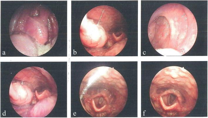
This composite of six clinical endoscopic images (a-f) captures various views of the upper airway and oropharynx obtained via laryngoscopy. Panels (a) through (d) demonstrate significant tonsillar hypertrophy, characterized by enlarged palatine tonsils that narrow the oropharyngeal space. The right tonsil displays grade 3 hypertrophy, while the left tonsil is grade 2. The images also reveal an elongated uvula and notable lymphoid hyperplasia along the posterior pharyngeal wall, appearing as a cobblestone-like, irregular mucosal texture. Panels (e) and (f) provide a clear view of the laryngeal inlet, showing the epiglottis, arytenoid cartilages, and the true vocal cords. The vocal cords appear as symmetrical, pearly-white bands in an abducted position, defining the glottic opening. These clinical findings are consistent with chronic tonsillitis and upper airway obstruction due to hypertrophic lymphoid tissue. The images serve as an educational tool for identifying grades of tonsillar enlargement and assessing pharyngeal lymphoid architecture.
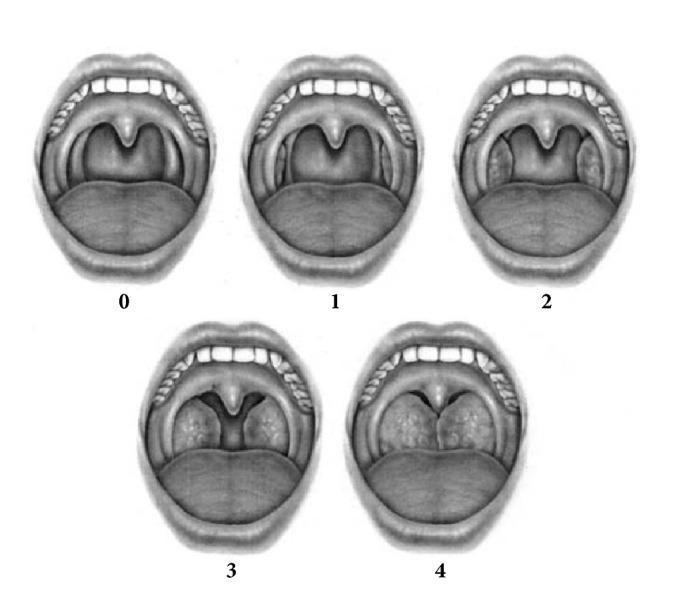
This Comparison Chart illustrates the Brodsky Grading Scale for tonsillar hypertrophy, a clinical assessment tool used in otolaryngology. The diagram features five grayscale anatomical illustrations of the open mouth and oropharynx, labeled 0 through 4, depicting the progressive obstruction of the pharyngeal space by the palatine tonsils. Key anatomical landmarks shown include the tongue, uvula, soft palate, and anterior tonsillar pillars. Grade 0 shows no visible tonsillar tissue (post-tonsillectomy). Grade 1 depicts tonsils confined within the tonsillar pillars (occupying <25% of the transverse oropharyngeal diameter). Grade 2 shows tonsils extending beyond the pillars, occupying 25%–50% of the space. Grade 3 demonstrates tonsils occupying 51%–75% of the space, significantly narrowing the airway. Grade 4 shows tonsils occupying >75% of the space, nearly meeting or touching at the midline (often called 'kissing tonsils'). This visual serves as an educational standard for diagnosing obstructive sleep apnea and planning surgical intervention in both pediatric and adult populations.
Grades of Tonsillitis / Tonsillar Enlargement
The Brodsky Grading Scale (Most Widely Used)
| Grade | Description | Oropharyngeal Obstruction | Clinical Notes |
|---|---|---|---|
| 0 | Tonsils absent / within the tonsillar fossa, not visible | None | Post-tonsillectomy or atrophic/absent tonsils |
| 1 | Tonsils just visible, extending to the anterior pillar | < 25% of oropharyngeal width | Minimal - often asymptomatic |
| 2 | Tonsils extend beyond the anterior pillar | 25 - 50% of oropharyngeal width | Mild obstruction; possible mild dysphagia |
| 3 | Tonsils extend well beyond the pillar, nearly to midline | 50 - 75% of oropharyngeal width | Moderate obstruction; snoring, sleep-disordered breathing |
| 4 | Tonsils touch/nearly touch at midline - "Kissing tonsils" | > 75% of oropharyngeal width | Severe obstruction; OSA, dysphagia, muffled voice |
Clinical Significance by Grade
| Grade | Clinical Category | Action |
|---|---|---|
| 0-1 | Non-obstructive | Observe; treat acute infection medically |
| 2 | Non-obstructive / borderline | Medical management; monitor |
| 3-4 | Obstructive | Strong consideration for tonsillectomy; evaluate for OSA with polysomnography |
- Grades 3-4 are significantly associated with a higher Apnea-Hypopnea Index (AHI) (correlation r = 0.76, p < 0.05) in children with sleep-disordered breathing
- "Kissing tonsils" (Grade 4) are a common indication for adenotonsillectomy in children
Types of Tonsillitis (Clinical Classification)
1. Acute Tonsillitis
- Short history of sore throat, fever, odynophagia
- Examination: erythema of tonsils and posterior pharyngeal wall; exudates may be present
- Tender jugulodigastric lymphadenopathy
- Causative organisms: GABHS (Group A beta-hemolytic Streptococcus) most common; also viruses, anaerobes
- Management: analgesia + hydration; antibiotics (benzyl-penicillin) if no improvement in 48-72 hours or severe disease
2. Recurrent Acute Tonsillitis
- Multiple discrete episodes of acute tonsillitis per year
- Paradise criteria for tonsillectomy: ≥7 episodes/year, ≥5/year for 2 years, or ≥3/year for 3 years (with documented fever, exudate, adenopathy, or positive strep culture)
3. Chronic Tonsillitis
- Persistent low-grade infection
- Symptoms: persistent sore throat, halitosis, foreign body sensation, tonsillar debris/crypts
- Tonsils may be enlarged or actually fibrosed and small
4. Peritonsillar Abscess (Quinsy) - Complication
- Pus collection lateral to the tonsil between tonsil and pharyngeal constrictor
- Key signs: trismus (best indicator), severe unilateral sore throat, odynophagia, "hot potato" voice, uvular deviation to opposite side
- Treatment: IV antibiotics (penicillin or cephalosporin) + needle aspiration or incision and drainage
Complications of Acute Tonsillitis
| Type | Examples |
|---|---|
| Local | Peritonsillar abscess (quinsy), parapharyngeal abscess, retropharyngeal abscess, otitis media |
| Systemic / Infective | Septicaemia, Lemierre syndrome (jugular vein thrombophlebitis) |
| Non-infective (post-streptococcal) | Rheumatic fever, acute post-streptococcal glomerulonephritis |
Indications for Tonsillectomy
- Grade 3-4 tonsillar hypertrophy with OSA
- Recurrent acute tonsillitis meeting Paradise criteria
- Peritonsillar abscess (more than one episode, or any episode with background recurrent tonsillitis)
- Suspected tonsillar malignancy (asymmetric tonsil, especially unilateral)
- Chronic tonsillitis not responding to medical treatment