
Denovo type 1 diabetes how to evaluate furthur
Reading File
De novo Type 1 Diabetes - Quick Overview
De novo (new-onset) Type 1 DM requires evaluation to confirm autoimmune etiology, assess residual beta-cell function, screen for complications, and rule out mimics (MODY, LADA, T2DM). Key workup includes autoantibodies (anti-GAD65, anti-IA2, anti-ZnT8, ICA), C-peptide, HbA1c, and metabolic panel.
Searching sources...
Finding Sources
Searching PubMed
"type 1 diabetes" AND "newly diagnosed" AND "evaluation"
Finding Sources
Searching PubMed
"type 1 diabetes" AND diagnosis AND autoantibody
Reading File
Reading File
Finding Sources
Reading File
Reading File
Finding Sources
Reading File
Finding Sources
Reading File
Finding Sources
Loading Image
Reading File
I now have comprehensive information to provide a thorough answer. Let me compile it.
Evaluation of De Novo (New-Onset) Type 1 Diabetes
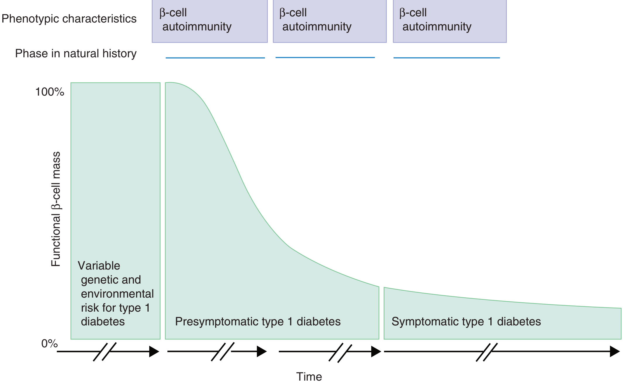
The figure below shows the natural history - by the time symptoms appear (Stage 3), up to 85-90% of functional beta-cell mass is already lost:
1. Confirm the Diagnosis of Diabetes
First, standard glycemic criteria must be met (ADA 2024):
| Test | Diagnostic Threshold |
|---|---|
| Fasting plasma glucose | ≥ 126 mg/dL (7.0 mmol/L) |
| 2-hour OGTT glucose | ≥ 200 mg/dL (11.1 mmol/L) |
| HbA1c | ≥ 6.5% |
| Random glucose + symptoms | ≥ 200 mg/dL |
2. Islet Autoantibody Panel (Confirm T1DM Etiology)
This is the cornerstone of confirming immune-mediated beta-cell destruction. Autoantibody markers are present in 85-90% of individuals with immune-mediated diabetes at the time of initial fasting hyperglycemia - (Tietz Textbook of Laboratory Medicine, p. 1728).
| Autoantibody | Notes |
|---|---|
| Anti-GAD65 (Glutamic Acid Decarboxylase) | Most sensitive marker; also elevated in LADA (but at much higher titers in stiff-person syndrome) |
| Anti-IA-2 / IA-2β (Tyrosine phosphatase) | High specificity; present in ~60-70% at diagnosis |
| Anti-ZnT8 (Zinc Transporter 8) | Present in 60-80% at new onset; detected in ~26% of those negative for other antibodies - (Tietz, p. 1727) |
| IAA (Insulin Autoantibodies) | More relevant in young children; may become negative after exogenous insulin started - must test BEFORE insulin initiation |
| ICA (Islet Cell Antibodies) | Older marker; risk approaches 100% for T1DM with even one positive ICA if accompanied by beta-cell dysfunction |
Key principle: The number of positive autoantibodies predicts risk - 1 antibody carries ~10% 10-year risk; multiple antibodies confer up to near 100% risk. - (Tietz, p. 1728)
When antibodies are especially useful: In obese adults, patients >40 years, those with DKA presentation but features of T2DM (strong family history, gestational DM history), or patients initially misclassified as T2DM. - (Goldman-Cecil Medicine, p. 2473)
3. Beta-Cell Reserve: C-Peptide
C-peptide (the connecting peptide cleaved from proinsulin) reflects endogenous insulin secretion:
- Fasting and stimulated C-peptide are used to measure residual beta-cell function
- Low/undetectable C-peptide confirms absolute insulin deficiency of T1DM
- C-peptide is preferred over insulin because it is not affected by exogenous insulin injections
- Glucagon stimulation test or Mixed Meal Tolerance Test (MMTT) can be used to provoke maximal C-peptide response - (Henry's Clinical Diagnosis, p. 619)
- Urine C-peptide:creatinine ratio is also useful in outpatient settings
- Note: C-peptide is elevated in renal failure (degraded renally, unlike insulin)
During the "honeymoon phase" (weeks to months after diagnosis), some beta-cell function persists - C-peptide may still be detectable. It progressively becomes undetectable within a few years.
4. HbA1c
- Reflects 2-3 month average glycemia
- Helps quantify the degree of hyperglycemia at presentation
- Used to monitor therapy going forward (target generally <7% in most adults)
5. Screen for Associated Autoimmune Conditions
T1DM is strongly associated with Autoimmune Polyglandular Syndrome Type 2 (APS-2) and other conditions:
| Condition | Screening Test | Frequency in T1DM |
|---|---|---|
| Autoimmune thyroid disease (Hashimoto's / Graves') | TSH, anti-TPO, anti-TG | 15-69% |
| Celiac disease | Anti-tTG IgA + total IgA (to rule out IgA deficiency) | 3-10% |
| Addison's disease (adrenal insufficiency) | Morning cortisol, 21-OH antibodies | Less common but life-threatening |
| Pernicious anemia | CBC, anti-parietal cell / anti-intrinsic factor antibodies, vitamin B12 | Screen in APS context |
| Vitiligo / alopecia | Clinical exam | - |
Harrison's Principles, p. 3134 notes APS-2 includes Addison's (50-70%), autoimmune thyroid disease (15-69%), and T1DM (40-50%).
6. Genetic Testing (Selected Cases)
- HLA typing (HLA-DQ/DR): HLA-DR4-DQ8 and DR3-DQ2 are present in ~90% of T1DM children. DR15-DQ6 is protective (found in only 1% with T1DM vs. 20% general population). - (Goldman-Cecil, p. 2472)
- Genetic testing is not routine clinical practice but is used in:
- Distinguishing T1DM from MODY (Maturity-Onset Diabetes of the Young)
- Research screening of first-degree relatives
- Counseling regarding recurrence risk in family members
7. Differentiate from Mimics
| Feature | T1DM | T2DM | LADA | MODY |
|---|---|---|---|---|
| Onset age | Usually <30 | >40 (but any age) | Adult (>30) | Any, often young |
| Autoantibodies | Positive (85-90%) | Negative | Positive (esp. GAD65) | Negative |
| C-peptide | Low/absent | Normal/high | Low (progressive) | Normal |
| BMI | Lean | Often obese | Variable | Lean/normal |
| Insulin dependence | Immediate | Delayed | Progressive | Variable |
| Genetics | HLA-DR3/DR4 | Polygenic | Similar to T1DM | Single gene (HNF, GCK) |
8. Baseline Complication Screening
Even at new diagnosis, complications may already be present (especially in late presenters). The ADA recommends screening starting at diagnosis for T2DM; for T1DM, screening is recommended after 5 years of disease - but at de novo presentation, baseline values should be established:
| Complication | Screening |
|---|---|
| Diabetic kidney disease (DKD) | Urine albumin:creatinine ratio (ACR), serum creatinine/eGFR |
| Retinopathy | Dilated fundus exam within 5 years of T1DM onset |
| Neuropathy | Clinical exam - monofilament, vibration |
| Cardiovascular risk | Lipid panel, blood pressure |
9. Acute Assessment at Presentation
- Arterial blood gas / venous blood gas - if DKA suspected (anion gap, pH)
- Serum electrolytes, BMP - Na, K, bicarbonate, BUN, creatinine
- Urine ketones / serum beta-hydroxybutyrate - confirm ketosis/ketoacidosis
- CBC - may show leukocytosis during DKA (stress response)
Between 13% and 80% of individuals worldwide present initially with DKA (higher in resource-limited settings and children under age 5). - (Goldman-Cecil, p. 2473)
Summary Checklist for De Novo T1DM
☐ Confirm diagnosis: FPG / OGTT / HbA1c
☐ Islet autoantibodies: GAD65, IA-2, ZnT8, IAA (before insulin started)
☐ C-peptide (fasting ± stimulated): assess residual beta-cell function
☐ HbA1c: degree of glycemic control
☐ Metabolic panel + ketones/pH: rule out DKA
☐ Thyroid function (TSH, anti-TPO): associated autoimmune screen
☐ Celiac serology (anti-tTG IgA + total IgA)
☐ Adrenal screen (morning cortisol) if features of APS-2
☐ Lipid panel, blood pressure: cardiovascular risk
☐ Urine ACR, eGFR: baseline renal function
☐ Ophthalmology referral: baseline retinal screen
☐ MODY genetics if: no autoantibodies + family history + lean young adult + mild fasting hyperglycemia
Recent evidence note: A 2024 Consensus Guidance in Diabetes Care (PMID 38912694) addresses monitoring of autoantibody-positive pre-Stage 3 individuals, reinforcing the value of serial autoantibody testing and C-peptide in risk stratification. A 2026 JAMA Review (PMID 41697686) and 2025 Diabetes Obes Metab review (PMID 40734585) provide updated guidance on T1DM classification and pathophysiology.
Find guidelines for treatment of type 1 diabetes mellitus
Reading File
Quick answer: T1DM treatment is built on insulin replacement (basal-bolus regimens), blood glucose monitoring, carbohydrate counting, and prevention/treatment of hypoglycemia and DKA. Modern guidelines (ADA 2025, ISPAD) emphasize continuous glucose monitoring (CGM), insulin pumps (CSII), and closed-loop systems as the preferred approach where available.
Searching sources...
Finding Sources
Searching PubMed
"type 1 diabetes" AND "management" AND "guidelines"
Finding Sources
Searching PubMed
"type 1 diabetes" AND insulin AND treatment
Searching the Web
ADA 2025 standards of medical care type 1 diabetes treatment guidelines
Finding Sources
Finding Sources
Reading Web Page
https://diabetesjournals.org/care/article/44/11/2589/138492/…
Reading File
Reading File
Finding Sources
Reading File
Finding Sources
Reading File
Finding Sources
Reading File
Finding Sources
Reading File
Reading File
I now have comprehensive information from multiple authoritative sources. Let me compile the full treatment guidelines.
Treatment of Type 1 Diabetes Mellitus - Evidence-Based Guidelines
Guideline sources: ADA Standards of Care in Diabetes 2025, ADA/EASD Consensus Report on T1DM Management in Adults, Goldman-Cecil Medicine (2024), Katzung's Pharmacology 16e, Textbook of Family Medicine 9e
1. Overarching Goals
| Goal | Target (ADA 2025) |
|---|---|
| HbA1c | <7.0% in most adults; individualize (e.g., <6.5% if safely achievable without hypoglycemia; <8.0% in elderly, hypoglycemia-prone) |
| Time in Range (CGM) | >70% of time at 70-180 mg/dL |
| Time below range | <4% of time <70 mg/dL; <1% of time <54 mg/dL |
| Time above range | <25% of time >180 mg/dL |
| Pre-meal glucose | 80-130 mg/dL |
| Post-meal (2h) glucose | <180 mg/dL |
The primary goal is maintaining physiologic glucose concentrations to prevent acute complications (DKA, hypoglycemia) and chronic complications (retinopathy, nephropathy, neuropathy, cardiovascular disease). - (Goldman-Cecil Medicine, p. 2474)
2. Insulin Therapy (The Cornerstone)
Everyone with T1DM requires exogenous insulin - there are no oral-only options.
2a. Insulin Types and Pharmacokinetics
| Insulin Type | Examples | Onset | Peak | Duration |
|---|---|---|---|---|
| Rapid-acting analogues (prandial) | Lispro, Aspart, Glulisine | 15-30 min | 30-90 min | 3-4 hours |
| Short-acting (Regular) | Human insulin U100/U500 | ~30 min | 2-4 hours | 5-8 hours |
| Intermediate-acting (NPH) | Human NPH | 2-4 hours | 6-10 hours | 16-20 hours |
| Long-acting analogues (basal) | Glargine U100 | 1.5-2 hours | Peakless | 16-20 hours |
| Detemir | 1.5-2 hours | Peakless | 16-20 hours | |
| Glargine U300 | 1.5-2 hours | Peakless | 30-36 hours | |
| Degludec U100/U200 | 1.5-2 hours | Peakless | 36-40 hours |
- (Goldman-Cecil Medicine, Table 210-4, p. 2474)
Long-acting analogues (glargine, detemir, degludec) produce less hypoglycemia and less weight gain than NPH, and have more predictable absorption. They are preferred over NPH.
2b. Multiple Daily Injection (MDI) - "Basal-Bolus" Regimen
The physiological insulin replacement strategy - most commonly used:
- Total Daily Dose (TDD): 0.3-1.0 unit/kg/day
- 50% as basal insulin (once or twice daily long-acting analogue)
- 50% divided as prandial boluses before each meal
Bolus dose calculation:
- 1 unit per 10-15 g carbohydrate consumed (insulin-to-carb ratio, varies by time of day)
- Plus a correction factor: 1 unit lowers blood glucose by ~20-50 mg/dL (individualized)
(Textbook of Family Medicine 9e, p. 987)
2c. Continuous Subcutaneous Insulin Infusion (CSII - Insulin Pump)
Delivers rapid-acting insulin 24/7 via a subcutaneous catheter, changed every 2-3 days.
Advantages over MDI:
- Better HbA1c control (reduces HbA1c by ~0.3-0.5%)
- Less hypoglycemia (especially nocturnal)
- Adjustable basal rates for exercise, illness, dawn phenomenon
- Better quality of life scores
Candidates for CSII (ADA/CSII criteria):
- Frequent hypoglycemia or hypoglycemia unawareness
- Dawn phenomenon causing AM hyperglycemia
- Poor glycemic control despite MDI
- Lifestyle flexibility needs (shift workers, athletes)
- Motivated, educated patients willing to self-monitor glucose 4-6x/day
(Katzung 16e, p. 1182; Textbook of Family Medicine 9e, p. 989)
2d. Closed-Loop Systems ("Artificial Pancreas") - Current Best Practice
Integrates CGM + insulin pump with an automated algorithm:
- CGM continuously measures interstitial glucose
- Algorithm automatically adjusts basal insulin delivery
- Patient still doses bolus insulin at meals (hybrid closed loop)
Examples: MiniMed 770G (Medtronic), Tandem Control-IQ, Omnipod 5
Evidence: Closed-loop systems reduce nighttime hypoglycemia and significantly lower HbA1c. Multiple RCTs and cross-over trials confirm superiority over MDI. - (Goldman-Cecil, Grade A References; Katzung 16e, p. 1182)
3. Glucose Monitoring
3a. Continuous Glucose Monitoring (CGM) - Preferred
- Real-time or flash CGM is now recommended for all T1DM patients regardless of treatment mode
- Studies show improved glucose control without increased hypoglycemia with CGM in adults ≥25 years on intensive insulin
- CGM alarms help detect and prevent hypoglycemia
- The CONCEPTT Trial (2017) showed CGM improved outcomes even in pregnant women with T1DM
(Tietz Laboratory Medicine, p. 2797)
3b. Self-Monitoring of Blood Glucose (SMBG)
- If CGM not available: monitor at minimum before meals, 2 hours post-meal, at bedtime, before exercise, and when symptomatic
- Test urine or capillary ketones if glucose persistently >250 mg/dL (>2+ = urgent care)
4. Diet and Lifestyle
- Carbohydrate counting is the preferred nutrition approach - allows flexible prandial dosing
- Carbohydrate consistency (fixed grams/meal) is an alternative for simpler dosing
- Learn to read food labels; consider a registered dietitian referral at diagnosis
- No specific carbohydrate target; generally 45-60% of calories for most
- Rapidly-absorbed carbohydrates (high GI foods) should be minimized
- Mediterranean diet recommended for cardiovascular benefit
- Caloric restriction only needed if overweight/obese
- Exercise: Regular aerobic and resistance exercise is recommended. Adjust insulin doses and carbohydrate intake around physical activity. Monitor glucose before, during, and after exercise. A 2025 EASD/ISPAD Position Statement (PMID 39657609) addresses automated insulin delivery around exercise. - (Goldman-Cecil, p. 2475)
5. Adjunctive Pharmacological Therapies
These are add-ons to insulin - not substitutes:
| Agent | Mechanism | HbA1c reduction | Notes |
|---|---|---|---|
| Pramlintide (amylin analogue) | Slows gastric emptying, suppresses glucagon | 0.3-0.5% | SC injection before meals; causes nausea; rarely used |
| SGLT2 inhibitors (dapagliflozin, sotagliflozin) | Glycosuria | 0.28-0.48% | Approved for T1DM in EU, not in US/UK; risk of euglycemic DKA (3-6%) |
| GLP-1 receptor agonists | Incretin effect | Variable | Under investigation for T1DM |
Important: SGLT2 inhibitors are not approved for T1DM in the United States due to DKA risk. They are available in some countries as adjuncts in adults who are overweight. If used, patients must be educated to monitor ketones. - (Goldman-Cecil, p. 2475)
6. Management of Acute Complications
6a. Hypoglycemia ("15-15 Rule")
- Mild/moderate (glucose <70 mg/dL, alert): 15 g fast-acting carbohydrate (4 glucose tablets, 150 mL juice); recheck in 15 min; repeat if still <70 mg/dL
- Severe (unconscious/unable to swallow): IM/SC glucagon (1 mg) OR IV dextrose 25 g (50 mL of 50% dextrose)
- Glucagon options: Traditional 1 mg IM kit; or nasal glucagon (Baqsimi) 3 mg intranasal - no reconstitution needed
- Hypoglycemia unawareness: raise glucose targets temporarily; structured hypoglycemia avoidance programs
6b. Diabetic Ketoacidosis (DKA)
- IV fluid resuscitation: Normal saline 1-2 L/hour initially, then 250-500 mL/hour
- Insulin: IV regular insulin infusion 0.1 unit/kg/hour (or 0.14 unit/kg/hour without bolus)
- Potassium replacement: Add K+ to IV fluids when K+ <5.5 mEq/L and adequate urine output; K+ must be ≥3.5 mEq/L before insulin started
- Bicarbonate: Only if pH <6.9
- Transition to SC insulin: When glucose <200-250 mg/dL, anion gap closed, patient tolerating oral intake - overlap SC and IV insulin by 1-2 hours before stopping drip
- Monitor glucose hourly; electrolytes every 2-4 hours
7. Technology Hierarchy (ADA 2025 Preferred Approach)
BEST: Closed-loop (hybrid/advanced hybrid)
         ↑
      CGM + CSII (open-loop)
         ↑
      CGM + MDI (basal-bolus)
         ↑
      SMBG + MDI (basal-bolus)  ← minimum standard
         ↑
AVOID: Single or twice-daily injections alone
8. Disease-Modifying Therapy (Prevention)
- Teplizumab (Tzield): Anti-CD3 monoclonal antibody, FDA approved (2022) for Stage 2 T1DM (2+ autoantibodies + impaired glucose tolerance in patients ≥8 years). 14-day IV infusion. Delays progression to Stage 3 (clinical) T1DM by ~25 months. - (Katzung 16e, p. 1174)
- Not a treatment for established T1DM - used in pre-symptomatic at-risk individuals
9. Monitoring and Follow-Up
| Parameter | Frequency |
|---|---|
| HbA1c | Every 3 months if not at target; every 6 months if stable and at goal |
| CGM metrics (TIR, TAR, TBR) | Review at every visit |
| Blood pressure | Every visit |
| Lipid panel | Annually |
| Urine ACR + eGFR | Annually (after 5 years of T1DM) |
| Ophthalmology | At 5 years, then annually |
| Foot exam | Annually |
| Thyroid function (TSH) | Annually or if symptoms |
| Celiac serology | At diagnosis, then every 2 years or if symptoms |
| Mental health screening | Annually (diabetes distress, depression, disordered eating) |
10. Multidisciplinary Team
Every patient with T1DM should ideally be managed by:
- Endocrinologist (primary management)
- Certified Diabetes Care and Education Specialist (CDCES)
- Registered Dietitian Nutritionist (RDN)
- Mental health professional
- Ophthalmologist / Podiatrist / Nephrologist as complications develop
Key Recent Guideline Updates (2024-2026)
- ADA Standards of Care 2025: Updated HbA1c targets, CGM as standard of care, closed-loop recommendation strengthened
- EASD/ISPAD Position Statement 2025 (PMID 39657609): Automated insulin delivery around exercise guidance
- International Consensus on CGM in Pregnancy with T1DM (2026, PMID 41421368): CGM + automated insulin delivery recommended during pregnancy
- Continuous Ketone Monitoring Expert Recommendations (2026, PMID 41381175): New technology for DKA prevention guidance
This is a shared conversation. Sign in to Orris to start your own chat.