Superior vena cava, Inferior vena cava, Portal vein: Their branches and anastomoses
Reading File
Finding Sources
Finding Sources
Finding Sources
Reading File
Reading File
Reading File
Finding Sources
Reading File
Reading File
Finding Sources
Loading Image
Loading Image
Loading Image
Finding Sources
Reading File
Finding Sources
Reading File
Reading File
Loading Image
I now have all the information needed for a comprehensive answer. Let me compile it.
Superior Vena Cava, Inferior Vena Cava & Portal Vein — Branches and Anastomoses
Source: Gray's Anatomy for Students
I. Superior Vena Cava (SVC)
Formation & Course
The SVC begins posterior to the lower edge of the right first costal cartilage, where the right and left brachiocephalic veins unite. It descends vertically and terminates at the lower edge of the right third costal cartilage, emptying into the right atrium. Its lower half lies within the pericardial sac (middle mediastinum).
Tributaries
The SVC itself receives only two direct tributaries:
- Azygos vein — arches over the root of the right lung at ~T4 to join the SVC just before it enters the pericardial sac
- Pericardial and mediastinal veins (minor)
Tributaries of the Brachiocephalic Veins (which form the SVC)
The right and left brachiocephalic veins each form at the junction of the internal jugular and subclavian veins on their respective sides.
| Tributaries | Right Brachiocephalic | Left Brachiocephalic |
|---|---|---|
| Vertebral veins | ✓ | ✓ |
| 1st posterior intercostal vein | ✓ | ✓ |
| Internal thoracic veins | ✓ | ✓ |
| Inferior thyroid veins | ✓ (may) | ✓ |
| Left superior intercostal vein | — | ✓ |
| Thymic & pericardial veins | may | may |
The Azygos System (key SVC tributary)
The azygos vein arises at L1/L2 from the junction of the right ascending lumbar vein and the right subcostal vein (or directly from the IVC). It enters the thorax through the aortic hiatus, ascends in the posterior mediastinum, and arches anteriorly at T4 to join the SVC.
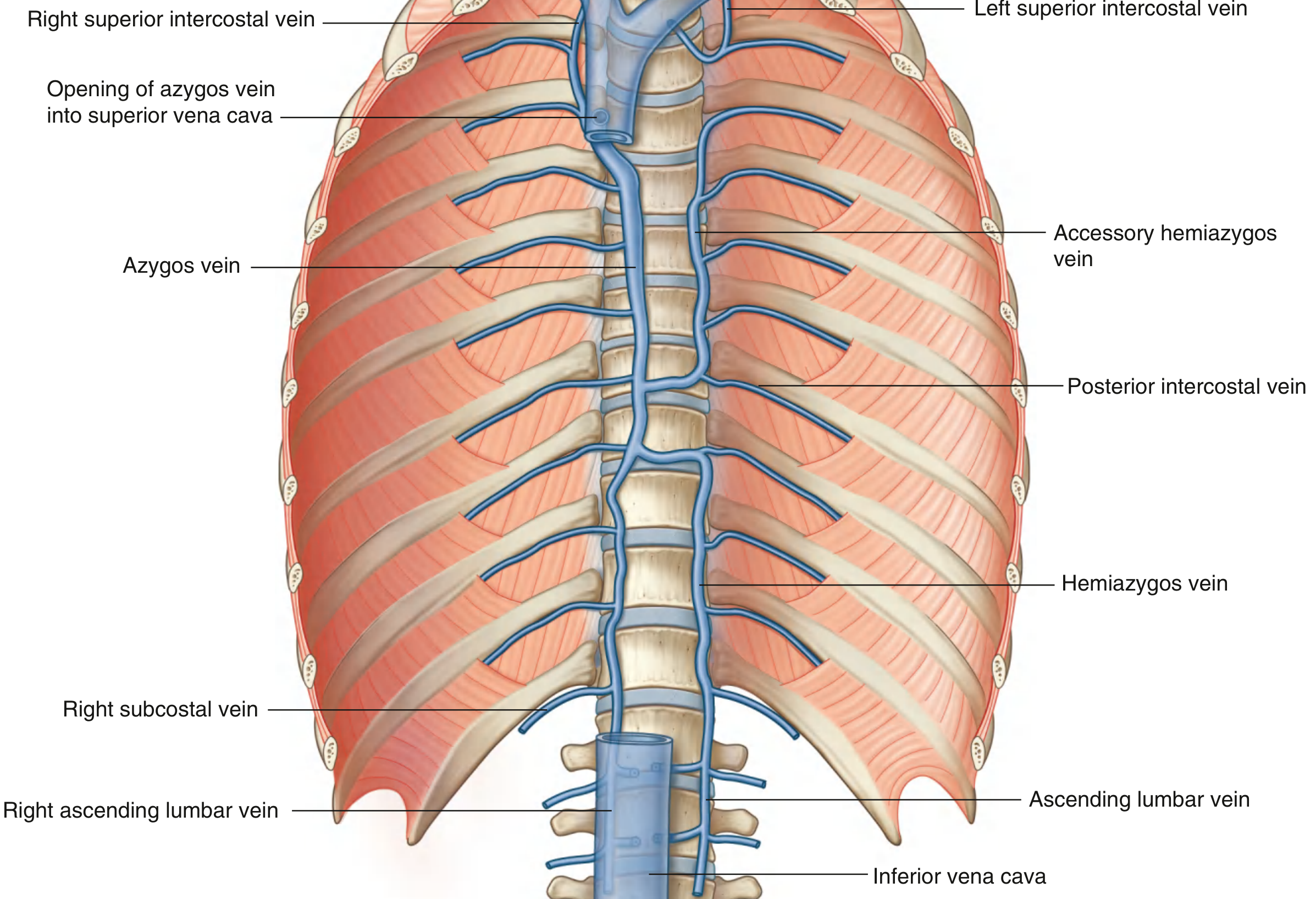
Tributaries of the azygos vein:
- Right superior intercostal vein (2nd–4th intercostal veins; the 1st drains into the right brachiocephalic vein)
- 5th–11th right posterior intercostal veins
- Hemiazygos vein (inferior hemiazygos) — arises from junction of left ascending lumbar + left subcostal veins; ascends on the left to ~T9, then crosses the vertebral column posteriorly to join the azygos. Receives the lowest 4–5 left posterior intercostal veins + esophageal and mediastinal veins
- Accessory hemiazygos vein (superior hemiazygos) — descends on the left from upper posterior mediastinum to ~T8, crosses to join azygos or hemiazygos; receives the 4th–8th left posterior intercostal veins; connects superiorly to the left superior intercostal vein
- Esophageal, mediastinal, pericardial, and right bronchial veins
II. Inferior Vena Cava (IVC)
Formation & Course
The IVC is formed at vertebral level LV by the union of the two common iliac veins, just to the right of the midline. It ascends anterior to the vertebral column, immediately to the right of the abdominal aorta, and exits the abdomen by piercing the central tendon of the diaphragm at T8 to drain into the right atrium.
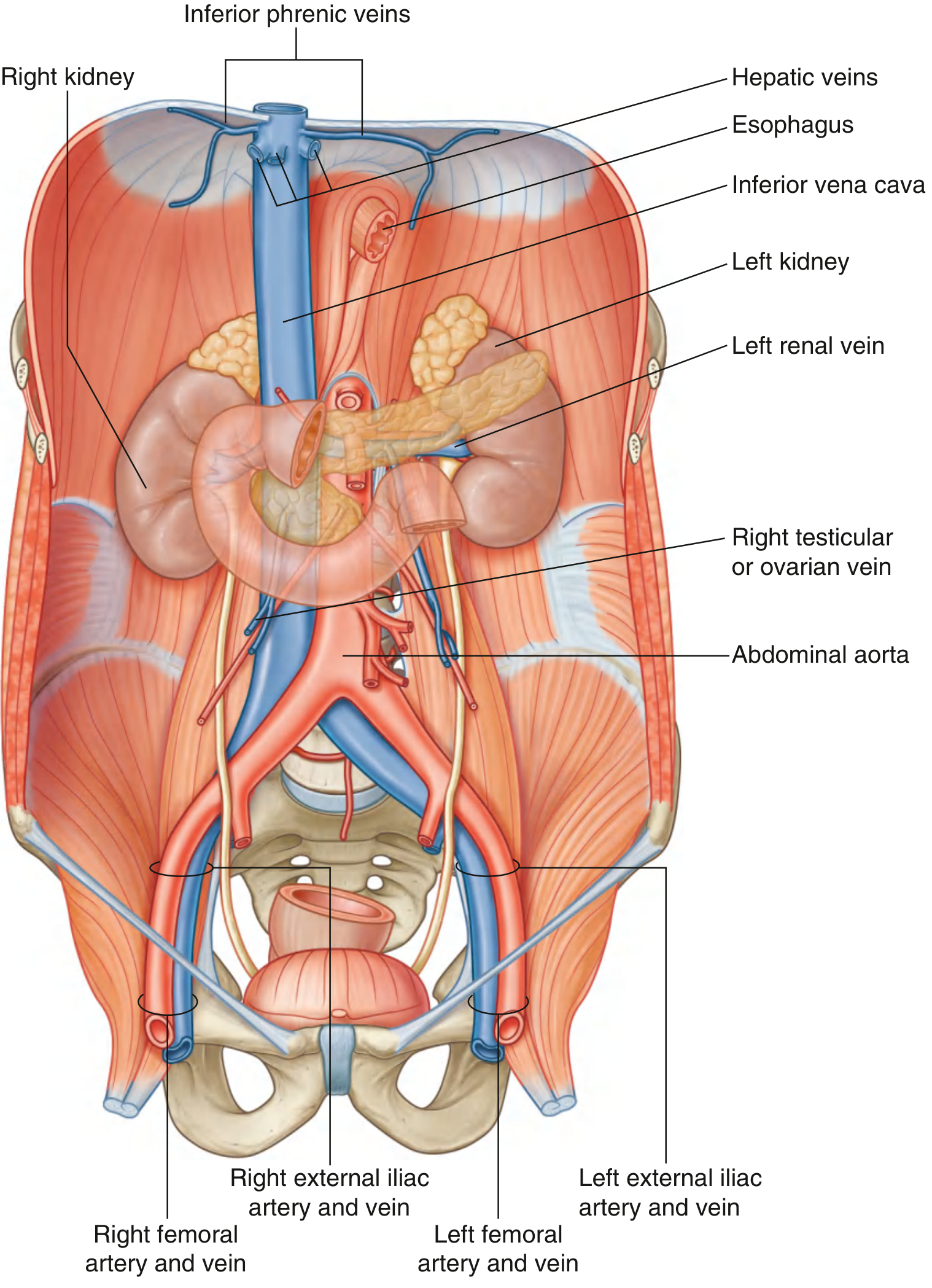
Tributaries (inferior → superior)
| Tributary | Level / Note |
|---|---|
| Common iliac veins | L5 — formation |
| Lumbar veins (3rd & 4th) | Drain directly into IVC; 1st & 2nd may go to ascending lumbar veins; 5th goes to iliolumbar vein → common iliac |
| Right testicular/ovarian vein | Right side drains directly; left side drains into left renal vein |
| Renal veins | Both sides |
| Right suprarenal vein | Right drains directly; left suprarenal drains into left renal vein |
| Inferior phrenic veins | Near the diaphragm |
| Hepatic veins | Just before piercing the diaphragm |
Note: Veins from the GI tract, spleen, pancreas, and gallbladder do not drain into the IVC directly — they form the portal system and pass through the liver first.
The Lumbar Veins and Their Anastomotic Role
The ascending lumbar veins connect the common iliac, iliolumbar, and lumbar veins below with the azygos and hemiazygos veins in the thorax above. This creates an important IVC–SVC collateral pathway: if the IVC becomes obstructed, the ascending lumbar veins serve as the major collateral channel between the lower and upper body.
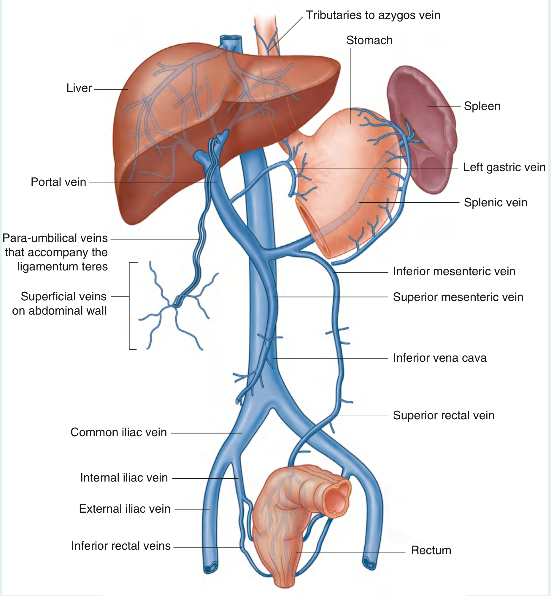
III. Portal Vein
Formation & Course
The portal vein is formed posterior to the neck of the pancreas at L2, by the union of the:
- Superior mesenteric vein
- Splenic vein
It ascends, passes posterior to the superior duodenum, enters the right free margin of the lesser omentum, and runs anterior to the omental (epiploic) foramen. In the lesser omentum it lies:
- Posterior to the bile duct (right) and hepatic artery proper (left)
- At the porta hepatis it divides into right and left branches entering the liver parenchyma

Tributaries of the Portal Vein
A. Splenic Vein
Formed from multiple vessels at the hilum of the spleen; passes through the splenorenal ligament with the splenic artery; runs along the posterior surface of the body of the pancreas.
Tributaries of the splenic vein:
- Short gastric veins (fundus + left greater curvature)
- Left gastro-omental vein (greater curvature)
- Pancreatic veins (body and tail)
- Inferior mesenteric vein (usually)
B. Superior Mesenteric Vein (SMV)
Drains the small intestine, cecum, ascending colon, and transverse colon. Begins in the right iliac fossa; ascends to the right of the superior mesenteric artery.
Tributaries of the SMV:
- Jejunal and ileal veins
- Ileocolic vein
- Right colic vein
- Middle colic vein
- Right gastro-omental vein (right greater curvature)
- Anterior and posterior inferior pancreaticoduodenal veins
C. Inferior Mesenteric Vein (IMV)
Drains rectum, sigmoid, descending colon, and splenic flexure. Begins as the superior rectal vein; ascends receiving sigmoid and left colic veins; usually joins the splenic vein (occasionally the junction of splenic + SMV, or SMV alone).
D. Direct Portal Vein Tributaries
- Right and left gastric veins — drain the lesser curvature of the stomach and abdominal esophagus
- Cystic veins — from the gallbladder
- Para-umbilical veins — associated with the obliterated umbilical vein (ligamentum teres); connect to veins of the anterior abdominal wall
IV. Portosystemic (Portocaval) Anastomoses
These are sites where portal tributaries communicate with systemic (caval) veins. In health they are small; in portal hypertension (e.g., cirrhosis) they dilate into varices.
| Site | Portal Side | Systemic Side | Varix Formed |
|---|---|---|---|
| Gastroesophageal junction | Left gastric vein + tributaries | Azygos system (SVC) | Esophageal varices — most dangerous; may bleed profusely |
| Anorectal junction | Superior rectal vein (IMV → portal) | Middle & inferior rectal veins → internal iliac → IVC | Anorectal varices |
| Umbilicus / anterior abdominal wall | Para-umbilical veins | Superficial veins of anterior abdominal wall → SVC (above) and femoral/external iliac (below) | Caput medusae |
| Retroperitoneum (minor) | Colic and mesenteric veins | Retroperitoneal veins → renal, suprarenal, gonadal (IVC) | Veins of Retzius (less clinically prominent) |
IVC–SVC Anastomoses (Caval Collaterals)
When the IVC itself is obstructed, blood finds alternative routes to the SVC via:
- Ascending lumbar veins → azygos/hemiazygos → SVC
- Vertebral venous plexus (Batson's plexus)
- Superficial epigastric / thoracoepigastric veins along the abdominal wall
Clinical Correlations at a Glance
| Finding | Mechanism | Vein System |
|---|---|---|
| SVC syndrome | Obstruction (usually tumor) → facial/arm swelling, engorged neck veins | SVC |
| Pulmonary embolism | DVT → thrombus via IVC → right heart → pulmonary artery | IVC |
| IVC filter placement | Via SVC → right atrium → IVC to catch emboli | SVC/IVC |
| TIPS procedure | Transjugular route: SVC → IVC → hepatic vein → portal vein | SVC/IVC/Portal |
| Esophageal varices | Portal hypertension → left gastric → azygos collaterals | Portal–SVC |
| Caput medusae | Portal hypertension → para-umbilical → abdominal wall veins | Portal–SVC/IVC |
| Anorectal varices | Superior rectal (portal) ↔ middle/inferior rectal (IVC) | Portal–IVC |
— Gray's Anatomy for Students, pp. 254–257, 268–269, 417–420, 453–454
This is a shared conversation. Sign in to Orris to start your own chat.