
Mri brain ango planing
MRI brain angiography planning sequences TOF MRA protocol
https://mrimaster.com/plan-mra-brain
MRI Brain Angiography (MRA Brain) - Planning & Protocol
What is MRA Brain?
Indications
- Intracranial aneurysm (detection/follow-up)
- Arteriovenous malformations (AVM)
- Ischemic stroke / TIA
- Arterial stenosis or occlusion
- Moyamoya disease
- Thunderclap headache (vascular cause suspected)
- Dural sinus thrombosis (MRV)
Sequences in MRA Brain Protocol
1. Three-Plane Localizer (Scout)
- Run first - takes < 25 seconds
- T1-weighted, low-resolution
- Used to localize anatomy and plan all subsequent sequences
2. T2 TSE Axial (Morphological Brain Sequence)
- Plan on the sagittal plane
- Angle the block parallel to the genu and splenium of the corpus callosum
- Check in coronal plane: perpendicular to the 3rd ventricle and brainstem
- Coverage: vertex to foramen magnum (entire brain)
3. 3D TOF (Time-of-Flight) - The KEY MRA Sequence
- T1-weighted gradient echo sequence
- Uses flow-related enhancement: flowing blood entering the slab gives bright signal; stationary tissue is saturated and suppressed
- Short TR deliberately saturates background tissue while incoming "fresh" blood remains bright
- No contrast needed
| Step | Action |
|---|---|
| Plane | Plan the axial 3D block on the sagittal localizer |
| Angulation | Angle the block parallel to the genu and splenium of the corpus callosum |
| Coverage | From 1 cm above the corpus callosum down to the foramen magnum - must cover the entire Circle of Willis |
| Coronal check | Ensure block is perpendicular to the 3rd ventricle and brainstem |
| Saturation band | Place a superior saturation band on top of the axial block to suppress venous inflow (preventing contamination from draining veins) |
| Multi-slab | Can use multi-slab acquisition to cover a larger volume while maintaining SNR |
4. Typical Scan Parameters (3D TOF)
| Parameter | Value |
|---|---|
| TR | 30-40 ms |
| TE | 5-9 ms |
| Flip angle | 25° |
| NEX | 1 |
| Slice thickness | 0.7 mm |
| Matrix | 256 x 256 |
| FOV | 250 mm |
| Phase direction | R > L |
| Gap | 33% |
| MTC (Magnetization Transfer Contrast) | Yes |
5. Post-Processing: Maximum Intensity Projection (MIP)
- After acquiring the 3D TOF volume, MIP reconstructions are generated
- MIP projects the brightest voxels along any viewing direction
- Creates angiogram-like images of the Circle of Willis
- Rotate in multiple planes (axial, coronal, oblique) for complete vascular assessment
Sequence Order Summary
- Localizer 3-plane (scout)
- T2 TSE Axial (brain morphology)
- TOF 3D Axial Multi-Slab (MRA - Circle of Willis)
Important Tips
- No contrast needed for standard TOF MRA (non-contrast technique)
- Contrast-enhanced MRA (CE-MRA) uses a fast 3D gradient echo (FLASH/SPGR) with gadolinium - used for follow-up after aneurysm coiling or when TOF is suboptimal
- Saturation bands are essential to suppress venous signal - place SUPERIOR to the slab
- On Siemens scanners: avoid excessive angulation of the slab to prevent signal loss or flow artifacts - keep the slab as straight (no tilt) as possible
- MRA brain is combined with MRA neck for full vascular workup in stroke patients
- MRV (MR Venography) uses 2D TOF in oblique sagittal + phase contrast sequences
Circle of Willis Coverage (Key Anatomy)
- Internal carotid arteries (ICA) and their branches
- Middle cerebral arteries (MCA) - M1, M2
- Anterior cerebral arteries (ACA) - A1, A2
- Posterior communicating arteries (PComm)
- Basilar artery
- Posterior cerebral arteries (PCA)
- Vertebral arteries (intracranial)
Image
- Generate a diagram showing MRA brain planning landmarks
- Search for reference images from radiology sources
Upload the image you want analyzed
- Analyze MRI/MRA brain scans
- Identify planning landmarks (corpus callosum, Circle of Willis, etc.)
- Explain what's shown in the image
- Answer any questions about the scan
Create a reference poster of MRA brain planning landmarks
A professional medical reference poster titled "MRA Brain Planning Landmarks" for MRI technologists. The poster shows two main diagrams side by side: LEFT SIDE - Sagittal brain MRI diagram (clean anatomical illustration) with labeled arrows pointing to: - Genu of corpus callosum (front) - Splenium of corpus callosum (back) - A horizontal planning line drawn parallel to the genu-splenium axis - 3D TOF slab block shown as a shaded rectangle covering from "1 cm above corpus callosum" down to "Foramen magnum" - Superior saturation band shown as a striped band above the slab - Label: "Coverage: Circle of Willis" RIGHT SIDE - Axial brain diagram (top-down view) showing the Circle of Willis with labeled vessels: - Internal Carotid Artery (ICA) - bilateral - Middle Cerebral Artery (MCA) - bilateral - Anterior Cerebral Artery (ACA) - bilateral - Anterior Communicating Artery (AComm) - Posterior Communicating Artery (PComm) - bilateral - Posterior Cerebral Artery (PCA) - bilateral - Basilar Artery - Vertebral Arteries - bilateral BOTTOM SECTION - A clean parameter table: "3D TOF Key Parameters: TR 30-40ms | TE 5-9ms | Flip Angle 25° | Slice 0.7mm | FOV 250mm | Matrix 256x256" BOTTOM RIGHT - Small inset box: "Saturation Band: Place SUPERIOR to slab (suppresses venous signal)" Color scheme: Dark navy blue background, white and cyan text, bright yellow annotation arrows, light blue anatomical structures. Clean, professional radiology department style. Medical poster layout.
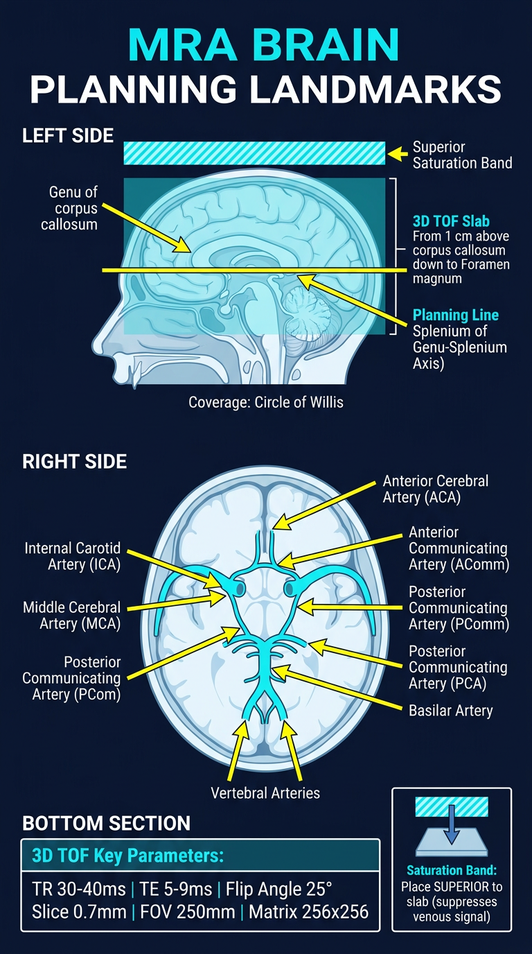
- Sagittal planning diagram - showing the 3D TOF slab positioned parallel to the genu-splenium of the corpus callosum, with the superior saturation band
- Circle of Willis axial diagram - all major vessels labeled (ICA, MCA, ACA, AComm, PComm, PCA, Basilar, Vertebrals)
- Key scan parameters - TR, TE, Flip angle, Slice thickness, FOV, Matrix
- Saturation band reminder - placement tip to suppress venous contamination