
Extensor mechanism of knee
extensor mechanism of the knee quadriceps patella patellar tendon anatomy
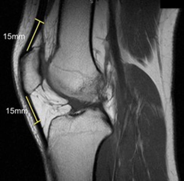
This diagnostic image is a sagittal proton-density magnetic resonance imaging (MRI) scan of the human knee joint, illustrating the anatomy and preoperative measurement protocol for ligament reconstruction. Key visible structures include the distal femur, the patella, the proximal tibia, and the extensor mechanism. The image specifically demonstrates a standardized method for assessing potential donor tissue thickness for autografts, such as Bone-Patellar Tendon-Bone (BPTB) or quadriceps tendon grafts. Two specific measurements are highlighted with yellow lines: a 15 mm segment extending proximally from the superior pole of the patella along the quadriceps tendon, and a 15 mm segment extending distally from the inferior pole of the patella along the patellar tendon. These markers indicate the anatomical locations where tendon thickness should be measured to evaluate graft suitability. The clinical focus of the image is orthopedic preoperative planning for Anterior Cruciate Ligament (ACL) reconstruction, providing a quantitative approach to individualizing surgical graft selection based on the patient's native anatomy.
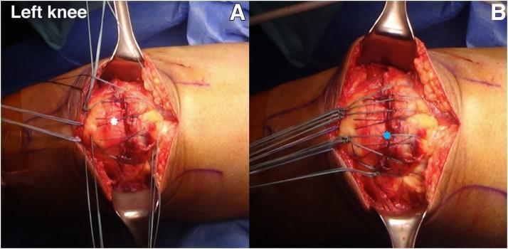
This clinical photograph displays a two-panel intraoperative view (A and B) of a surgical repair for a quadriceps tendon rupture in a left knee. The patient is in a supine position with the surgical site exposed via a midline longitudinal incision. In panel A, the peritendinous quadriceps tendon is being repaired using a figure-of-8 suture technique with No. 1-0 Vicryl sutures, indicated by a white star. The sutures approximate the anterior soft tissue and retinaculum over the superior pole of the patella. Panel B demonstrates the 'suture bridge' technique, where multiple suture limbs originating from the quadriceps tendon are gathered and secured at the inferior pole of the patella, marked by a blue star. This construct ensures the full thickness of the tendon is reattached to the patellar bone, restoring the extensor mechanism. Surgical retractors are visible, providing visualization of the patellar anatomy and the distribution of high-strength suture material across the repair site. The images illustrate key steps in transosseous tendon-to-bone fixation.
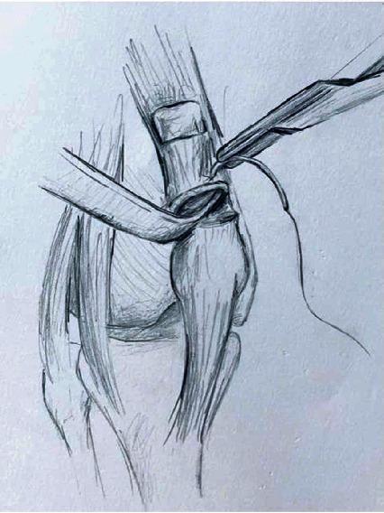
This medical illustration depicts a surgical technique for quadriceps tendon reconstruction using a synthetic mesh graft. The drawing shows the knee joint anatomy including the patella, patellar tendon, and the remnant of the quadriceps tendon. A tubularized mesh, such as a polypropylene mesh, is seen being secured to the proximal quadriceps tendon stump. A surgical needle holder or forceps is shown in the superior-right field, applying a running suture—specifically a Krackow suture technique—to achieve intrasubstance fixation. The mesh is integrated into a longitudinal tunnel within the tendon, providing a scaffold for repair in cases of chronic or recurrent rupture. The illustration focuses on the proximal fixation stage, emphasizing the mechanical anchoring of the graft to the native soft tissue to restore the extensor mechanism of the knee. This pedagogical diagram is intended for orthopaedic surgical education, demonstrating salvage procedures for complex tendon repairs.
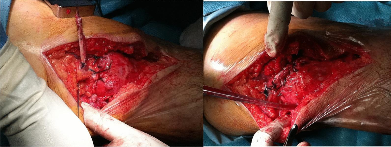
This clinical photograph set depicts an intraoperative view of an orthopedic surgical procedure, specifically a quadriceps tendon rupture repair. The image shows a large, open surgical site on the anterior aspect of the knee. The exposed anatomy includes the subcutaneous adipose tissue, the patella, and the proximal retracted stump of the quadriceps tendon. Key visual features involve the application of a tendon graft secured with multiple black, non-absorbable sutures. These sutures are visible anchoring the graft material to both the periosteum at the patellar tunnel exit holes and to the proximal tendon stump using a series of interrupted knots. Hemostasis is partially managed, with visible areas of clotted and fresh blood within the wound bed. The procedural focus illustrated is the terminal stage of a figure-of-eight graft augmentation used to bridge and reinforce the primary quadriceps tendon repair, demonstrating the method of attachment and suture patterns required for functional stability of the extensor mechanism.
quadriceps muscle anatomy knee vastus rectus femoris diagram
Extensor Mechanism of the Knee
Components
- Quadriceps muscle group (4 muscles)
- Quadriceps tendon
- Medial and lateral retinacula
- Patella (sesamoid bone)
- Patellar tendon (ligament)
- Tibial tubercle (distal bony attachment)
1. Quadriceps Muscle Group
| Muscle | Origin | Note |
|---|---|---|
| Rectus femoris | Anterior inferior iliac spine (AIIS) | Only bi-articular head; crosses hip and knee |
| Vastus lateralis | Lateral lip of linea aspera, greater trochanter | Largest head |
| Vastus medialis | Medial lip of linea aspera | VMO fibers critical for patellar tracking |
| Vastus intermedius | Anterior shaft of femur | Deepest head |
2. Quadriceps Tendon
- Formed by the convergence of all four quadriceps heads
- Has a tri-laminar structure: superficial layer (rectus femoris), middle layer (vastus medialis + lateralis), deep layer (vastus intermedius)
- Inserts on the superior pole (base) of the patella
- The medial and lateral retinacula are expansions of the vastus medialis and lateralis that bypass the patella and insert directly into the tibia - providing additional stabilization
3. The Patella
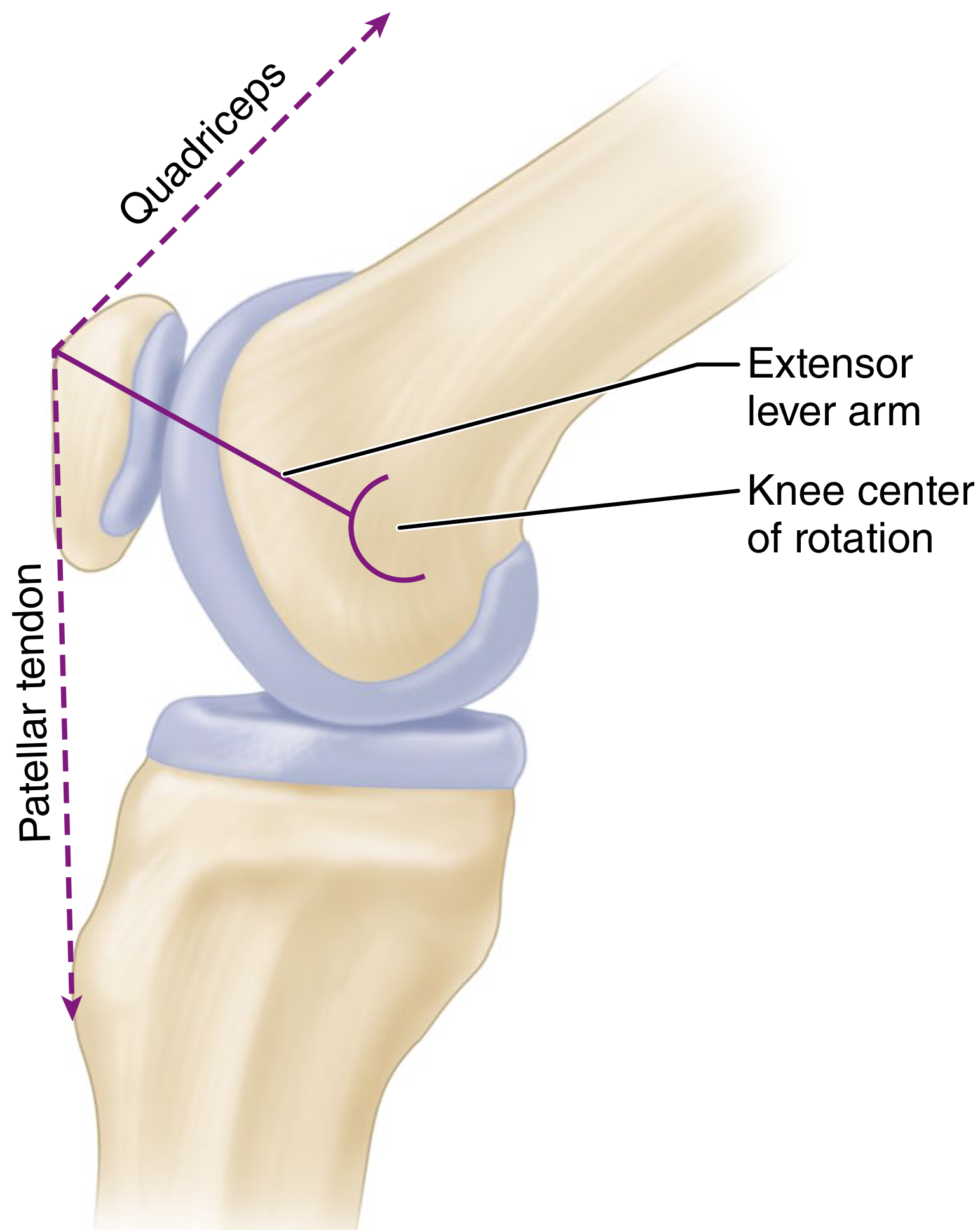
- Increases the mechanical lever arm of the extensor mechanism by displacing the quadriceps and patellar tendon force vectors anteriorly away from the knee's center of rotation
- This lever arm effect is greatest at 20-30 degrees of flexion
- Quadriceps force required for extension increases significantly in the last 20 degrees of extension (less patella in contact with trochlear groove)
4. Patellar Tendon (Ligament)
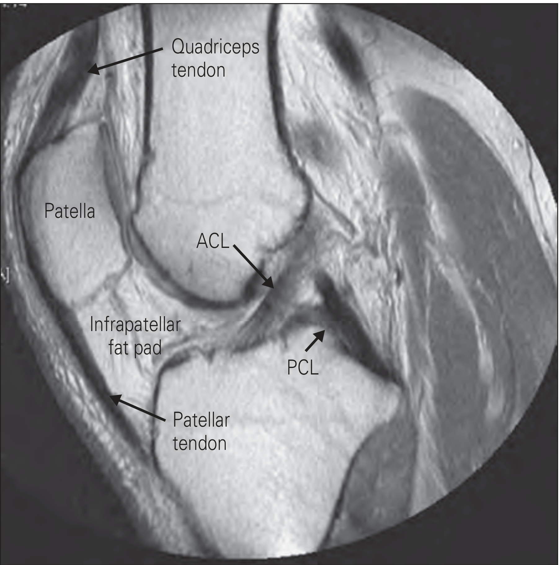
- Runs from the inferior pole (apex) of the patella to the tibial tubercle
- Approximately 4-5 cm long, 3 cm wide
- Strictly speaking it is a ligament (bone to bone), but commonly called "patellar tendon"
- The infrapatellar fat pad (Hoffa's fat pad) lies posterior to the patellar tendon and anterior to the tibia; it is covered by synovial membrane
5. Retinacula
- Medial retinaculum: expansion of vastus medialis; resists lateral patellar displacement
- Lateral retinaculum: expansion of vastus lateralis; tightness can cause lateral patellar tilt
- They serve as secondary patellar stabilizers and help transmit extensor forces even when the central tendon is disrupted
Biomechanics
Q Angle
- Line from ASIS to center of patella (line of quadriceps pull)
- Line from center of patella to tibial tubercle (line of patellar tendon)
Patellofemoral Joint Reaction Force
- During normal walking: ~0.5 x body weight
- Stair climbing: ~3-4 x body weight
- Squatting (>90° flexion): 7-8 x body weight
- These forces are distributed over the patellofemoral contact area, which changes throughout the range of motion
Patellofemoral Contact Areas
- At ~20° flexion: inferior facets contact trochlea
- At 90° flexion: mid-patella contacts trochlea
- At full flexion: only the odd facet (most medial) contacts the medial femoral condyle
VMO and Patellar Stability
- The vastus medialis obliquus (VMO) fibers insert at ~55° to the patellar axis
- VMO is the primary dynamic medial stabilizer of the patella
- Resists lateral subluxation especially in early flexion (0-30°) before bony trochlear constraint becomes dominant
- In deep flexion, bony trochlear groove geometry becomes the primary constraint
Clinical Significance
Disruption of the Extensor Mechanism
- Patellar fracture - most common
- Quadriceps tendon rupture - more common in patients >40 years, associated with systemic diseases (SLE, diabetes, gout, hyperparathyroidism, uremia, obesity), corticosteroid use, and fluoroquinolone antibiotics
- Patellar tendon rupture - more common in patients <40 years, especially athletes
- Inability to actively extend the knee or perform straight leg raise
- Palpable gap in the quadriceps tendon or at the patellar poles
- Patella alta (high-riding patella) = patellar tendon rupture
- Patella baja/infera (low-riding patella) = quadriceps tendon rupture
Common Pathologies
| Condition | Location | Population |
|---|---|---|
| Quadriceps tendon rupture | Suprapatellar | >40 yr, systemic disease |
| Patellar tendon rupture/avulsion | Infrapatellar | <40 yr, athletes |
| Patellar fracture | Patella | Direct blow / eccentric load |
| Patellar tendinopathy ("Jumper's knee") | Inferior pole | Athletes, jumping sports |
| Patellofemoral pain syndrome | PF joint | Young active individuals |
| Osgood-Schlatter disease | Tibial tubercle | Adolescents |
- Rosen's Emergency Medicine, 9th Ed. - "Extensor Mechanism," p. block 7
- Campbell's Operative Orthopaedics, 15th Ed. 2026 - "Patellofemoral Joint Biomechanics and Functional Anatomy," p. 498; "Rupture of Extensor Mechanism of Knee," p. 2970
- Rheumatology, 2-Volume Set, 2022, Elsevier - "Extensor mechanism," p. block 10