
Cheyne stokes
Reading File
Now let me search the medical library and PubMed simultaneously.
Finding Sources
Finding Sources
Searching PubMed
"Cheyne-Stokes respiration"[MeSH Terms]
Excellent results. Now let me read the most relevant sections in parallel.
Reading File
Reading File
Reading File
Reading File
I have excellent material from multiple authoritative textbooks. Let me also fetch the key diagram.
Loading Image
Reading File
I now have comprehensive information from multiple authoritative sources. Here is the full answer:
Cheyne-Stokes Respiration
Definition and Pattern
Cheyne-Stokes respiration (CSR) is a pattern of periodic breathing characterized by rhythmic, cyclical alternation between hyperpnea and apnea. The hallmark is a smooth crescendo-decrescendo (waxing and waning) change in tidal volume, which peaks and then tapers down to a central apnea lasting typically 15-60 seconds, after which the cycle repeats.
- Apneic phase: 15-60 seconds; arterial CO2 is at its lowest at apnea onset
- Hyperpneic phase: usually lasts longer than the apneic phase
- CO2 tension varies by ~14 mmHg and SpO2 by up to 18% across each cycle
The pattern was first graphically described by William Stokes (1854), based on an earlier account by John Cheyne.
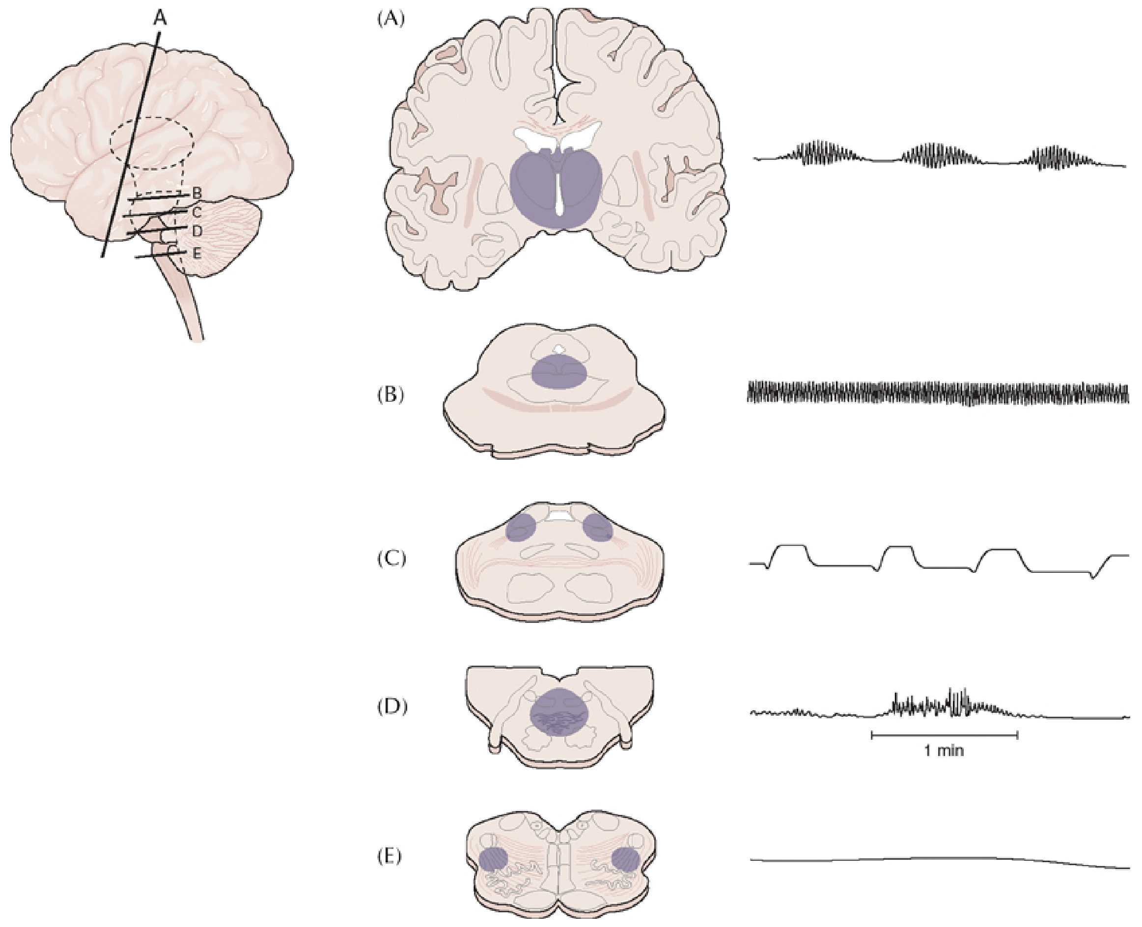
Pathophysiology / Mechanism
CSR arises from instability in the respiratory control loop, not depression of it. The respiratory response to CO2 is actually heightened (not blunted) in CSR.
Two main mechanisms operate:
1. Prolonged Lung-to-Brain Circulation Time (main mechanism in heart failure)
- In heart failure, slowed circulation means pulmonary capillary blood gases reach the medullary chemoreceptors with an abnormal delay - roughly 180 degrees out of phase with current alveolar gas tensions.
- Hyperventilation lowers alveolar CO2, but before this low-CO2 blood reaches the brain, more low-CO2 blood has accumulated. When it finally arrives, the brain sees a sudden fall in CO2, suppresses breathing (apnea), then the cycle resets.
- The feedback loop is too long - it oscillates.
2. Enhanced CO2 Chemosensitivity (neurologic/general)
- Patients with increased chemosensitivity overrespond to CO2: small rises drive marked hyperventilation, which overshoots, driving CO2 below the apnea threshold.
- Neural reflexes from extravascular lung fluid and elevated left atrial pressure further amplify the ventilatory drive.
- During waking, descending cortical pathways prevent post-hyperventilation apnea. During sleep or forebrain impairment, this "damping" is removed and oscillations emerge.
(Plum and Posner's Diagnosis and Treatment of Stupor and Coma; Ganong's Review of Medical Physiology; Fishman's Pulmonary Diseases and Disorders)
Causes and Associations
| Category | Examples |
|---|---|
| Cardiac | Congestive heart failure (most common - occurs in 20-40% of LV systolic dysfunction) |
| Neurologic | Metabolic encephalopathy (uremia, hepatic failure), bilateral forebrain or diencephalic lesions, cerebral infarcts, forebrain mass with diencephalic shift, pontine tegmentum lesions |
| Physiologic | Normal infants, healthy elderly, high altitude |
| Drugs/toxins | Respiratory depressants (e.g., morphine), raised intracranial pressure |
| Sleep | Predominantly NREM sleep; resolves in REM sleep |
Risk factors in heart failure: male sex, advanced age, low baseline PaCO2, atrial fibrillation.
CSR represents about 5-10% of all sleep apnea cases and is uncommon outside heart failure.
Neurological Localization
CSR is classically associated with lesions at the forebrain/diencephalic level (see panel A in the figure above). As lesions descend:
- Pons: apneustic breathing (inspiratory pauses)
- Pontomedullary junction: cluster/ataxic breathing
- Ventrolateral medulla: apnea
This makes CSR a useful localizing sign in comatose patients - it implies the brainstem itself is intact, with supratentorial or diencephalic dysfunction.
(Plum and Posner's; Localization in Clinical Neurology; Bradley and Daroff's Neurology in Clinical Practice)
Clinical Features and Diagnosis
- Patients may report fatigue, daytime sleepiness, and paroxysmal nocturnal dyspnea (PND - a classic heart failure symptom, often caused by underlying CSR)
- Arousals occur during the hyperpneic phase (unlike OSA where arousals correlate with airflow obstruction)
- Bedside diagnosis: careful observation of breathing pattern; crescendo-decrescendo respirations visible
- Sleep lab (PSG): gold standard; CSR resolves in REM sleep; arousals on EEG during hyperpnea
- CSR does not resolve immediately with CPAP (distinguishing it from OSA)
- Mental status fluctuations and EEG changes may occur due to swings in cerebral blood flow
Treatment
- Treat the underlying cause first - optimizing heart failure management (diuretics, ACE inhibitors, beta-blockers, etc.) often resolves CSR
- CPAP: improves breathing indices but not superior to optimal medical therapy for mortality; does not work immediately
- Adaptive servo-ventilation (ASV): improves CSR and quality of life but the SERVE-HF trial showed increased mortality in heart failure with reduced EF - avoid ASV in HFrEF
- Transvenous phrenic nerve stimulation (remedē System): FDA-approved implantable device; normalizes central apnea indices and improves SpO2, but mortality data still pending
- Supplemental O2: can reduce hypoxic oscillations
- Theophylline (respiratory stimulant): occasionally used but limited evidence
(Goldman-Cecil Medicine; Murray & Nadel's Respiratory Medicine)
Key Distinguishing Points: CSR vs. OSA
| Feature | CSR | OSA |
|---|---|---|
| Pattern | Smooth crescendo-decrescendo | Absent airflow with effort |
| Arousals | During hyperpneic phase | End of apnea |
| REM sleep | Resolves | Persists or worsens |
| Response to CPAP | Gradual, incomplete | Immediate |
| Type | Central apnea | Obstructive apnea |
Sources: Plum and Posner's Diagnosis and Treatment of Stupor and Coma; Goldman-Cecil Medicine; Fishman's Pulmonary Diseases and Disorders; Ganong's Review of Medical Physiology, 26th ed.; Murray & Nadel's Textbook of Respiratory Medicine; Localization in Clinical Neurology, 8e.
This is a shared conversation. Sign in to Orris to start your own chat.