
I am medical student, suun har cheez detail mai explain Krna and first pointers mai explain Krna then paragraph mai then image ka thorugh
I'd love to help you as a medical student! Let me understand what topic you want to study.
Aap kis topic ke baare mein padhna chahte hain?
Asking for Preferences
Aap kaunsa medical topic padhna chahte hain? (Which medical topic do you want to study?)
Anatomy! Great choice. Aur thoda specify karein:
Asking for Preferences
Anatomy ka kaunsa specific area / topic padhna hai?
Asking for Preferences
Head & Neck mein specifically kaunsa topic chahiye?
Bilkul! Parotid & Salivary Glands - ek important aur favorite exam topic. Ab main textbooks se detail mein dhundh ke aapko explain karta hoon.
Searching sources...
Reading File
Finding Sources
Finding Sources
Gray's Anatomy for Students aur K.J. Lee's mein best content hai. Ab main parallel mein read karta hoon:
Reading File
Reading File
Excellent! Bahut acha content mila. Ab submandibular aur sublingual ke baare mein aur inn images ko fetch karta hoon:
Reading File
Loading Image
Loading Image
Excellent images! Ab innervation image aur submandibular/sublingual image bhi fetch karta hoon:
Loading Image
Reading File
Ab mere paas saara zaruri content hai. Main ab ek detailed, structured answer likhta hoon.
🦷 Salivary Glands - Complete Anatomy
Source: Gray's Anatomy for Students
OVERVIEW - Salivary Glands Kya Hain?
Quick Pointers:
- Teen pairs of major salivary glands hoti hain: Parotid, Submandibular, Sublingual
- Inke alawa bahut saari minor salivary glands bhi hain (tongue, palate, cheeks, lips mein)
- Minor glands directly oral cavity mein ya small ducts ke through open hoti hain
- Parotid = Sabse badi | Submandibular = Middle | Sublingual = Sabse choti
1. PAROTID GLAND - Sabse Badi Gland
🔵 Pointers (Key Facts)
- Sabse badi major salivary gland hai
- Position: Ear ke neeche aur saamne, mandible ke ramus ke posterior mein
- Shape: Roughly triangular / wedge shaped
- Upar tak zygomatic arch tak extend karti hai
- Neeche mandible ke lower border tak
- Peeche sternocleidomastoid muscle ko cover karti hai
- Aage masseter muscle ke halfway tak jaati hai
📝 Paragraph (Detail mein)
Parotid gland ek shallow, triangular-shaped trench mein position hoti hai jo teen structures se banti hai: peeche sternocleidomastoid muscle, saamne mandible ka ramus, aur upar external acoustic meatus aur zygomatic arch ka posterior aspect. Ye gland masseter muscle ke upar anterior direction mein bhi extend karti hai, aur neeche posterior belly of digastric muscle ke upar.
🎯 Parotid Duct (Stensen's Duct)
Pointers:
- Gland ke anterior edge se nikalta hai
- Zygomatic arch aur corner of mouth ke beech mein (midway)
- Face par transversely cross karta hai
- Masseter ke medial border par turn leke deep jaata hai
- Buccinator muscle ko pierce karta hai
- Oral cavity mein 2nd upper molar tooth ke paas open hota hai
📸 Image 1: Parotid Gland - Lateral View aur Cross Section
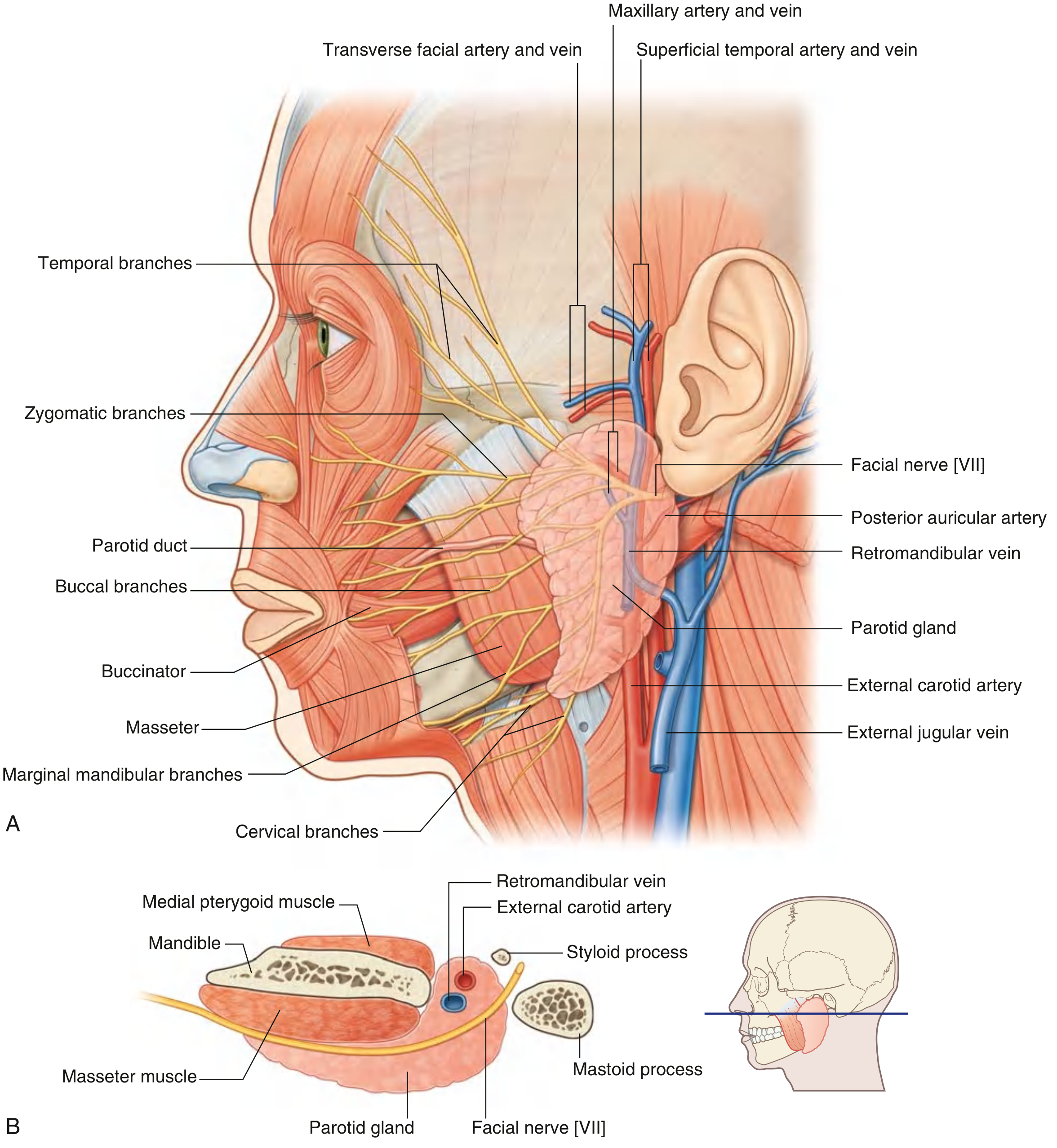
Fig. 8.65 (Gray's Anatomy) - (A) Lateral view mein Facial nerve [VII] ki 5 terminal branches clearly dikhti hain - Temporal, Zygomatic, Buccal, Marginal Mandibular, Cervical. (B) Cross section mein Parotid gland ke andar ka content: External carotid artery, Retromandibular vein, Facial nerve [VII].
📸 Image 2: Parotid Gland ka Position - Surface Anatomy
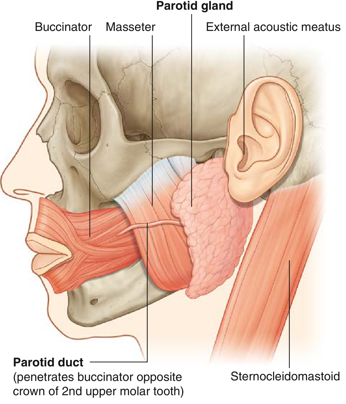
Fig. 8.267 - Clearly dikhta hai: Parotid gland, Parotid duct buccinator ko pierce karta hua, Masseter, Sternocleidomastoid aur External Acoustic Meatus se relations.
⚡ Important Relationships - Gland ke Andar Kya Hai?
Pointers:
- Facial Nerve [VII] - gland ke andar se guzarta hai
- External Carotid Artery - gland mein enter ya deep surface par
- Retromandibular Vein - gland se guzarta hai
Facial Nerve [VII] - EXAM IMPORTANT ⭐
Pointers:
- Stylomastoid foramen se skull exit karta hai
- Parotid gland mein enter karke upper aur lower trunk mein divide hota hai
- Aage aur anastomosis ke baad 5 terminal branches banti hain
- Ye branches gland ke upper, anterior, aur lower borders se bahar nikalti hain
| Branch | Direction se Nikalta Hai |
|---|---|
| Temporal | Upper border |
| Zygomatic | Upper/Anterior border |
| Buccal | Anterior border |
| Marginal Mandibular | Lower border |
| Cervical | Lower border |
Clinical Importance: Parotid surgery (Parotidectomy) mein Facial Nerve [VII] ko damage se bachana bahut mushkil hota hai - isliye surgeon ko ye anatomy perfectly pata honi chahiye!
External Carotid Artery
Pointers:
- Gland ke inferior border mein enter karta hai (ya deep surface se guzarta hai)
- Upar jaate hue Posterior Auricular Artery deta hai
- Phir 2 terminal branches mein divide hota hai:
- Maxillary Artery - horizontally, mandible ke deep
- Superficial Temporal Artery - upar jaata hai, pehle Transverse Facial Artery deta hai phir gland ke upper border se bahar nikalta hai
2. SUBMANDIBULAR GLAND
🔵 Pointers
- Parotid se choti lekin sublingual se badi
- Shape: Hook-shaped (do arms hain)
- Superficial (larger) part: Mylohyoid muscle ke neeche, mandible ke submandibular fossa mein
- Deep (smaller) part: Mylohyoid ke posterior margin ke around loop karke oral cavity mein enter karti hai - hyoglossus muscle ki lateral surface par
📝 Paragraph
Submandibular gland hook-shaped hoti hai. Iska bada arm horizontal plane mein mylohyoid muscle ke neeche forward direction mein hota hai aur mandible mein ek shallow impression (submandibular fossa) ke against direct contact mein hota hai jo mylohyoid line ke inferior mein hoti hai. Chota arm (deep part) mylohyoid ke posterior margin ke around loop karke oral cavity mein ghus jaata hai jahan ye hyoglossus muscle ki lateral surface par tongue ke root ke lateral mein hoti hai.
Submandibular Duct (Wharton's Duct)
Pointers:
- Deep part ke medial side se emerge hota hai
- Oral cavity mein forward pass karta hai
- Sublingual caruncle (papilla) par open hota hai - tongue ke frenulum ke base ke paas
Lingual Nerve ka Important Relation
- Lingual nerve submandibular duct ke neeche se loop karti hai
- Pehle duct ki lateral side cross karti hai
- Phir duct ki medial side cross karti hai
- Phir tongue mein ascend karti hai
Mnemonic: "Lingual nerve wraps around the duct like a rope" - lateral se medial jaata hai!
3. SUBLINGUAL GLAND - Sabse Choti
🔵 Pointers
- Sabse choti major salivary gland
- Shape: Almond shaped
- Location: Oral cavity ke floor mein - submandibular duct aur lingual nerve ke lateral mein
- Mandible ke medial surface ke against - sublingual fossa banati hai (mylohyoid line ke superior, anterior 1/3 mein)
- Sublingual fold raise karti hai - oral cavity floor mein elongate fold of mucosa
Sublingual Gland ka Drainage - Multiple Ducts
- Minor sublingual ducts (bahut saari) - sublingual fold ke crest par open hoti hain
- Kabhi kabhi major sublingual duct - submandibular duct ke saath sublingual caruncle par open hota hai
📸 Image 3: Submandibular aur Sublingual Glands - All Views
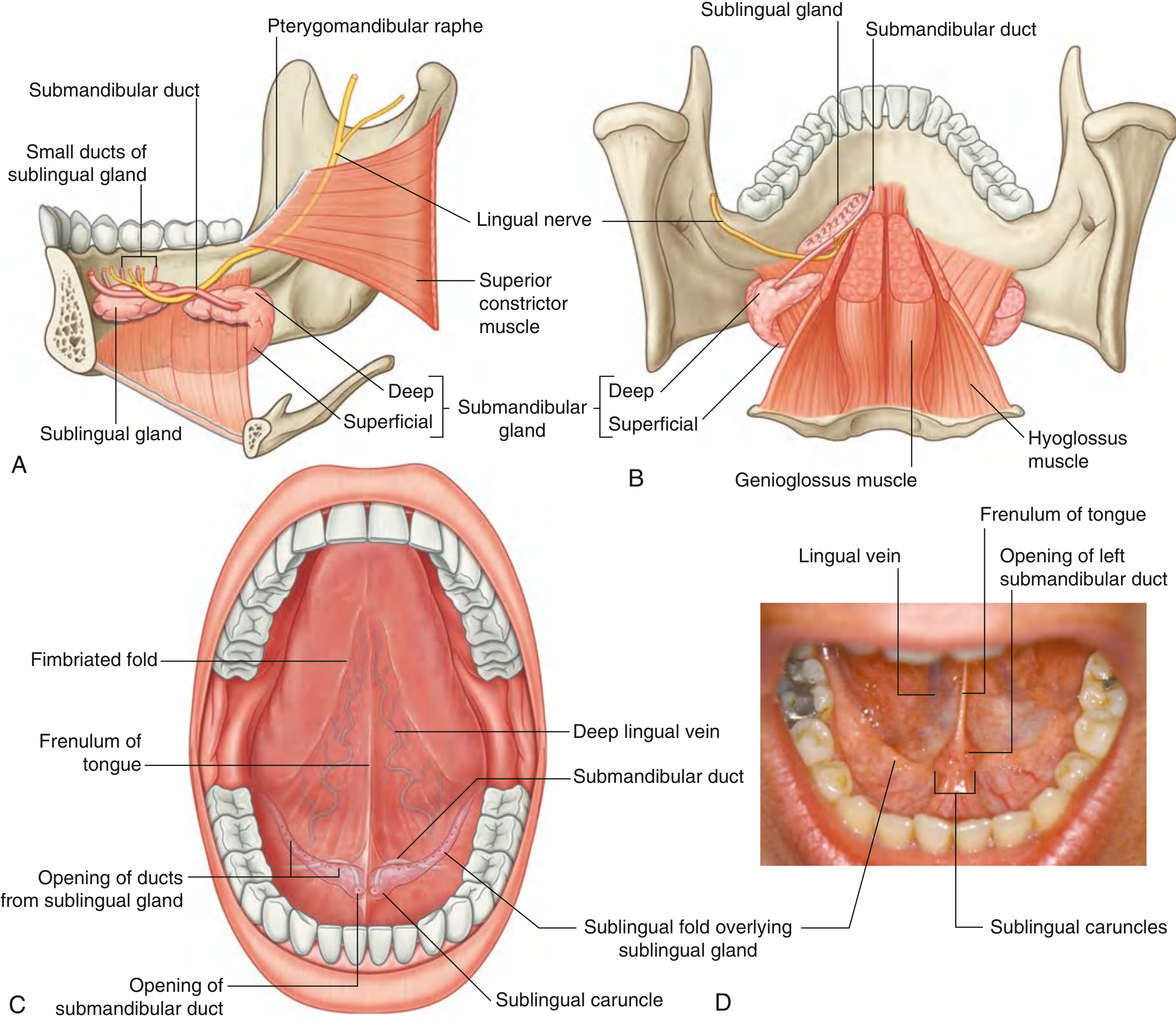
Fig. 8.268 - (A) Medial view: Submandibular gland ka hook shape, deep aur superficial parts, sublingual gland, Wharton's duct, Lingual nerve. (B) Posterior view: Hyoglossus muscle ke relation. (C) Anterior view: Oral cavity ka floor - sublingual fold, sublingual caruncle, opening of ducts. (D) Real photo: Tongue ke neeche submandibular duct ka actual opening sublingual caruncle par.
4. BLOOD SUPPLY - Vessels
Parotid Gland
- Arterial supply: External carotid artery aur uski branches
- Venous drainage: External jugular vein mein
- Lymphatics: Pehle parotid nodes mein (jo gland ke upar ya andar hain) → phir superficial aur deep cervical nodes mein
Submandibular & Sublingual Glands
- Arterial supply: Facial aur lingual arteries ki branches
- Venous drainage: Lingual aur facial veins mein
- Lymphatics: Submandibular nodes → deep cervical nodes (specially jugulo-omohyoid node)
5. INNERVATION - Nerve Supply ⭐⭐ (Exam Favourite!)
PARASYMPATHETIC (Secretomotor) - Saliva Banata Hai
Ye 2 pathways hain based on oral fissure level:
| Parotid Gland | Submandibular + Sublingual | |
|---|---|---|
| Cranial Nerve | CN IX (Glossopharyngeal) | CN VII (Facial) |
| Branch | Lesser Petrosal Nerve | Chorda Tympani |
| Ganglion | Otic Ganglion | Submandibular Ganglion |
| Reaches via | Auriculotemporal nerve (V3) | Lingual nerve (V3) |
Parotid Gland ka Parasympathetic Pathway (Step by Step)
- CN IX → Inferior Petrosal Sinus → Jugular Foramen se exit
- Lesser Petrosal Nerve → Foramen Ovale se → Infratemporal fossa
- Otic Ganglion mein synapse (V3 ke medial mein)
- Postganglionic fibers → Auriculotemporal Nerve (V3 ki branch) ke saath → Parotid gland
Submandibular + Sublingual ka Parasympathetic Pathway
- CN VII → Facial Canal → Chorda Tympani branch
- Chorda Tympani → Infratemporal fossa mein Lingual Nerve (V3) se join
- Oral cavity mein enter → Submandibular Ganglion mein synapse
- Postganglionic fibers → directly submandibular gland ko
- Kuch fibers → lingual nerve ke saath → sublingual aur oral cavity glands ko
SYMPATHETIC
Pointers:
- Superior cervical ganglion se postganglionic fibers
- Blood vessels ke along travel karte hain
- Role: Saliva secretion inhibit karta hai (reduce karta hai)
Clinical Tip: Anticholinergic drugs (atropine) → saliva dry ho jaata hai (dry mouth = xerostomia). Cholinomimetics → excess saliva production.
QUICK COMPARISON TABLE - Teeno Glands
| Feature | Parotid | Submandibular | Sublingual |
|---|---|---|---|
| Size | Largest | Medium | Smallest |
| Shape | Irregular/Triangular | Hook-shaped | Almond-shaped |
| Duct | Stensen's duct | Wharton's duct | Multiple minor ducts |
| Opens at | 2nd upper molar | Sublingual caruncle | Sublingual fold |
| Nerve (Para) | CN IX via Otic gang | CN VII via Subm. gang | CN VII via Subm. gang |
| Artery | External carotid | Facial + Lingual | Facial + Lingual |
| Vein | External jugular | Lingual + Facial | Lingual + Facial |
| Lymph | Parotid → Cervical | Submandibular → Cervical | Submandibular → Cervical |
CLINICAL MNEMONICS 🧠
- "Stensen's = Side tooth" - Parotid duct 2nd upper molar ke paas opens (S for Side, S for Second)
- "Wharton's = Underneath tongue" - Submandibular duct sublingual caruncle par tongue ke neeche
- "Ten, Zygote, Bite, My Chin" = Temporal, Zygomatic, Buccal, Marginal Mandibular, Cervical branches of facial nerve
- "IX for Parotid, VII for rest" - CN9 parotid ko, CN7 submandibular aur sublingual ko
Source: Gray's Anatomy for Students, Chapter 8 (Head & Neck), pp. 1051, 1242-1244
Aapko koi specific topic aur deep mein chahiye? Jaise parotidectomy, parotid tumors, mumps, ya koi aur salivary gland disease? Batao! 🎓
This is a shared conversation. Sign in to Orris to start your own chat.