
from Guyton and Hall textbook of medical physiology, 4th South Asia edition, write a 10-mark long answer on the Gastric Glands-enumerate,secretion , function; Treatment of Peptic Ulcer
Reading File
Finding Sources
Finding Sources
Finding Sources
Reading File
Reading File
Reading File
Reading File
Reading File
Reading File
Reading File
Loading Image
Loading Image
I now have all the information needed from Guyton and Hall. Let me compose the complete 10-mark long answer.
Gastric Glands: Enumeration, Secretions, Functions; and Treatment of Peptic Ulcer
(Source: Guyton and Hall Textbook of Medical Physiology, 4th South Asia Edition - Chapters 65 & 67)
I. GASTRIC GLANDS - ENUMERATION
The stomach mucosa contains two major types of tubular glands, plus a continuous surface lining of mucous cells:
A. Oxyntic Glands (Gastric Glands)
- Located in the inner surface of the fundus and body - the proximal 80% of the stomach.
- These are the primary acid-secreting glands of the stomach.
B. Pyloric Glands
- Located in the antral portion - the distal 20% of the stomach.
- Primarily mucus-secreting; also secrete the hormone gastrin.
C. Surface Mucous Cells
- Cover the entire stomach surface between the glands.
- Not glands per se, but form a continuous mucus-secreting sheet across the mucosa.
II. CELLS OF THE OXYNTIC (GASTRIC) GLAND AND THEIR SECRETIONS
Figure 65.4 (Guyton & Hall) - Gastric (oxyntic) gland from the body of the stomach:
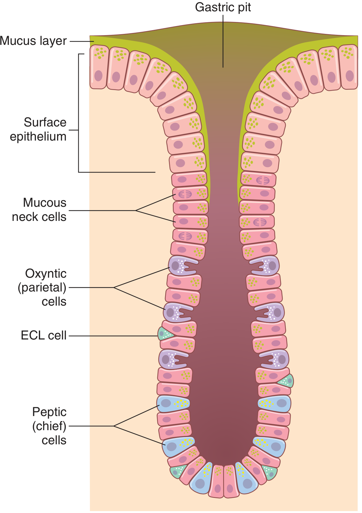
The oxyntic gland is composed of three main cell types, plus additional specialised cells:
1. Mucous Neck Cells
- Secrete: Mucus (thin, less viscid than surface mucus)
- Function: Lubrication and protection of the gland lining; also secrete small amounts of pepsinogen
- Located in the neck (upper portion) of the gland
2. Peptic (Chief) Cells
- Secrete: Pepsinogen (the zymogen precursor of pepsin)
- Located at the base (deeper portion) of the gland
- Activation: Pepsinogen is inactive when secreted. On contact with HCl, the 42,500 molecular weight pepsinogen is cleaved to the active enzyme pepsin (mol. wt. ~35,000). Pepsin is optimally active at pH 1.8-3.5 and is inactivated above pH 5.
- Regulation of pepsinogen secretion:
- Acetylcholine released from vagus nerves or gastric enteric plexus
- Acid in the stomach (indirectly via enteric reflexes)
- Secretin released from the duodenum (moderate stimulation)
3. Parietal (Oxyntic) Cells
- Secrete: (a) Hydrochloric acid (HCl) and (b) Intrinsic factor
- These are the only gastric cells that secrete HCl
- The secreted acid solution contains ~160 mmol/L HCl, nearly isotonic with body fluids; pH ~0.8, making H⁺ concentration approximately 3 million times that of arterial blood
- Energy required: more than 1500 cal/L of gastric juice
Mechanism of HCl Secretion (H⁺-K⁺ ATPase pump):
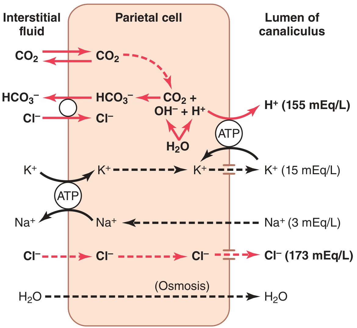
The parietal cell contains branching intracellular canaliculi where HCl is formed. The steps are:
- Water inside the cell dissociates into H⁺ and OH⁻
- H⁺ is actively secreted into the canaliculus in exchange for K⁺, via the H⁺-K⁺ ATPase (the "proton pump")
- OH⁻ combines with CO₂ (via carbonic anhydrase) to form HCO₃⁻, which is transported out of the cell's basolateral membrane in exchange for Cl⁻
- Cl⁻ enters the cell and is secreted through chloride channels into the canaliculus, forming HCl
- Water follows by osmosis; the final secretion contains HCl ~150-160 mEq/L, KCl ~15 mEq/L, and a small amount of NaCl
Intrinsic Factor: Secreted alongside HCl by parietal cells. It is a glycoprotein that combines with vitamin B₁₂ in the stomach and protects it from digestion, enabling its absorption in the terminal ileum. Destruction of parietal cells (e.g., chronic gastritis) causes both achlorhydria and pernicious anemia.
Stimulation of Parietal Cells (Three stimuli):
- Acetylcholine - from vagal or enteric nerve endings
- Gastrin - hormone released from pyloric gland G-cells
- Histamine - released by ECL cells (see below)
4. Enterochromaffin-Like (ECL) Cells
- Secrete: Histamine
- Lie in the deep recesses of the oxyntic glands, in direct contact with parietal cells
- Histamine acts on H₂ receptors of parietal cells to strongly stimulate HCl secretion
- Primary activators of ECL cells: gastrin and acetylcholine
III. PYLORIC GLANDS
- Structurally similar to oxyntic glands but contain few peptic cells and almost no parietal cells
- Contain mostly mucous cells (identical to mucous neck cells of oxyntic glands)
- Secretions:
- Thin mucus - lubricates food movement and protects pyloric mucosa from gastric acid
- Small amounts of pepsinogen
- Gastrin (from G-cells) - the key hormone controlling gastric secretion
IV. SURFACE MUCOUS CELLS
- Cover the entire mucosal surface between glands
- Secrete large quantities of viscid, alkaline mucus forming a gel layer often >1 mm thick over the stomach mucosa
- The alkalinity of this mucus prevents the underlying stomach wall from direct exposure to the highly acidic, proteolytic secretions
- Even slight irritation directly stimulates these cells to produce additional mucus (protective reflex)
V. PHASES OF GASTRIC SECRETION (Summary)
| Phase | Stimulus | Contribution |
|---|---|---|
| Cephalic | Sight, smell, taste, thought of food - via vagus | ~30% |
| Gastric | Food in stomach - vagovagal reflexes, enteric reflexes, gastrin | ~60% |
| Intestinal | Food in duodenum - small gastrin release from duodenal mucosa | ~10% |
VI. PEPTIC ULCER
Definition
A peptic ulcer is an excoriated area of stomach or intestinal mucosa caused by the digestive action of gastric juice or upper small intestinal secretions. Most frequent sites: within a few centimeters of the pylorus, lesser curvature of the antral stomach, and lower esophagus.
Basic Cause
Peptic ulceration results from an imbalance between:
- Aggressive factors: rate of gastric acid and pepsin secretion
- Defensive factors: the gastroduodenal mucosal barrier and neutralisation by duodenal secretions
A peptic ulcer can be caused by:
- Excess secretion of acid and pepsin by the gastric mucosa, OR
- Diminished ability of the gastroduodenal mucosal barrier to protect against digestion
Specific Causes
1. Helicobacter pylori infection (most common - ≥75% of cases)
- H. pylori causes chronic infection of the terminal gastric mucosa and initial duodenal mucosa
- Mechanism: H. pylori penetrates the mucosal barrier physically and by releasing ammonium that liquefies the barrier and stimulates excess HCl secretion
- Infection can last a lifetime unless eradicated with antibacterial therapy
2. Excess acid secretion
- Many duodenal ulcer patients secrete up to twice normal gastric acid - partly from bacterial infection, partly from other causes (e.g., psychic disturbances)
3. Other predisposing factors:
- Smoking - increased nervous stimulation of secretory glands
- Excess alcohol - breaks down the mucosal barrier
- Aspirin and NSAIDs - strong propensity for breaking down the mucosal barrier
VII. TREATMENT OF PEPTIC ULCER
Since the discovery that most peptic ulceration has an infectious basis (H. pylori), therapy has changed dramatically. Almost all patients can be treated effectively by two measures:
1. Antibiotics (to eradicate H. pylori)
- Used along with other agents to kill the infectious bacteria
- Standard regimen involves combinations such as amoxicillin + clarithromycin + a proton pump inhibitor (triple therapy)
2. Acid-Suppressant Drugs
The goal is to reduce gastric acid secretion to allow mucosal healing:
(a) H₂-Receptor Antagonists (Antihistamines)
- Example: Ranitidine
- Mechanism: Blocks the stimulatory effect of histamine on H₂ receptors of gastric gland parietal cells
- Effect: Reduces gastric acid secretion by 70-80%
(b) Proton Pump Inhibitors (PPIs)
- Example: Omeprazole
- Mechanism: Directly blocks the H⁺-K⁺ ATPase pump (the proton pump) in parietal cells
- Effect: Can almost completely block gastric acid secretion
- PPIs are more potent than H₂ blockers
Additional/Supportive Measures
- Antacids - neutralise existing stomach acid
- Mucosal protective agents (e.g., sucralfate, bismuth compounds) - reinforce the mucosal barrier
- Avoidance of aggravating factors: stopping smoking, avoiding NSAIDs, avoiding excess alcohol
SUMMARY TABLE
| Gland/Cell | Location | Secretion | Function |
|---|---|---|---|
| Oxyntic gland - Mucous neck cells | Fundus/body (80%) | Thin mucus, small pepsinogen | Lubrication, protection |
| Oxyntic gland - Chief (peptic) cells | Fundus/body (80%) | Pepsinogen | Activated to pepsin → protein digestion |
| Oxyntic gland - Parietal cells | Fundus/body (80%) | HCl, Intrinsic factor | Protein digestion (acid), B₁₂ absorption |
| Oxyntic gland - ECL cells | Fundus/body (80%) | Histamine | Stimulates parietal cells to secrete HCl |
| Pyloric glands (G-cells) | Antrum (20%) | Mucus, pepsinogen, Gastrin | Mucosal protection, control of gastric secretion |
| Surface mucous cells | Entire stomach mucosa | Viscid alkaline mucus | Gastric mucosal protection (>1 mm gel layer) |
References: Guyton and Hall Textbook of Medical Physiology, 4th South Asia Edition - Chapter 65 (Secretory Functions of the Alimentary Tract), pp. 801-804; Chapter 67 (Physiology of Gastrointestinal Disorders), p. 825
This is a shared conversation. Sign in to Orris to start your own chat.