
Bone
Reading File
The query "Bone" is quite broad. Let me clarify what you're looking for before diving in.
Asking for Preferences
What aspect of bone are you interested in?
Bone histology - covering cellular components, matrix, types, and microscopic organization.
Searching sources...
Finding Sources
Finding Sources
Finding Sources
Finding Sources
Good - found the Histology textbook as the primary source. Let me read the relevant sections now.
Finding Sources
Reading File
Reading File
Reading File
Excellent content found. Let me read the detailed cell descriptions and bone types, and fetch the key diagram.
Reading File
Loading Image
Excellent - got the comprehensive bone diagram. Now let me read the detailed cell descriptions from the histology textbook.
Reading File
Reading File
Reading File
I now have very rich content. Let me also grab the osteoclast description and bone matrix details, and fetch one more key image.
Reading File
Loading Image
I now have comprehensive, detailed information from the histology textbooks. Here is the full, organized answer on bone histology:
Bone Histology
Bone is a specialized connective tissue with a mineralized extracellular matrix, distinguishing it from all other connective tissues. The mineral is calcium phosphate in the form of hydroxyapatite crystals [Ca₁₀(PO₄)₆(OH)₂].
Structure of a Long Bone
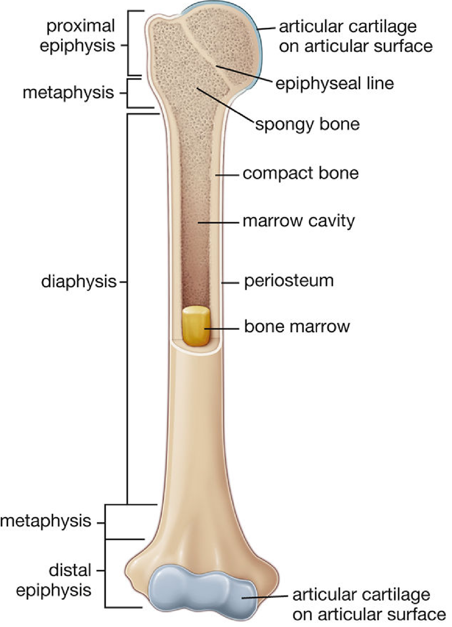
A long bone has three regions:
- Diaphysis - the shaft, composed mainly of compact bone surrounding the marrow cavity
- Epiphysis - the expanded ends, mostly spongy bone with a thin compact outer shell; covered by articular cartilage (hyaline)
- Metaphysis - the flared zone between diaphysis and epiphysis; contains the epiphyseal line (remnant of the growth plate)
Microscopic Organization
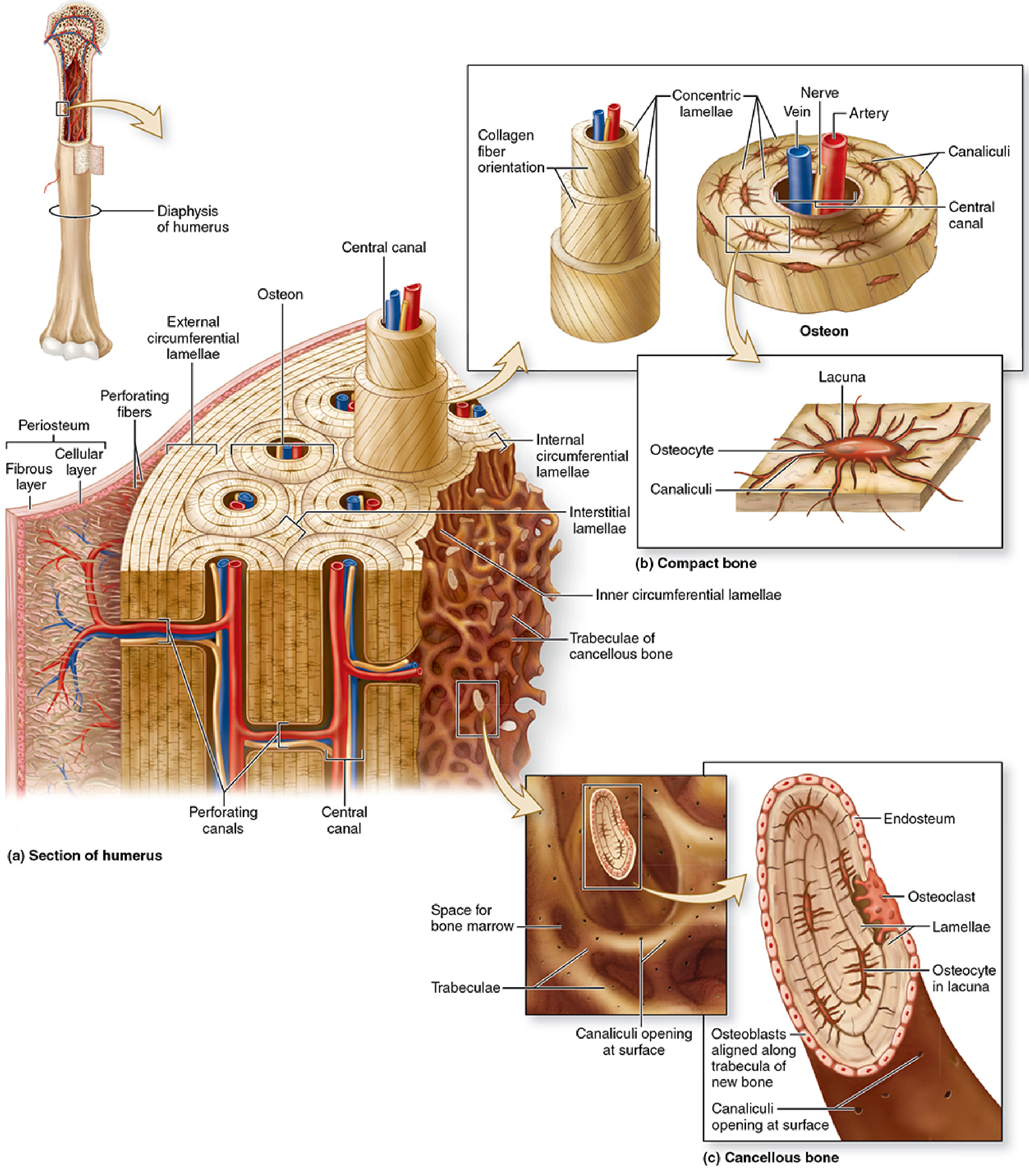
Compact (Cortical) Bone
The structural unit is the osteon (Haversian system) - a series of concentric cylindrical lamellae surrounding a central (Haversian) canal containing blood vessels and nerves. Key components:
| Structure | Description |
|---|---|
| Osteon / Haversian system | Concentric lamellae around a central canal |
| Central (Haversian) canal | Contains artery, vein, nerve, and loose CT |
| Perforating (Volkmann) canals | Run perpendicular to central canals; connect them to the periosteum |
| Lamellae | Layers of calcified matrix; collagen fibers in adjacent lamellae run in alternating directions for strength |
| Lacunae | Small spaces housing osteocytes, between lamellae |
| Canaliculi | Tiny channels radiating from lacunae; carry osteocyte processes and allow nutrient/waste exchange |
| Circumferential lamellae | Outer and inner layers encircling the entire bone shaft |
| Interstitial lamellae | Remnants of old, remodeled osteons filling gaps between complete osteons |
Spongy (Cancellous / Trabecular) Bone
Consists of a network of interconnecting trabeculae (bony spicules) enclosing spaces filled with bone marrow. It lacks osteons; osteocytes receive nutrients directly by diffusion from marrow vasculature through canaliculi.
Bone Matrix
The matrix has two components:
Organic (~35%):
- Type I collagen = 90% of all bone protein; provides tensile strength
- Non-collagenous proteins: osteocalcin, osteonectin, osteopontin (BSP-1), bone sialoprotein-2 (BSP-2), thrombospondins, proteoglycans
Inorganic (~65%):
- Hydroxyapatite crystals deposited along collagen fibrils; provides compressive strength and rigidity
- Also stores calcium, phosphate, carbonate, magnesium, and fluoride
The unmineralized bone matrix (freshly secreted by osteoblasts) is called osteoid. Mineralization is initiated by osteoblast-secreted matrix vesicles (50-250 nm).
Bone Cells (4 Types)
1. Osteoprogenitor Cells
- Derived from mesenchymal stem cells in bone marrow
- Flat/squamous appearance, pale elongated nucleus, scanty cytoplasm
- Found on periosteal and endosteal surfaces
- Key transcription factor: CBFA1/RUNX2 triggers differentiation into osteoblasts
- Stimulated by IGF-1, IGF-2, and BMPs
2. Osteoblasts
- The bone-forming cell; cuboidal to polygonal, basophilic cytoplasm (rich rER and Golgi), mononuclear
- Located on bone surfaces (forming a single layer)
- Secretes type I collagen and bone matrix proteins (osteocalcin, osteonectin, BSP)
- Initiates mineralization via matrix vesicles
- Expresses alkaline phosphatase (TNAP) - a clinical marker of osteoblast activity
- Also secretes RANKL (stimulates osteoclasts) and osteoprotegerin (OPG) (inhibits osteoclasts - acts as a decoy RANKL receptor)
- Life span: ~12 days
- When trapped in matrix → becomes an osteocyte
3. Osteocytes
- The mature bone cell, the most numerous bone cell (~95% of all bone cells)
- Trapped within lacunae; communicate via ~50 cell processes in canaliculi
- Connect to neighboring cells via gap junctions (connexins) at process tips
- Transformation from osteoblast to osteocyte takes ~3 days; cell volume shrinks ~70%
- In H&E sections: small, dark nucleus, scant cytoplasm, often shrunken by decalcification artifact
- Functions: maintain bone matrix, sense mechanical loading, regulate calcium homeostasis, secrete sclerostin (inhibits bone formation) and FGF-23 (regulates phosphate)
- Life span: 10-20 years
4. Osteoclasts
- The bone-resorbing cell; derived from monocyte/macrophage precursors (CFU-GM) in bone marrow - hematopoietic origin, NOT mesenchymal
- Large, multinucleated (up to 50 nuclei), acidophilic cytoplasm
- Located in Howship's lacunae (resorption pits on bone surface)
- Have a ruffled border (amplified membrane facing bone) and a sealing zone (actin ring that isolates the resorption compartment)
- Mechanism of resorption:
- Secrete H⁺ (via carbonic anhydrase II + proton pump TCIRG1) → acidifies sealed compartment → dissolves hydroxyapatite
- Secrete lysosomal enzymes including cathepsin K → digests collagen and organic matrix
- Regulated by RANK-RANKL signaling (RANKL on osteoblasts/T-cells binds RANK on osteoclasts → activates resorption); OPG blocks this
- PTH indirectly increases osteoclast activity (osteoclasts have no PTH receptors; PTH acts on osteoblasts/T-cells which upregulate RANKL)
- Calcitonin directly inhibits osteoclasts (they do have calcitonin receptors)
Types of Bone Tissue
Lamellar Bone (Mature / Secondary Bone)
- Adult bone - highly organized, collagen fibers run in alternating directions between adjacent lamellae
- Includes both compact and spongy bone in the adult
- Remodeled continuously throughout life
Woven Bone (Immature / Primary Bone)
- First bone formed: during fetal development, fracture repair, or pathological states (e.g., Paget's disease, osteosarcoma)
- Collagen fibers run randomly (haphazardly) in all directions - hence also called fiber bone
- Weaker than lamellar bone; normally replaced by lamellar bone
Periosteum and Endosteum
- Periosteum: dense fibrous connective tissue covering all external bone surfaces (except articular cartilage). Has two layers:
- Outer fibrous layer: dense CT with fibroblasts; anchored by Sharpey's (perforating) fibers into bone matrix
- Inner cellular (cambium) layer: osteoprogenitor cells and osteoblasts; responsible for appositional bone growth and fracture repair
- Endosteum: thin layer of cells (osteoprogenitor + bone-lining cells) lining all internal cavities - marrow cavity, Haversian canals, Volkmann canals, and trabecular surfaces
Summary Table: Bone Cells
| Feature | Osteoblast | Osteocyte | Osteoclast |
|---|---|---|---|
| % of bone cells | <5% | ~95% | <1-2% |
| Origin | Mesenchymal (osteoprogenitor) | Osteoblast | Hematopoietic (monocyte) |
| Nuclei | 1 (mononuclear) | 1 (mononuclear) | Multiple (multinucleated) |
| Cytoplasm | Basophilic | Pale/scanty | Acidophilic (foamy) |
| Location | Bone surface | Lacunae in matrix | Howship's lacunae |
| Function | Deposits matrix; initiates mineralization | Maintains matrix; mechanosensing | Resorbs bone |
| Key marker | Alkaline phosphatase, osteocalcin | Sclerostin, FGF-23 | TRAP (tartrate-resistant acid phosphatase) |
| Life span | ~12 days | 10-20 years | Weeks |
Sources: Histology: A Text and Atlas with Correlated Cell and Molecular Biology (Pawlina & Ross, 9781975181512); Junqueira's Basic Histology (9781264930395); Gray's Anatomy for Students (9780323934237)
This is a shared conversation. Sign in to Orris to start your own chat.