
Dupuytrens contracture
Reading File
Dupuytren's contracture is a fibroproliferative disorder of the palmar fascia causing progressive finger flexion contractures. Let me search the medical library and recent literature in parallel.
Searching sources...
Finding Sources
Searching PubMed
Dupuytren contracture treatment
Good - I have excellent sources. Let me now read the most comprehensive textbook sections in parallel.
Reading File
Reading File
Excellent content. Let me read more of the rheumatology textbook (which has 5 detailed sections) and also get the orthopaedics extended sections:
Reading File
Reading File
Let me get the remaining rheumatology sections and fetch the key clinical images:
Reading File
Loading Image
Loading Image
I now have a comprehensive picture. Let me compile the full answer.
Dupuytren's Contracture (Dupuytren Disease)
Definition and Pathophysiology
Dupuytren's contracture is a benign fibroproliferative disorder of the palmar fascia (aponeurosis) causing progressive thickening, nodule formation, and eventually flexion contractures of the fingers. The key cellular player is the myofibroblast - the predominant cell type found histologically in contracted Dupuytren fascia. These cells drive:
- An increase in the type III : type I collagen ratio
- Contraction of palmar fascia cords pulling digits into flexion
- Skin pitting from local fibroblastic invasion (though the process is non-malignant)
- The tendons and joints themselves are not directly involved with Dupuytren tissue
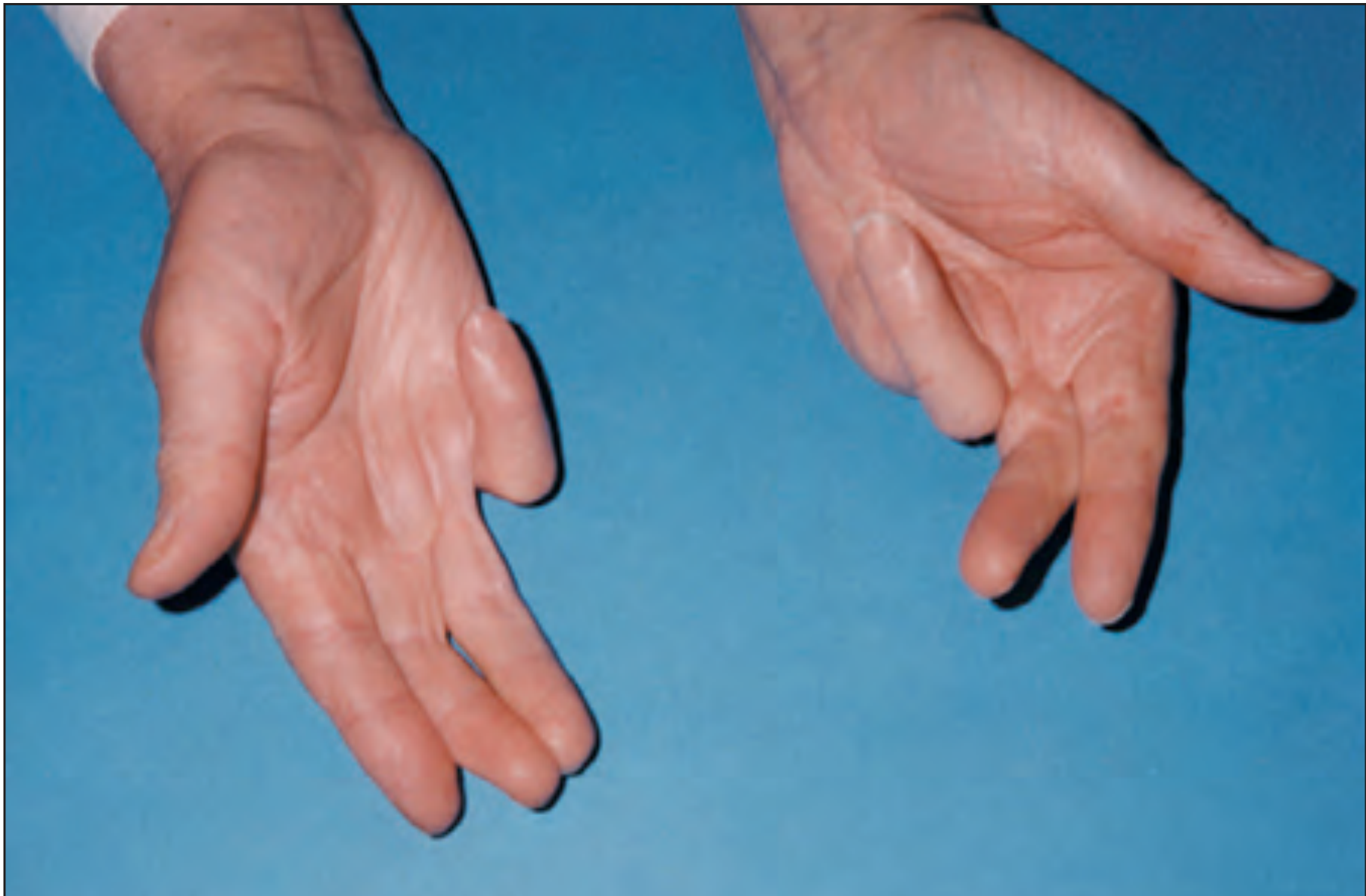
Clinical manifestation of Dupuytren disease: bilateral hand contractures. Note the amputated little finger on the left hand from advanced disease. (Rheumatology 2-Volume Set, 2022)
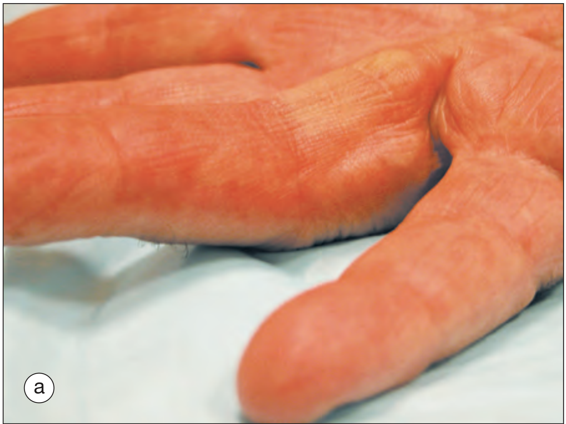
Pitting of the skin and a longitudinal cord causing MCP joint flexion in early-stage Dupuytren contracture. (Rheumatology 2-Volume Set, 2022)
Epidemiology and Risk Factors
- Most common in White patients of Northern European descent
- Men affected ~7x more often than women
- Rarely seen before age 40
- Autosomal dominant with variable penetrance (patients often identify an affected family member)
- Associated with: tobacco use, epilepsy (and anti-epileptic drugs), chronic pulmonary disease, HIV, diabetes, alcohol use, liver disease
- Trauma has not been proven as a causative factor
- Occupation is NOT an established risk factor
Anatomy - Key Points
| Structure | Involvement |
|---|---|
| Cleland (dorsal) ligaments | NOT involved |
| Grayson (volar) ligaments | Involved |
| Spiral cord | Causes PIP contracture; displaces neurovascular bundle centrally and superficially (at risk during surgery) |
| Palmar fascia | Primary site of disease |
Clinical Features
- Fingers affected (in order of frequency): ring finger > small finger > long finger (bilateral in many patients)
- Progression: palmar nodules (early) → longitudinal cords → flexion contractures at MCP and PIP joints
- Usually painless (except early nodule stage can be tender)
- The "tabletop test" (Hueston test): inability to flatten the hand against a flat surface is a classic functional finding
- Patients also struggle putting a hand into a pocket or glove
Dupuytren Diathesis
A more severe, aggressive form. Features:
- Earlier onset (younger patients)
- Bilateral disease
- Ectopic fibrosis at other sites: plantar fascia (Ledderhose disease), penile fascia (Peyronie disease), knuckle pads (Garrod pads), popliteal fascia
Staging
Tubiana Classification (based on total flexion contracture of MCP + PIP + DIP):
- Stage 0: No deformity
- Stage N: Nodule only, no contracture
- Stage I: 0-45° total contracture
- Stage II: 45-90°
- Stage III: 90-135°
- Stage IV: >135°
Treatment
Conservative (mild disease)
- Local heat application, stretching exercises, protective gloves
- Splinting does not alter disease progression
- Intralesional corticosteroid injections may relieve pain from tender nodules
Indications for Intervention
Intervention is indicated when:
- MCP joint flexion contracture > 30 degrees
- Any PIP joint flexion contracture
- Functional impairment (inability to flatten hand - positive Hueston test)
Treatment Options
| Method | Description | Outcomes | Notes |
|---|---|---|---|
| Limited fasciectomy (open) | Surgical excision of diseased fascia; preferred technique | Best long-term correction | Risk: digital nerve injury, wound breakdown, CRPS |
| Total palmar fasciectomy | Wider excision ± skin grafting | For severe disease | Higher morbidity |
| Needle aponeurotomy (NA) / Percutaneous needle fasciotomy | Percutaneous division of cords | Effective short-term; smaller incisions | High recurrence rate |
| Collagenase clostridium histolyticum (CCH) injection | Enzymatic digestion of cord followed by manipulation | Comparable to surgery for MCP correction; up to 85% MCP correction, ~60% PIP correction | Skin tears more common complication than tendon rupture; growing first-line adoption |
- Recurrence is the most common complication after fasciectomy (also the most common complication overall)
- Other surgical complications: digital nerve injury, wound breakdown, complex regional pain syndrome (CRPS), hematoma
Important Notes on Collagenase
- Adverse effects of CCH injection: pain, swelling, bruising (temporary)
- Skin tears > flexor tendon rupture as complication
- Risk of recurrence remains significant regardless of method, especially in: younger patients, bilateral disease, strong family history, fibrotic lesions elsewhere (Dupuytren diathesis)
When Full Extension May Not Be Achievable
In long-standing disease, MCP or PIP joint capsules become contracted independently of the Dupuytren tissue. In these cases, full digital extension requires extensive joint release, which carries additional neurovascular risk.
Recent Evidence (2023-2026)
Recent systematic reviews provide updated guidance:
- A 2023 meta-analysis (Zhang et al., PMID 37725027) comparing collagenase vs. needle fasciotomy for single-digit contractures found similar efficacy for MCP correction but collagenase superior for PIP joints.
- A 2024 network meta-analysis (Nann et al., PMID 37246411) of surgical approaches found no single technique definitively superior, highlighting shared decision-making.
- A 2025 GRADE-based systematic review (Shaheen et al., PMID 39451139) assessed all treatments, noting limited high-quality evidence overall.
- A 2026 review (Pistone et al., PMID 40811833) examined needle fasciotomy with and without corticosteroids, suggesting adjunctive steroids may reduce early recurrence.
Key Exam Points Summary
| Fact | Answer |
|---|---|
| Predominant cell type | Myofibroblast |
| Collagen ratio change | Type III : I increased |
| Ligament involved | Grayson (volar) - NOT Cleland |
| Most common finger affected | Ring finger |
| Surgical indication (MCP) | >30° contracture |
| Any PIP contracture | Indication for surgery |
| Gold standard surgery | Open limited fasciectomy |
| Most common complication | Recurrence |
| Test for indication | Hueston (tabletop) test |
| Structure at risk in surgery | Neurovascular bundle (displaced by spiral cord) |
- Miller's Review of Orthopaedics, 9th Edition, p. block5
- Rheumatology, 2-Volume Set (2022, Elsevier), p. 725
This is a shared conversation. Sign in to Orris to start your own chat.