
Pelvic organ prolapse management
"pelvic organ prolapse" AND management
pelvic organ prolapse classification stages diagram

This clinical photograph demonstrates a severe case of stage IV pelvic organ prolapse (procidentia) in a patient. The image shows a large, cylindrical, pinkish-red mass of tissue protruding entirely beyond the vaginal introitus and lying on the vulva. The visible structures include the everted anterior and posterior vaginal walls and the uterine cervix, which occupies the most distal portion of the prolapsed mass. The surface texture of the prolapsed tissue appears moist and slightly lobulated with visible rugae. This visual representation illustrates maximal descent of pelvic organs as defined by the Pelvic Organ Prolapse Quantification (POP-Q) system, where the entire vaginal length is essentially everted. The clinical context suggests a profound failure of the pelvic floor support structures, including the cardinal and uterosacral ligaments. This material is highly relevant for medical education in gynecology, specifically for identifying advanced stages of pelvic floor dysfunction and understanding the anatomical landmarks involved in total vault prolapse.
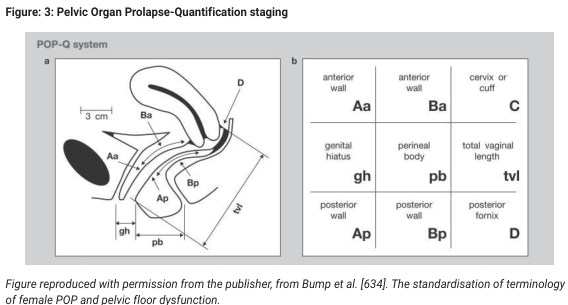
Summary : This figure illustrates the Pelvic Organ Prolapse Quantification (POP-Q) system, which is used for staging and standardizing the assessment of pelvic organ prolapse in women. It includes both an anatomical diagram and a terminology grid for reference points used in the POP-Q system. illustration: # POP-Q System Diagram : • Sagittal anatomical illustration of the female pelvis showing the vagina, bladder, and rectum. • Key reference points are labeled: Aa, Ba, Ap, Bp, C, D, gh, pb, tvl. • Measurements such as "3 cm" and distances for genital hiatus (gh) and perineal body (pb) are indicated with double-headed arrows. • The diagram visually demonstrates where each POP-Q point is located along the vaginal walls and perineal structures. # POP-Q Terminology Grid : • 3x3 grid listing anatomical reference points and their abbreviations: – Anterior wall: Aa, Ba – Cervix or cuff: C – Genital hiatus: gh – Perineal body: pb – Total vaginal length: tvl – Posterior wall: Ap, Bp – Posterior fornix: D • Each cell contains the abbreviation and the corresponding anatomical term. # Technical Details : • Figure is adapted from Bump et al., referencing the standardization of terminology for female pelvic organ prolapse and pelvic floor dysfunction. • No scale bar, but a 3 cm reference is shown for anatomical context. # Spatial Relationships : • The anatomical diagram is oriented in the sagittal plane, with the anterior wall (bladder side) on the left and the posterior wall (rectum side) on the right. • The grid provides a cross-reference for the anatomical points shown in the diagram. # Analysis : • The figure provides a comprehensive visual guide to the POP-Q system, clarifying the location and terminology of key reference points used in clinical staging of pelvic organ prolapse. • The combination of diagram and grid supports both anatomical understanding and standardized clinical communication.
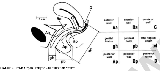
Summary : This figure illustrates the Pelvic Organ Prolapse Quantification (POP-Q) System, showing anatomical landmarks and measurement points used to assess pelvic organ prolapse in women. It includes a labeled sagittal diagram of the female pelvis and a corresponding table summarizing the anatomical sites and their abbreviations. illustration: # Sagittal Diagram : • Depicts the female pelvis in sagittal section, showing the vagina, cervix, bladder, and rectum. • Key anatomical landmarks are labeled: Aa, Ba, C, D, Ap, Bp, gh, pb, tvl. • Measurement lines and distances are indicated, such as a 3 cm segment and the total vaginal length (tvl). • The diagram shows the anterior and posterior vaginal walls, genital hiatus, perineal body, and posterior fornix. # Table of Measurement Points : • Lists abbreviations and their corresponding anatomical sites: – Aa: anterior wall – Ba: anterior wall – C: cervix or cuff – gh: genital hiatus – pb: perineal body – tvl: total vaginal length – Ap: posterior wall – Bp: posterior wall – D: posterior fornix • Table is organized by anatomical region (anterior wall, posterior wall, etc.). # Technical Details : • No scale bar, but a 3 cm reference is shown on the diagram. • All labels are clear and correspond to standard POP-Q terminology. • The figure is black-and-white, schematic, and designed for educational/clinical reference. # Spatial Relationships : • The diagram shows the spatial arrangement of the vagina, cervix, and surrounding structures. • Measurement points are distributed along the vaginal walls and perineal body. • The table provides a cross-reference for each labeled point in the diagram. Analysis : • The figure provides a comprehensive visual guide to the POP-Q system, clarifying the location and meaning of each measurement point. • The combination of diagram and table enables accurate identification and quantification of pelvic organ prolapse sites. • No data trends or anomalies are present; the figure is purely anatomical and instructional.
Pelvic Organ Prolapse (POP) - Management
Definition
Classification: POP-Q System
| Stage | Description |
|---|---|
| Stage 0 | No prolapse |
| Stage I | Most distal point > 1 cm above hymen |
| Stage II | Most distal point between -1 cm and +1 cm of hymen |
| Stage III | Most distal point > 1 cm beyond hymen but < total vaginal length - 2 cm |
| Stage IV | Complete eversion; most distal point ≥ total vaginal length - 2 cm |
Three Compartments of Prolapse
| Compartment | Defect | Condition |
|---|---|---|
| Anterior | Bladder herniation through anterior wall | Cystocele |
| Apical | Uterine/cervical descent or vaginal vault after hysterectomy | Uterine prolapse / Vault prolapse |
| Posterior | Rectal mucosa herniation through posterior wall | Rectocele; enterocele (bowel through pelvic floor) |
Evaluation
- Symptoms: Vaginal bulge (cardinal symptom), pelvic pressure (worse in afternoon/after activity), urinary symptoms (voiding difficulty, urgency, incontinence), defecatory dysfunction, sexual dysfunction, and the need to manually reduce the prolapse to void/defecate
- Physical examination: Assess all three compartments with maximal Valsalva; note position of uterus/cuff
- POP-Q staging for documentation and longitudinal follow-up
- Pelvic muscle function assessment (levator ani integrity)
- Bladder/bowel evaluation for co-existing dysfunction
- Imaging (MRI/dynamic defecography) when functional disorders coexist
Management
1. Non-Surgical (Conservative) Treatment
a. Pelvic Floor Muscle Training (PFMT)
- First-line for mild-to-moderate symptomatic POP
- Structured Kegel exercises reduce prolapse symptoms and may slow progression
- Most beneficial when initiated early, ideally with physiotherapist supervision
b. Pessary Therapy
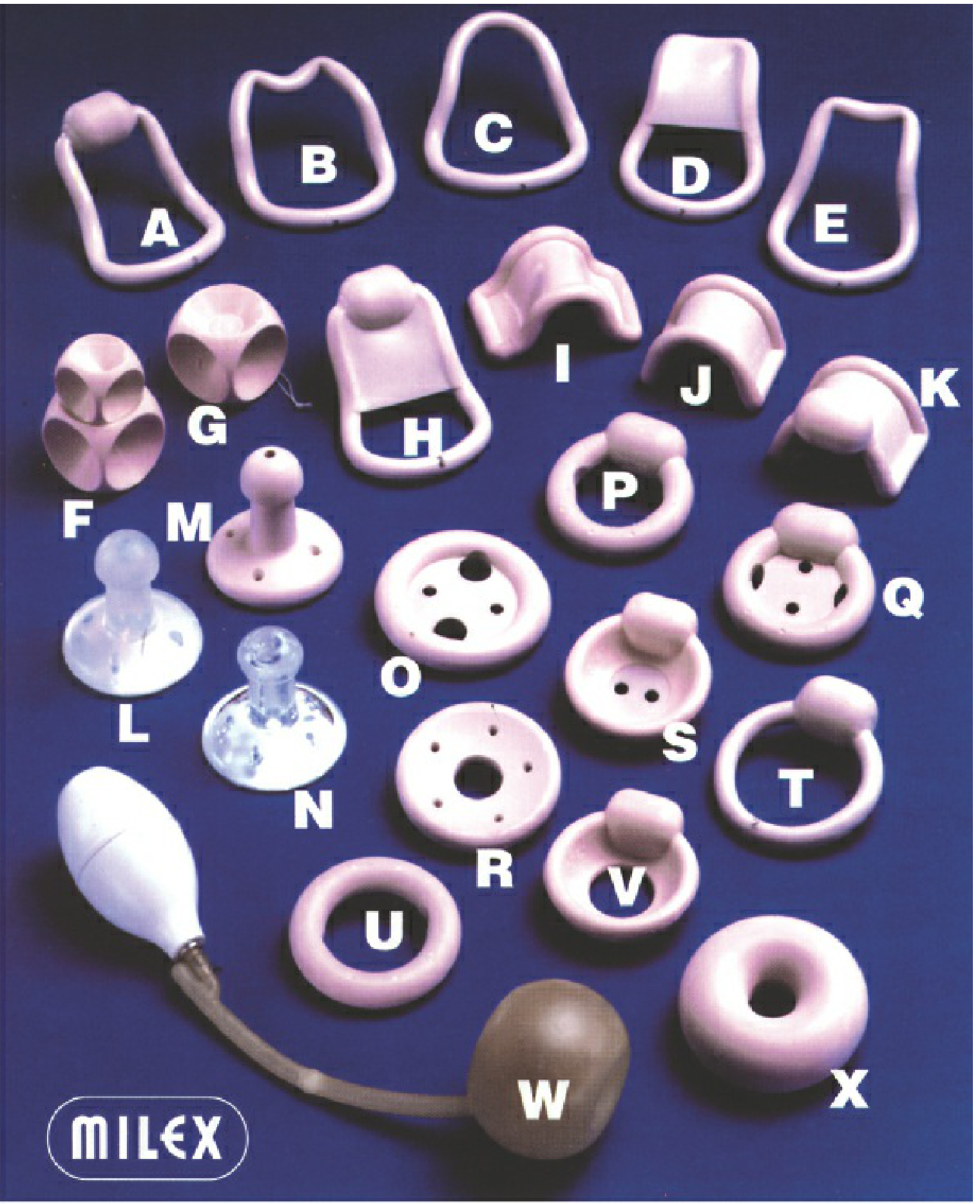
- Supportive pessaries (ring with/without support, Shaatz, Regula) - for 1st and 2nd degree prolapse
- Space-occupying pessaries (Gellhorn, cube, donut, Inflatob) - for 3rd degree / severe prolapse; use suction or pressure mechanism
- For cystocele/rectocele: Gehrung pessary; Gehrung with knob for cystocele + SUI
- Trial-and-error fitting - changing size or type at least once after initial fitting is common
- Correctly sized pessary: patient cannot feel it; too small = falls out; too large = discomfort or erosion
- Reassess at 1-2 weeks, then 4-6 weeks post-fitting; thereafter every 6-12 months
- Improves quality of life, body image, and urinary/bulge symptoms
- Long-term success factors: age ≥72 years, careful fitting, clear patient instructions
- Concomitant vaginal estrogen helps prevent tissue erosion and infection
2. Surgical Treatment
- Approach: Vaginal vs. abdominal (open/laparoscopic/robotic)
- Type: Reconstructive vs. obliterative
- Material: Native tissue vs. graft (biologic or synthetic mesh)
A. Apical Compartment
- Sacrospinous ligament suspension (SSLS) - most widely used vaginal apical repair; sutures fix vaginal apex to sacrospinous ligament
- Uterosacral ligament suspension (USLS) - sutures placed at ischial spine level; bilateral technique
- No significant difference in composite success (absence of apical descent, bothersome symptoms, retreatment)
- 2-year success: 59% (USLS) vs. 61% (SSLS)
- 5-year success declined to 44% (USLS) and 33% (SSLS) - Berek & Novak's Gynecology, p. 1628
- Sacrospinous hysteropexy
- Manchester procedure (with cervical amputation)
- Considered in women desiring uterine preservation
- Abdominal sacrocolpopexy - polypropylene mesh sutured from vaginal apex to sacral promontory; considered the gold standard for apical prolapse repair, especially vault prolapse
- Laparoscopic/robotic sacrocolpopexy - equivalent outcomes with less morbidity; preferred approach in many centers
- Abdominal uterosacral suspension - open/laparoscopic fixation of cuff to uterosacral ligaments
B. Anterior Compartment (Cystocele)
- Anterior vaginal colporrhaphy - standard repair; midline plication of pubocervical fascia; recurrence rates 3-92% (wide due to varying definitions)
- Paravaginal repair - restores lateral attachment of anterior vaginal wall to arcus tendineus fascia pelvis (ATFP); useful for lateral/displacement cystocele
- Mesh augmentation (transvaginal or transabdominal) - improves anatomic outcomes but increases risk of mesh erosion and dyspareunia; requires careful patient counseling
- Midurethral sling can be added simultaneously if stress incontinence is present
C. Posterior Compartment (Rectocele / Enterocele)
- Posterior colporrhaphy - most common repair; plication of rectovaginal septum ± perineorrhaphy; anatomic cure 76-96%; ineffective for constipation; de novo dyspareunia 7-26% (less with avoiding levator plication)
- Site-specific (defect-directed) repair - identifies and repairs discrete fascial tears; may have better functional outcomes
- Transanal repair - colorectal approach; avoided by most gynecologists due to higher dyspareunia
- Abdominal sacral colpoperineopexy - for perineal descent syndrome
- Culdoplasty - performed when enterocele is present (McCall, Halban, Moschowitz techniques)
Rectocele repair is indicated for bothersome prolapse bulge symptoms, NOT for the purpose of correcting disordered defecation. Literature does not support rectovaginal reinforcement procedures for improving constipation. - Berek & Novak's Gynecology, p. 1723
D. Obliterative Procedures
- Le Fort partial colpocleisis / total colpocleisis - suitable for elderly, sexually inactive women with high surgical risk; very high success rates; irreversible
- Not appropriate for women who wish to maintain penetrative sexual function
3. Management of Concomitant Urinary Symptoms
- Occult stress incontinence (unmasked after prolapse reduction) should be assessed pre-operatively with a pessary reduction test
- Midurethral sling (tension-free vaginal tape / transobturator tape) can be placed simultaneously with prolapse repair
- Voiding dysfunction with high post-void residual: urodynamic evaluation first; patient counseled on potential for continued problems post-surgery
Surgical Approach Comparison
| Factor | Vaginal | Abdominal/Laparoscopic |
|---|---|---|
| Recovery | Faster | Longer |
| Apical success | Moderate (44-61% at 2-5 yr) | Higher (sacrocolpopexy) |
| Mesh erosion | Higher (transvaginal mesh) | Lower (abdominal mesh) |
| Access | Better for elderly/frail | Better for younger, sexually active |
| Obliterative option | Yes (colpocleisis) | No |
Special Populations
- POP is particularly prevalent due to tissue changes (progressive loss of smooth/striated muscle, increased collagen)
- Pessaries are very well tolerated; long-term success higher in age ≥72
- Surgery is feasible and most women return to baseline function
- Visiting nurse services can assist with pessary maintenance if cognitive/motor impairment present
- Campbell-Walsh-Wein Urology, p. 3860
Recent Evidence (2024-2026)
Summary Algorithm
Symptomatic POP confirmed on POP-Q
        |
        |--> Mild-Moderate (Stages I-II, no symptoms beyond hymen)
        |         --> PFMT + lifestyle modification
        |         --> Pessary if PFMT insufficient
        |
        |--> Moderate-Severe (Stage II-IV, bothersome symptoms)
        |         --> Pessary (all patients; especially elderly, high surgical risk)
        |         --> Surgery if pessary fails/declined/not suitable
        |               |
        |               |--> Sexually active + want reconstruction
        |               |     --> Native tissue repair (vaginal or abdominal)
        |               |     --> Abdominal sacrocolpopexy (best apical outcomes)
        |               |
        |               |--> Elderly + sexually inactive + unfit
        |                     --> Colpocleisis (obliterative)
        |
        |--> Concomitant SUI --> Add midurethral sling
        |--> Concomitant bowel --> Rectocele repair (bulge only, not defecation)
Medical ethics in pediatrics
"medical ethics" AND pediatrics AND "informed consent"
medical ethics pediatrics AAP guidelines consent assent best interests 2024 2025
https://www.openanesthesia.org/keywords/pediatric-consent-an…
pediatric end of life ethics withholding treatment neonates AAP 2024
Medical Ethics in Pediatrics
Why Pediatric Ethics Is Distinct
1. Core Ethical Principles in Pediatrics
| Principle | Pediatric Application |
|---|---|
| Autonomy | Limited in young children; developing in adolescents; must be actively fostered via assent |
| Beneficence | "Best interests of the child" is the operative standard |
| Non-maleficence | Includes protecting from harmful parental decisions as well as over-medicalization |
| Justice | Equal access to care, equitable treatment regardless of disability, resource allocation in NICUs |
2. Consent, Parental Permission, and Assent
Informed Consent vs. Parental Permission vs. Child Assent
- Informed consent - a legal and ethical standard requiring disclosure, comprehension, voluntariness, and decisional capacity. Legally, minors below the age of majority (18 in most jurisdictions) cannot provide it.
- Parental permission - the proxy authorization given by parents or legal guardians on the child's behalf. The underlying ethical value is not parental autonomy or rights - it is serving the child's welfare. Parental permission is held to the same standard of disclosure as adult informed consent but is not equivalent to it.
- Child assent - the child's affirmative understanding and agreement to a procedure or treatment. It does not carry the same informational and decision-making standards as adult consent, and it does not legally protect the child (that is the function of parental permission). Its value lies in respecting the child as a developing person and promoting moral growth and self-determination.
"The assent process allows children to obtain needed practice in making decisions, contributes to their perception that they have some control over their lives, and results in a greater sense of self-esteem and competence, while reducing anxiety." - PMC10075240, Children and bioethics, J Bioeth Inq 2023
The "Rule of 7s" (Developmental Stages for Assent)
| Age | Capacity |
|---|---|
| < 7 years | Generally lack capacity for assent; clinician focuses on parental permission; child may be informed in age-appropriate language |
| 7-14 years | Can provide meaningful assent; should be engaged, informed, and their dissent considered seriously |
| 14-18 years | Often have adult-equivalent decision-making capacity; assent approaches full informed consent; dissent carries increasing weight |
When Can a Minor Consent Independently?
- Emancipated minors - legally independent from parents (married, in military, financially self-supporting, living independently); can consent as adults
- Mature minor doctrine - adolescents who demonstrate sufficient intelligence and maturity to understand the nature and consequences of a treatment may consent independently, even without emancipation
- Gillick competence (UK concept, widely influential) - a child below 16 who has sufficient understanding and intelligence to understand the treatment may consent; the clinician determines competence on a case-by-case basis
- State-specific exceptions - in most jurisdictions, minors can consent independently for: sexual health services (STI testing/treatment), contraception, pregnancy, mental health outpatient care, substance abuse treatment
3. The Best Interests Standard
- Expected benefits and burdens of treatment
- Quality of life (current and projected)
- Child's expressed preferences and values (age-appropriately)
- Family and cultural context
Approaches to Decision-Making Surrogacy
| Framework | Description |
|---|---|
| Best interest standard | Parents must choose what a reasonable person would conclude is best for the child; most widely used |
| Harm principle | A "harm threshold" is established; parental decisions are generally accepted as long as they do not cross this threshold into clear harm |
| Constrained parental autonomy | Parents have broad discretion, constrained only by reasonable limits set by clinicians and the state |
| Shared family-centered decision-making | Collaborative model involving child, family, and clinical team; recognizes that parents usually know their child best |
4. When Parental Decisions Are Contested
Parental Refusal of Life-Saving Treatment
- Religious or cultural objection (e.g., Jehovah's Witnesses refusing blood transfusion for a child) - courts in most jurisdictions will override parental refusal when treatment is life-saving and the child has not personally expressed a competent, mature refusal
- Parental authority is not absolute - it is a responsibility, not a right; the state (via parens patriae doctrine) has authority to protect children from decisions that place them in serious danger
- The harm threshold guides this: minor inconveniences do not justify overriding parents; imminently life-threatening situations do
Parental Demands for Non-Beneficial Treatment
- Ethically, clinicians are not obligated to provide treatment that offers no realistic benefit
- Communication, palliative care involvement, and ethics committee consultation are the first steps
- Unilateral withdrawal is rarely appropriate without exhausting mediation
Child Maltreatment and Mandatory Reporting
- Report on reasonable suspicion - proof is not required
- Failure to report is legally and ethically actionable
- The report transfers investigative authority to child protective services
5. Adolescent Confidentiality
- Fostering the therapeutic relationship by maintaining confidentiality
- Parental rights to information about their minor child's health
- Mandatory limits on confidentiality (harm to self or others, abuse, certain communicable diseases)
- Establish a confidentiality agreement with the adolescent and parents at the start of care (typically around age 12-14)
- Disclose only when there is imminent, serious risk
- Most health systems and professional guidelines recommend a "limited confidentiality" model: information is kept private unless the adolescent is in danger
6. End-of-Life Ethics in Pediatrics
Withholding and Withdrawing Life-Sustaining Medical Treatment (LSMT)
- Withholding and withdrawing LSMT are ethically and morally equivalent - the same ethical justification applies to both; emotional and psychological differences do not make withdrawal less permissible than non-initiation
- Forgoing LSMT is ethically supportable when the burdens of treatment outweigh the benefits to the child
- A useful guiding question: "Are we doing this to the child or for the child?"
Shared Decision-Making in End-of-Life Scenarios
- Most pediatric deaths in the U.S. follow decisions to withhold, not escalate, or withdraw life-sustaining treatments
- ~20% of pediatric deaths occur in the emergency department - often without prior preparedness
- The AAP emphasizes the role of the medical home and trusted longitudinal relationships in end-of-life planning
Neonates With Uncertain Prognosis
- Extremely preterm infants (< 25 weeks gestation)
- Infants after profound birth asphyxia
- Life-threatening congenital anomalies
- Multiple organ system failure
Palliative Sedation
- Rapid escalation of analgesics/sedatives to deep sedation is ethically permissible under the doctrine of double effect
- The overriding goal must be symptom relief, not hastening death
- This is distinct from physician-assisted death, which is not sanctioned in pediatrics
Do-Not-Resuscitate (DNR) Orders in Schools
7. Research Ethics in Pediatrics
| Category | Requirement |
|---|---|
| Minimal risk research | Parental permission + child assent |
| Greater than minimal risk with direct benefit | Parental permission + child assent; IRB must justify anticipated benefit |
| Greater than minimal risk without direct benefit | Requires both parents' permission + assent; must yield generalizable knowledge about children's conditions |
| Serious illness, no alternative | IRB may waive some requirements under strict conditions |
- Children cannot advocate for themselves the way adults can - exploitation risk is higher
- Excluding children from trials protects them in the short term but deprives them of evidence-based treatments ("therapeutic orphan" problem)
- Precision medicine and genomic research raise new questions about consent for future use of data, incidental findings, and disclosure to families
8. Special Ethical Topics
Gender-Diverse Children and Adolescents
- No reliable indicators exist to distinguish children whose gender dysphoria (GD) will persist into adulthood from those who will desist
- The ethics of social transition in young children remain contested - risks of harm apply both to early transition (in children who later desist) and to blocking transition (in those who persist)
- Puberty-blocking agents: the Tavistock/GIDS case in the UK became a landmark legal moment (2020 ruling required court authorization for minors under 16; overturned on appeal in 2021)
- Multiple U.S. states have enacted legislation restricting or criminalizing gender-affirming care for minors - federal courts have challenged these as unconstitutional
Children With Disabilities
- The equal moral worth of children with disabilities is a foundational principle
- Quality-of-life judgments by others (including parents) about children with disabilities are ethically suspect and should not alone determine treatment intensity
- Disability rights frameworks emphasize that decisions should not be made based on the clinician's projection of a "normal" life
Childhood Vaccination
Summary of Core Principles
| Ethical Issue | Key Principle | Practical Action |
|---|---|---|
| Consent | Legal authority rests with parents | Obtain parental permission; document appropriately |
| Assent | Child's developing autonomy deserves respect | Engage child age-appropriately from ~7 years |
| Best interests | Child's welfare is primary, not parental preference | Apply harm threshold; involve ethics committee if disputed |
| Treatment refusal | Parental refusal can be overridden when life-threatening | Court order if needed; state parens patriae |
| Confidentiality | Adolescents have qualified confidentiality rights | Define limits upfront; disclose only for serious risk |
| End of life | Withholding = withdrawing; burden-benefit framework | Shared decision-making; involve palliative care; ethics consult |
| Abuse | Mandatory reporting obligation | Report on reasonable suspicion; proof not required |
| Research | Extra protections for child subjects | Two-tier: parental permission + child assent |