
development of mandible in detail
development of mandible Meckel's cartilage ossification embryology diagram
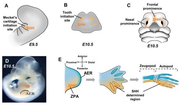
Educational medical diagram illustrating the spatial expression of Sonic Hedgehog (SHH) during murine craniofacial and limb morphogenesis at embryonic days E9.5 and E10.5. Panels A-C depict craniofacial development: (A) SHH signaling in the first branchial arch at E9.5 identifying the Meckel's cartilage initiation site; (B) expression in the mandible at E10.5 marking tooth initiation sites; and (C) localization in the frontal and nasal prominences at E10.5 guiding upper facial structure formation. Panels D and E illustrate limb bud development. (D) A whole-mount embryo image showing the Apical Ectodermal Ridge (AER) marked by Dlx2/LacZ expression at E10.5. (E) Schematic of the limb bud showing SHH localization in the Zone of Polarizing Activity (ZPA) at the posterior margin. The progression from limb bud to autopod demonstrates how SHH signaling determines the posterior skeletal elements, including the zeugopod and autopod (digits). The diagram highlights the crucial role of SHH in proximodistal and anteroposterior axial patterning during skeletal embryogenesis.
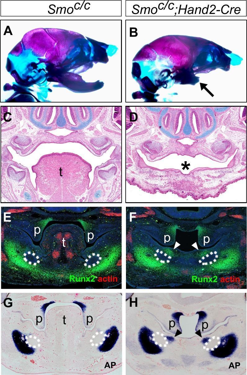
This composite image presents a comparative developmental study of craniofacial morphogenesis in mouse embryos, focusing on the Smoc/c control (left) versus Smoc/c;Hand2-Cre mutants (right). (A-B) Whole-mount skeletal preparations stained with Alizarin Red and Alcian Blue show lateral views of the head. The mutant (B) exhibits significant mandibular hypoplasia (arrow) while the cranium remains intact. (C-D) H&E-stained frontal sections at E16.5 demonstrate complete tongue agenesis (asterisk) in the mutant compared to the well-developed tongue ('t') in the control. (E-F) Immunofluorescence staining for Runx2 (green) and muscle alpha-actin (red) at E12.5 reveals ectopic ossification (arrowheads) in the mutant's oral mandibular region and absence of tongue musculature. Dashed lines delineate the Meckel’s cartilage primordia. (G-H) Alkaline phosphatase (AP) activity staining (blue) confirms this ectopic ossification in the oral mesenchyme of the mutant (H, arrowheads). This panel illustrates the role of hedgehog signaling in patterning the oral-aboral axis of the mandible and tongue development, demonstrating that disruption leads to mandibular truncation and tongue agenesis.
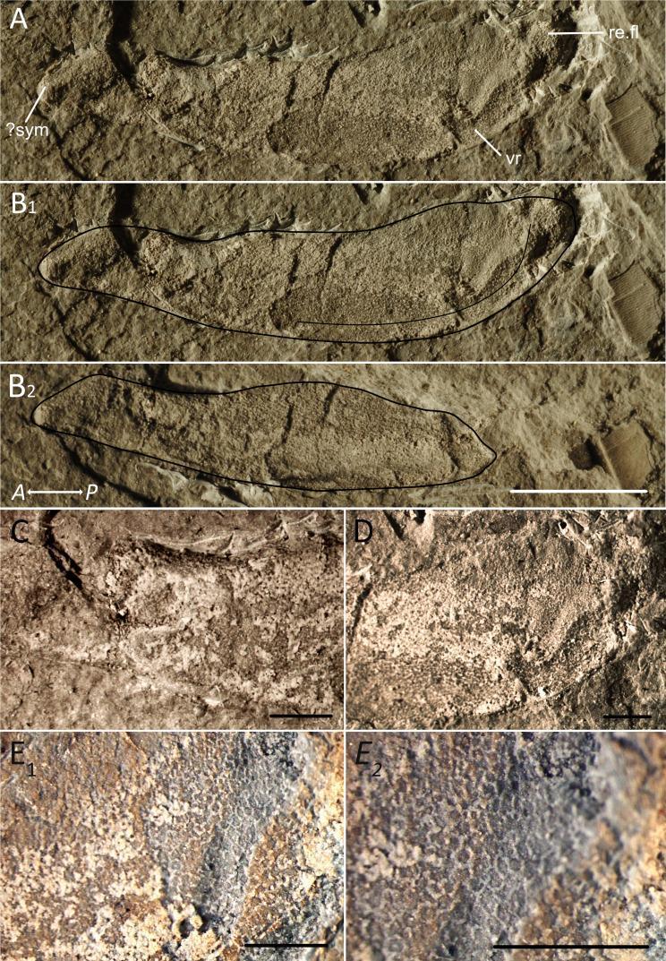
This composite educational image displays the morphological and histological features of a fossilized Meckel's cartilage, an embryonic precursor to the mandible. (A) and (B1-B2) present a lateral view and traced outlines of the elongated cartilage, showing a gently convex ventral edge, a thickened anterior symphysis, and a posterior retroarticular flange. A distinct ventral ridge (vr) is visible along the jaw's middle-to-posterior aspect. (C) and (D) provide high-magnification views of the anterior and posterior regions, respectively, highlighting differences in mineralization. (E1) and (E2) demonstrate the microstructure of the calcified cartilage, characterized by a tessellated polygonal pattern. These individual tesserae are separated by distinct, slightly rounded borders, representing intertesseral fibers. This tessellated prismatic calcified cartilage is a key diagnostic feature in chondrichthyan anatomy, used here to demonstrate the evolution and structural integrity of cartilaginous skeletal elements. The specimen serves as a model for understanding early jaw development and the histological transition of skeletal tissues.
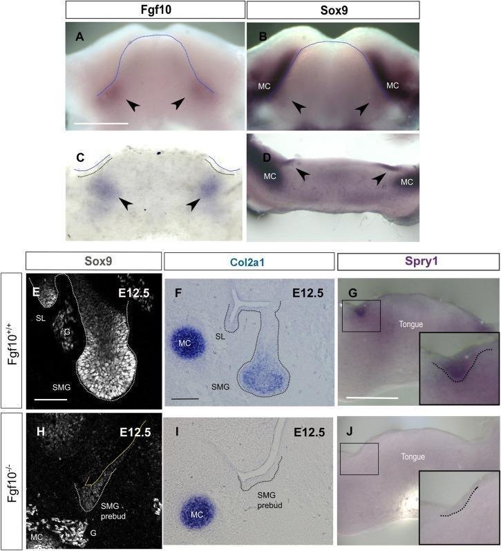
This composite educational graphic illustrates the regulatory role of Fgf10 in Sox9 expression during embryonic salivary gland development. Panels A-D use in situ hybridization to demonstrate the spatial overlap of Fgf10 and Sox9 mRNA in the E11.0 murine mandible and frontal mandibular slices, with arrowheads highlighting expression at the site of submandibular gland (SMG) initiation. Meckel’s cartilage (MC) is noted as a landmark. Panels E-J provide a comparison between wild-type (Fgf10+/+) and mutant (Fgf10-/-) embryos at E12.5. Immunofluorescence (E, H) and in situ hybridization (F, G, I, J) show that loss of Fgf10 results in a significant reduction of Sox9 protein, Col2a1 mRNA, and Spry1 mRNA within the SMG epithelium. While the wild-type (Fgf10+/+) samples exhibit well-defined epithelial buds with robust expression of these markers, the Fgf10-/- mutants display arrested 'pre-bud' morphology and diminished signaling. These findings establish Fgf10 as a critical upstream regulator of the Sox9-Col2a1 pathway necessary for epithelial branching morphogenesis.
Development of the Mandible
1. Embryological Origin
- Dorsal: maxillary process - gives rise to maxilla, zygomatic bone, and part of temporal bone
- Ventral: mandibular process - contains Meckel's cartilage and gives rise to the mandible
- The Developing Human: Clinically Oriented Embryology, p. 466-468
- Langman's Medical Embryology, p. 170
2. Meckel's Cartilage - The First Arch Cartilage
| Region | Fate |
|---|---|
| Dorsal end (proximal) | Breaks away - forms malleus and incus (middle ear ossicles) |
| Middle portion | Regresses; its perichondrium forms the sphenomandibular ligament and anterior ligament of the malleus |
| Ventral end (distal) | Guides early morphogenesis of the mandible body; eventually disappears |
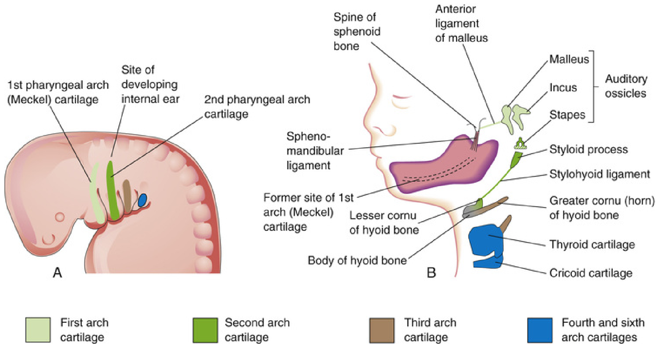
3. Mode of Ossification - Intramembranous
- Mesenchyme surrounding Meckel's cartilage condenses and differentiates into osteoblasts
- Osteoblasts secrete osteoid (unmineralized bone matrix), which then mineralizes
- Bone first appears as small, irregularly shaped spicules and trabeculae that interconnect and take on the three-dimensional shape of the mandible
- Developing teeth, Meckel's cartilage, and oral cavity are all visible in adjacent relation in early histological sections
- Histology: A Text and Atlas (Pawlina), p. 637-638
4. Ossification Center and Timeline
- The primary ossification center appears at approximately 6 weeks IUL (some sources say the 6th-7th week) near the bifurcation of the inferior alveolar nerve into the mental and incisive branches - lateral to Meckel's cartilage
- The ossification center in the mandible appears at the 2nd month of intrauterine life (slightly later than the clavicle, which is the very first bone in the body to ossify at 4-5 weeks)
- Each half of the mandible develops from a single primary ossification center
- The ventral parts of Meckel's cartilages form the horseshoe-shaped primordium, and by keeping pace with its growth, they guide early morphogenesis
5. Secondary Cartilages of the Mandible
| Secondary Cartilage | Location | Fate |
|---|---|---|
| Condylar cartilage | Head of condyle | Most important; persists as growth cartilage until ~20 years; replaced by endochondral ossification |
| Coronoid cartilage | Coronoid process | Appears and disappears during fetal life |
| Symphyseal cartilages | Chin region (bilateral) | Unite to form the symphysis menti; fuse during 2nd year of life |
6. Parts of the Mandible and Their Development
- Body and mental region: intramembranous ossification of mesenchyme lateral to ventral Meckel's cartilage
- Ramus: proximal part of Meckel's cartilage is remodeled and contributes to the ramus
- Condylar process: secondary condylar cartilage, later replaced by endochondral ossification
- Coronoid process: secondary coronoid cartilage (transient)
- Symphysis menti: bilateral symphyseal cartilages fuse in the 2nd year of life - the two halves of the mandible remain separate at the symphysis at birth and unite by the 2nd year
7. Muscles and Nerve - First Arch Derivatives
- Muscles of mastication (temporalis, masseter, medial and lateral pterygoids)
- Mylohyoid and anterior belly of digastric
- Tensor tympani
- Tensor veli palatini
8. Fate of Meckel's Cartilage - Summary Table
| Part of Meckel's Cartilage | Adult Derivative |
|---|---|
| Proximal dorsal end | Malleus (head & neck) + Incus (body & short process) |
| Intermediate portion | Regresses; perichondrium → sphenomandibular ligament + anterior ligament of malleus |
| Ventral distal portion | Acts as template, disappears; site marked by sphenomandibular ligament origin |
9. Postnatal Growth of the Mandible
- Growth occurs mainly at the condylar cartilage (secondary cartilage) - endochondral mechanism
- Surface remodeling occurs by periosteal deposition and osteoclastic resorption
- The ramus grows posteriorly and superiorly while the symphysis ossifies
- The condylar growth plate is sensitive to functional stimuli (masticatory forces, bite) - basis for functional appliance therapy in orthodontics
- Growth largely ceases around 18-20 years with fusion of condylar cartilage
10. Clinical Correlates
- Treacher Collins syndrome (mandibulofacial dysostosis): failure of first arch neural crest cell migration - leads to hypoplastic mandible (micrognathia), malar hypoplasia
- Pierre Robin sequence: severe micrognathia + glossoptosis + cleft palate - mandible fails to develop adequately in early fetal life
- Hemifacial microsomia: unilateral underdevelopment of first arch derivatives
- Bifid mandible / cleft mandible: failure of the two halves to fuse at the symphysis
- Exostosis (tori mandibularis): benign bony overgrowth on lingual surface; developmental variant
- The Developing Human: Clinically Oriented Embryology (Moore & Persaud), p. 466-469
- Langman's Medical Embryology, p. 170
- Cummings Otolaryngology: Head and Neck Surgery, p. 3505
- Histology: A Text and Atlas (Pawlina), p. 637-638
- The Essentials of Forensic Medicine and Toxicology (2026), p. 7532
Explain the condylar cartilage's role in postnatal growth and orthodontic implications
condylar cartilage mandibular growth orthodontic functional appliance
mandibular condyle growth functional appliance
PMID: 41054808
mandibular growth modification Twin Block Herbst appliance
PMID: 40008555
condylar cartilage zones histology mandibular growth
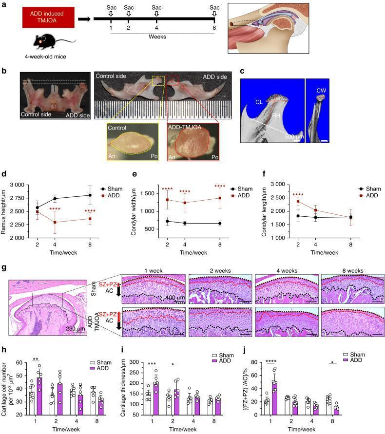
This composite educational image illustrates the effects of Anterior Disc Displacement (ADD) on mandibular growth and temporomandibular joint (TMJ) cartilage in a mouse model. (a) A schematic shows the timeline of ADD induction and sacrifice at 1, 2, 4, and 8 weeks. (b) Clinical photographs compare a smooth, uniform control condyle to an irregular, rough ADD condyle. (c-f) Micro-CT reconstructions and line graphs demonstrate growth retardation in the ADD group, specifically showing significantly reduced ramus height (RH) and increased condylar width (CW) and length (CL) compared to Sham controls. (g) H&E stained histological sections reveal TMJ condylar cartilage changes; ADD groups show disarranged cell distribution and increased thickness in the fibrocartilage (FZ) and proliferative zones (PZ) compared to Sham. (h-j) Bar graphs quantify cellularity, total cartilage thickness, and the ratio of superficial layers (FZ+PZ) to articular cartilage (AC), highlighting pathological remodeling over time. This visual resource is designed for dental and orthopedic research, illustrating the link between mechanical disc displacement and secondary osteoarthritic changes (TMJOA) in a growing mandible.
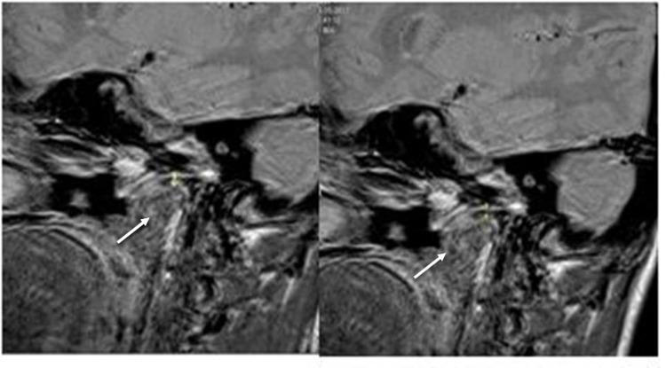
This diagnostic image consists of two sagittal MRI scans of the human temporomandibular joint (TMJ), comparing a baseline state (T0, left) with a follow-up state (T1, right) after functional appliance therapy. The scans clearly depict the mandibular condyle, the articular eminence of the temporal bone, and the intervening joint space. White arrows in both images highlight the condylar cartilage layer covering the superior-posterior aspect of the mandibular condyle. A comparison between T0 and T1 reveals a visible increase in the thickness and signal intensity of the condylar cartilage at T1. This finding serves as a clinical marker for orthopedic remodeling and growth stimulation of the cartilage following the use of a twin-block appliance in orthodontic treatment. The surrounding soft tissues, including the joint capsule and lateral pterygoid muscle attachments, are also visible, providing anatomical context for the assessment of functional jaw orthopedics.
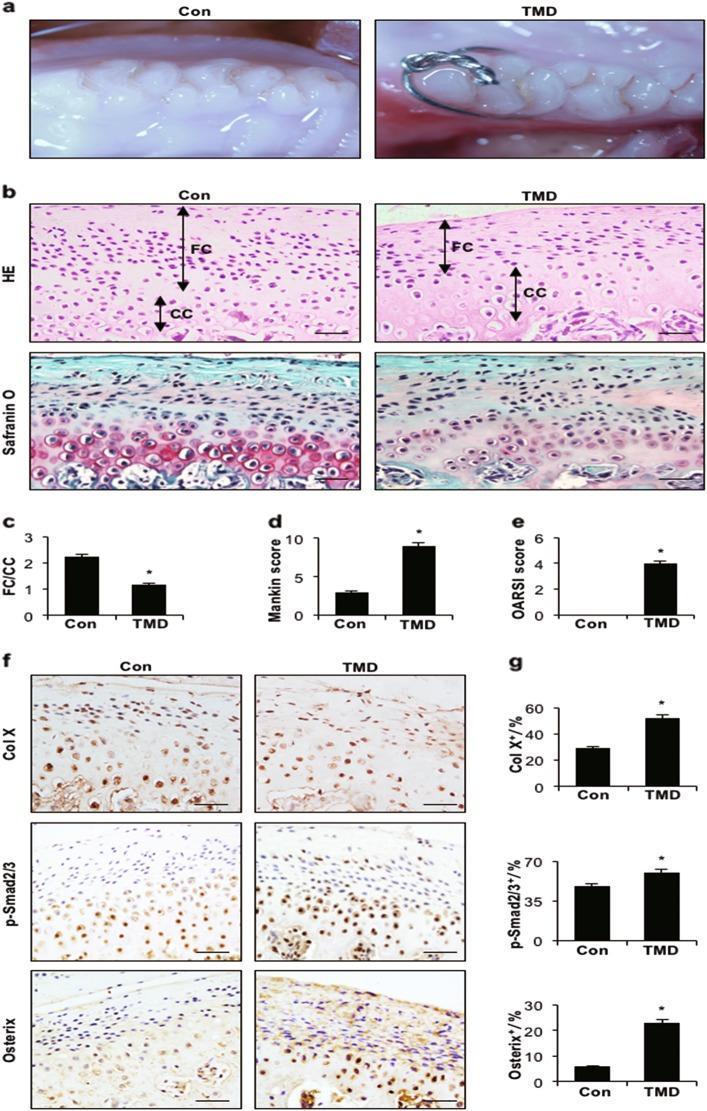
Educational figure illustrating cartilage degeneration in a rat model of temporomandibular disorder (TMD). Panel A shows clinical photographs of the molar occlusion relationship in control vs. TMD groups. Panel B presents histological sections of mandibular condylar cartilage: Haematoxylin and Eosin (HE) staining highlights a significantly decreased thickness of the fibrocartilage (FC) and calcified cartilage (CC) layers in TMD rats; Safranin O and fast green staining demonstrate a marked loss of red-stained proteoglycans in the TMD group, indicating cartilage degradation. Quantitative bar graphs (C-E) show a decreased FC/CC ratio and significantly elevated Mankin and OARSI scores in TMD samples. Panel F provides immunohistochemical (IHC) analyses of Col X, p-Smad2/3, and Osterix markers, where the TMD group exhibits increased brown chromogen intensity and higher positive cell counts (quantified in G). These findings demonstrate that abnormal occlusion triggers pathological remodeling and TGF-̢β/Smad pathway activation in the temporomandibular joint.
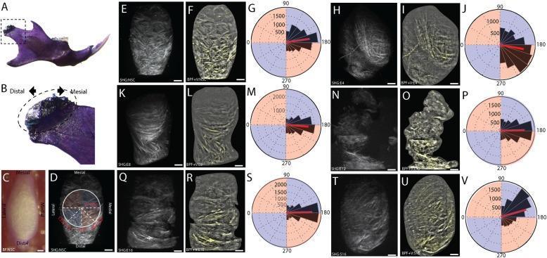
This composite educational image illustrates quantitative orientation analysis of the mandibular condylar cartilage (MCC) using Second Harmonic Generation (SHG) microscopy to track the progression of Temporomandibular Joint Osteoarthritis (TMJ-OA). Panels A-D define the anatomical orientation, showing the mesiodistal and mediolateral axes of the mandibular condyle. The main data displays (E-V) present a longitudinal comparison between non-surgical controls, a 16-week sham-control, and post-injury time points (4, 8, 12, and 16 weeks). Each time point features three representations: raw SHG scans (E, H, K, N, Q, T) showing fibrillar collagen; band-pass filtered SHG reconstructions (F, I, L, O, R, U) providing enhanced visualization of extracellular matrix (ECM) organization; and rose plots (G, J, M, P, S, V) quantifying vector orientations. The series demonstrates early-stage ECM remodeling at 4-8 weeks, followed by severe structural degradation at 12-16 weeks, characterized by visible fibrillations, fissures, and clefts in the superficial layer. The rose plots specifically show shifts in mean collagen fiber orientation (red line) and variance, with 180° indicating a mediolateral vector orientation.
condylar cartilage zones histology mandibular growth mechanism orthodontics
https://pmc.ncbi.nlm.nih.gov/articles/PMC12755033
Condylar Cartilage: Postnatal Growth & Orthodontic Implications
1. What is the Condylar Cartilage?
2. Histological Zones of the Condylar Cartilage
| Zone | Cell Type | Function |
|---|---|---|
| Articular/Fibrous zone | Fibrocartilage (type I collagen), flattened cells | Weight-bearing articular surface; unlike hyaline cartilage in other joints |
| Proliferative (prechondroblastic) zone | Undifferentiated mesenchymal stem cells | Source of new chondrocytes; site of active mitosis; responds to mechanical stimuli |
| Chondroblastic/Hypertrophic zone | Maturing chondrocytes (type II collagen) | Cartilage matrix production; cells enlarge progressively |
| Calcified cartilage / Ossification zone | Dying hypertrophic chondrocytes, mineral deposition | Replaced by bone via endochondral ossification |
- Epiphyseal plates: stem cells sit deep; growth is unidirectional (longitudinal)
- MCC: stem cells sit superficially (in the fibrous/proliferative zone); growth is multidirectional - posterosuperiorly and laterally
- This haphazard cell arrangement means the condyle can adapt rapidly to altered position or function
3. Growth Direction and Mechanism
- New cartilage is formed in the proliferative zone of the condyle
- Cartilage undergoes endochondral ossification at its base, converting to bone
- This process effectively pushes the condyle upward into the glenoid fossa, which accommodates by remodeling
- The ramus simultaneously lengthens posteriorly and superiorly
- Pre-occlusal phase - endochondral ossification similar to long-bone growth plate, PTHrP-driven
- Occlusal establishment phase - mechanical loading from occlusion triggers and modifies endochondral ossification
- Post-growth phase - endochondral ossification ceases; MCC transitions to purely articular function but retains remodeling capacity
4. Growth Completion and Timing
- MCC growth is active during adolescence, peaks at the pubertal growth spurt
- Girls: growth largely complete by ~15-17 years
- Boys: growth complete by ~18-20 years
- Activity can be assessed with bone scintigraphy (Tc-99m) - radionuclide imaging demonstrates whether condylar growth is still active before surgical correction
5. Key Biological Property: Mechanosensitivity
- Stimulated by anterior positioning of the mandible (stretching the lateral pterygoid, increasing joint loading)
- Inhibited by abnormal loading, disc displacement, or restricted function
6. Orthodontic Implications
A. The Scientific Rationale for Functional Appliances
- Stretches the retrodiscal tissues and joint capsule
- Stimulates the proliferative zone of the MCC via increased proprioceptive/mechanical input
- Promotes increased cell division in the prechondroblastic zone
- Accelerates cartilage formation and endochondral ossification at the condyle
- Simultaneously promotes glenoid fossa remodeling to accommodate condylar displacement
B. Common Functional Appliances and Their Effects
| Appliance | Type | Mechanism | Effect |
|---|---|---|---|
| Twin Block | Removable | Inclined bite blocks advance mandible | Mandibular growth stimulation, SNB increase |
| Herbst | Fixed | Rigid telescoping mechanism | Continuous 24h advancement; dental + skeletal effects |
| Activator | Removable | Mandibular posturing | Class II correction, vertical control |
| Forsus | Fixed | Spring-loaded, attached to archwire | Late adolescent/early adult use |
| Functional clear aligners | Removable | Advancing bite in aligner sequence | Emerging evidence; comparable to traditional functionals |
C. Evidence on Functional Appliance Efficacy (2025 Systematic Review)
- Anterior condylar shifts
- Altered condylar shape (increased height and width)
- Retracted articular discs and increased joint spaces
- Glenoid fossa remodeling
D. Cephalometric Changes Targeted
- Skeletal: ↑ SNB angle, ↓ ANB angle, ↑ mandibular length (Co-Gn), ↑ ramal height
- Dental: ↓ overjet, correction of molar relationship (Class I)
- Soft tissue: lip profile improvement
E. Timing and the "Window of Opportunity"
- Confirmed by cervical vertebral maturation (CVM) staging - CS3/CS4 represents peak mandibular growth velocity
- Treatment outside this window yields predominantly dental movement rather than skeletal adaptation
- Bone scintigraphy can confirm active condylar growth if clinical timing is uncertain
F. Clinical Sequelae of Condylar Cartilage Pathology
| Condition | Mechanism | Result |
|---|---|---|
| Condylar hyperplasia | Autonomous continued growth after normal cessation | Progressive facial asymmetry, open bite |
| Condylar hypoplasia | Arrested growth (radiation, trauma, infection) | Micrognathia, facial asymmetry |
| TMJ disc displacement | Anterior disc displacement disrupts condylar loading | Growth retardation, asymmetric condylar development |
| Juvenile idiopathic arthritis | Inflammatory destruction of condylar cartilage | Mandibular undergrowth, anterior open bite, micrognathia |
Summary Points
- The condylar cartilage is a secondary cartilage that serves as both the articular surface of the TMJ and the primary growth center of the mandible
- Growth is driven by endochondral ossification within the MCC, predominantly in a posterior-superior direction
- Its unique mechanosensitivity (proliferative zone at the surface, multidirectional cell arrangement) is what makes the mandible modifiable by functional forces
- Functional appliances exploit this by anteriorly posturing the mandible during the pubertal growth window, stimulating condylar cartilage proliferation
- Evidence for genuine skeletal change from functional appliances exists but is of low to moderate quality, and the effect is modest
- After growth cessation, the MCC transitions to pure articular function but retains lifelong remodeling capacity in response to mechanical load
- Scott-Brown's Otorhinolaryngology Head & Neck Surgery, Vol. 2 (TMJ section)
- Grainger & Allison's Diagnostic Radiology (condylar growth assessment)
- Tsutsumi-Arai et al. (2025) - PMC12755033 - MCC in development and diseases
- Mbyayingabo et al. (2025, PMID 41054808) - Systematic review: condylar morphology changes with functional appliances
- Ghorbani et al. (2025, PMID 40008555) - Meta-analysis: functional clear aligners for Class II correction