
My father is 65 years old he works in animal hospital he has very working schedule and he is complaining pain near elbow and on shoulder from may and it doesn't pain in relaxing position but when he lifts his arm for removing vest or picking something he feels pain sensation and near elbow it is hard lump of muscle not like a swelling so what it is?
My father is 65 years old he works in animal hospital he has very working schedule and he is complaining pain near elbow and on shoulder from may and it doesn't pain in relaxing position but when he lifts his arm for removing vest or picking something he feels pain sensation and near elbow it is hard lump of muscle not like a swelling so what it is?
- Pain only with movement (lifting, overhead reaching) but not at rest = classic tendon/muscle pathology
- Hard lump near the elbow = the biceps muscle belly has retracted downward ("Popeye sign") after the tendon tears at the top
- Shoulder pain with arm elevation = rotator cuff strain or tear, often occurs alongside biceps tendon issues
biceps tendon rupture Popeye sign shoulder pain hard lump near elbow 65 year old
biceps tendon rupture Popeye sign bulge upper arm
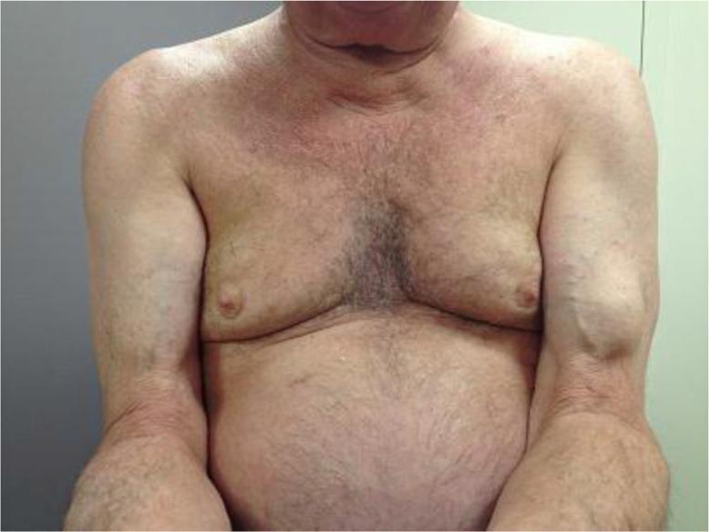
This clinical photograph shows the upper torso and bilateral arms of an elderly male patient. The primary finding is a prominent, well-defined, rounded mass located on the anterior aspect of the left mid-upper arm, creating a significant contour deformity compared to the contralateral side. This visual manifestation is known as the 'Popeye sign' (or Popeye deformity), which indicates a rupture of the long head of the biceps brachii tendon. The rupture causes the muscle belly to retract distally toward the elbow, forming the characteristic bulge. The patient displays age-related skin changes, including fine wrinkling, thinning of the dermis, and prominent superficial veins in the forearms. No acute ecchymosis or inflammatory skin changes are visible, suggesting a chronic or non-acute presentation. This image is used in medical education to demonstrate classic physical examination findings in musculoskeletal pathology and orthopedic trauma.
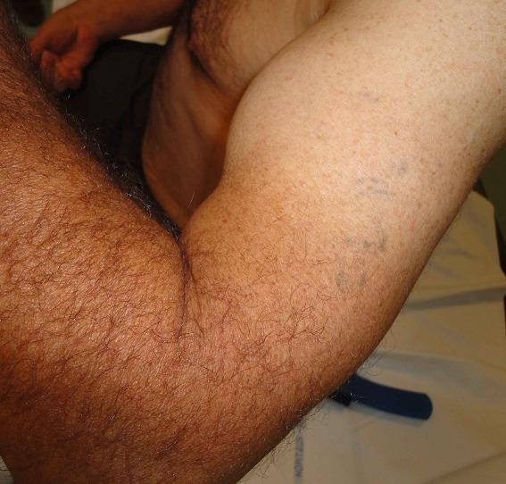
This clinical photograph displays a lateral view of a flexed left upper extremity, demonstrating a classic 'Popeye deformity' or 'Popeye sign.' The visual finding consists of a prominent, rounded, and distal bulge of the biceps brachii muscle belly, which has retracted toward the mid-to-distal humerus. This characteristic contour abnormality is pathognomonic for a rupture of the biceps tendon (proximal or distal, though in this context distal is specified). Associated findings include visible ecchymosis and bruising on the medial aspect of the arm, indicating acute soft tissue trauma and hematoma formation. The deformity is highlighted by the elbow being held in a flexed position, which accentuates the muscular bunching. This image is a primary educational resource for orthopedic physical examination findings in musculoskeletal injuries, specifically for medical students and residents learning to identify tendon ruptures through visual inspection and surface anatomy.
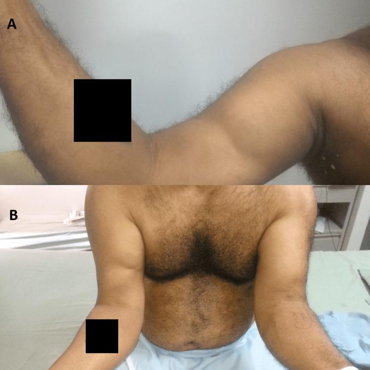
Clinical photograph of a 48-year-old male demonstrating a 'reverse Popeye sign' characteristic of a distal biceps tendon rupture. Panel A shows a lateral view of the flexed right arm, where the biceps brachii muscle belly is retracted proximally toward the shoulder, creating a prominent bulge in the upper arm and an abnormal depression near the antecubital fossa. Panel B provides a comparative frontal view of both upper extremities. The right arm exhibits significant muscular asymmetry compared to the normal left arm; the right biceps muscle mass is bunched and shifted superiorly, whereas the left arm shows normal muscle distribution along the humerus. This visual finding indicates a complete avulsion of the distal biceps tendon from its insertion on the radial tuberosity, leading to proximal retraction of the muscle. The image is a classic representation of soft tissue orthopedic trauma and is used to teach clinical diagnostic signs for tendon injuries in the musculoskeletal system.
Most Likely Diagnosis: Proximal Biceps Tendon Rupture ("Popeye Sign") + Rotator Cuff Involvement
What Has Happened
Why Does the Shoulder Also Hurt?
- Pain when lifting the arm overhead (removing a vest, reaching up)
- Pain on shoulder movement but not at rest
- No pain when the arm is simply hanging down
Why Did It Happen?
| Risk Factor | How It Applies |
|---|---|
| Age 65+ | Tendons naturally degenerate and weaken with age |
| Heavy workload | Repetitive overhead tasks at an animal hospital (lifting, restraining animals) |
| Gradual onset since May | Suggests progressive degeneration, not a single trauma |
| No pain at rest | Consistent with tendon pathology, not bone or joint disease |
What You Should Do
- Ultrasound - quick, cheap, and very good at visualizing tendons
- MRI of the shoulder - gold standard to see the full extent of any rotator cuff tear alongside the biceps
- Rest and activity modification (reduce heavy lifting)
- Anti-inflammatory medications (like ibuprofen/diclofenac) for a short course
- Physiotherapy to strengthen the rotator cuff and shoulder muscles
- The cosmetic bulge (Popeye lump) typically remains but does not cause functional problems
What This Is NOT
Bottom Line
Important note: This is an educational assessment based on the symptoms described. Only a doctor examining your father in person can make a definitive diagnosis. Please do not delay seeing a specialist.
It is on the backside of elbow not in front and does it can coz freeze of arm?
hard lump back of elbow posterior shoulder pain arm stiffness frozen shoulder 65 year old worker
olecranon bursitis posterior elbow swelling lump
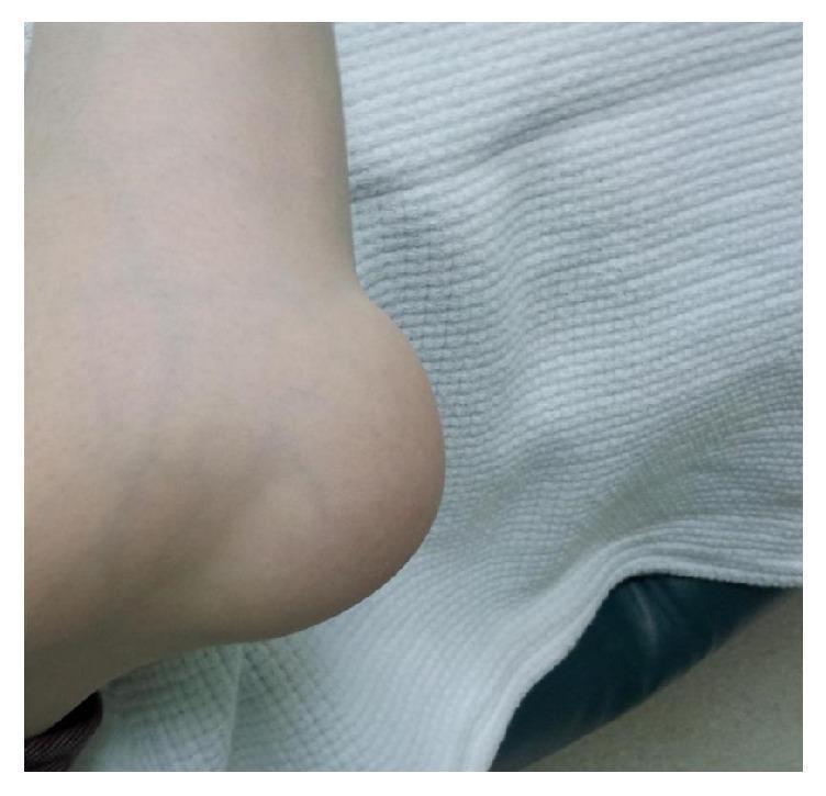
This clinical photograph displays a prominent, localized soft tissue swelling over the posterior aspect of the right elbow joint, consistent with olecranon bursitis. The swelling is large, hemispherical, and well-demarcated, significantly distorting the normal anatomical contour of the elbow. The overlying skin appears intact and relatively normal in color, without gross evidence of erythema, ulceration, or acute inflammatory changes. Bony landmarks of the elbow, such as the olecranon process, are obscured by the mass. This visual finding is characteristic of fluid accumulation within the olecranon bursa, which can occur due to trauma, repetitive pressure, or inflammatory and infectious conditions. The image serves as an educational example for musculoskeletal examination and the differential diagnosis of joint-related swellings.
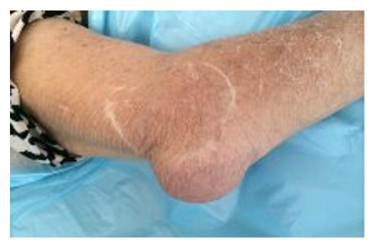
This clinical photograph shows the posterior aspect of a right elbow, demonstrating significant localized pathology. The primary finding is a prominent, well-demarcated swelling over the olecranon process, consistent with olecranon bursitis. The overlying skin exhibits diffuse erythema and mild distension, signs indicative of an acute inflammatory or infectious process (such as septic bursitis or rheumatoid-associated inflammation). The surrounding skin on the forearm and upper arm appears xerotic with fine scaling. This image is an educational example of joint-adjacent soft tissue inflammation and bursal distension, relevant to rheumatology, orthopedics, and emergency medicine for differentiating between superficial bursal inflammation and deep joint space involvement.
Revised Picture: Two Separate Issues Happening Together
1. Hard Lump at the Back of the Elbow - Olecranon Bursitis
- It feels like a "golf ball" or firm lump right at the tip of the elbow (posterior/back side)
- Often painless or only mildly sore - fits your description of pain mainly on movement
- Caused by repetitive friction, pressure, or physical work - exactly your father's situation
- It is not dangerous in most cases
2. Shoulder Pain on Lifting/Overhead Movement - Likely Rotator Cuff or Early Frozen Shoulder
If the shoulder pain is ignored and the arm is not kept moving, frozen shoulder (adhesive capsulitis) can develop or worsen. The shoulder joint capsule thickens, scar tissue forms, and movement becomes progressively more restricted - eventually the arm feels "locked" or "frozen." This process can take months and is harder to reverse if left untreated.
"Adhesive capsulitis or frozen shoulder presents with marked limitation of shoulder joint movement such that the scapula moves en bloc with the arm and is associated with movement-evoked pain."
Summary: What Your Father Has (Most Likely)
| Problem | Location | Cause | Urgency |
|---|---|---|---|
| Olecranon bursitis | Back of elbow - hard lump | Repetitive work/pressure on elbow | Low - see a doctor, not emergency |
| Rotator cuff tendinitis OR early frozen shoulder | Shoulder pain on lifting arm | Overuse + age-related degeneration | Moderate - needs treatment soon to prevent worsening |
What To Do
- See an orthopedic doctor - both problems can be examined together
- For the elbow lump: a doctor may drain it with a needle (aspiration) if large, or give a steroid injection. It responds well to compression and ice
- For the shoulder: physiotherapy is essential and must start soon - the longer it is left, the greater the risk of the shoulder truly freezing up
- He must keep moving his arm - resting it completely will accelerate frozen shoulder
- Short course of anti-inflammatory tablets (ibuprofen/diclofenac) can help both problems
Reminder: This is educational guidance only. A doctor must examine your father in person for a proper diagnosis and treatment plan.
So what it is management? How long it will take to cure? And the hard lump is not to much it will only when we palpate it it feels like something hard on that side but it doesn't have any shape
About the Hard Lump - Revised Understanding
- A small bone spur (osteophyte) at the tip of the olecranon - very common in older manual workers, caused by years of stress on the elbow
- Thickened triceps tendon insertion - the tendon that attaches to the back of the elbow can become calcified and firm with age and use
- Tiny chronic bursitis - a very mild, early-stage olecranon bursitis with minimal fluid
Full Management Plan
For the Elbow (hard lump at the back)
| Step | What to Do |
|---|---|
| Rest the elbow | Avoid leaning on hard surfaces, resting elbow on tables/counters |
| Protective padding | Use an elbow pad to cushion the area during work |
| Anti-inflammatory gel | Apply diclofenac gel (Voltaren) directly to the area twice daily |
| Doctor visit | Get an X-ray to confirm if it is a bone spur or calcification |
For the Shoulder (the more important problem)
- Ibuprofen 400mg or Diclofenac 50mg after food, twice daily for 2-3 weeks (reduces inflammation)
- Pantoprazole tablet to protect the stomach while taking these
- Do NOT take on an empty stomach
- Begin as soon as possible with a qualified physiotherapist
- Key exercises include:
- Pendulum exercises - letting the arm hang and swing gently in circles
- Towel stretch - holding a towel behind the back and gently pulling the affected arm upward
- Wall climbing - walking fingers up a wall to gradually increase overhead reach
- Cross-body stretch - pulling the arm across the chest gently
- Goal is to maintain and restore range of motion and prevent the shoulder from freezing
- A doctor can inject a corticosteroid (like triamcinolone) directly into the shoulder joint
- This is very effective at reducing inflammation and pain quickly
- Allows the patient to do physiotherapy with less pain
- To check for a rotator cuff tear, which may need more targeted treatment or rarely, surgery
How Long Will It Take to Cure?
| Stage | Duration | What Happens |
|---|---|---|
| Painful stage (current likely stage) | 2 - 9 months | Pain with movement, shoulder stiffness starting |
| Freezing stage (if untreated) | 4 - 12 months | Motion severely restricted, arm feels locked |
| Thawing stage (recovery) | Several months to 1 year | Pain reduces, movement slowly returns |
- Pain should improve within 4 - 8 weeks of physiotherapy + medication
- Good functional movement should return within 3 - 6 months
- Complete recovery in most cases by 6 - 12 months
- The shoulder can freeze and the total course stretches to 12 - 18 months or longer, and according to Campbell's, about 10% of patients have long-term problems even after recovery. Only 51% of untreated patients become fully pain-free.
The Most Important Advice
Your father must NOT rest his arm completely. The natural instinct is to avoid using a painful shoulder, but this is exactly what causes it to freeze. He must keep gently moving it every day, even if there is mild discomfort. This is the single most important thing to prevent the arm from freezing up.
Reminder: This is educational guidance only and not a substitute for professional medical examination and diagnosis.
It is from may
What 3 Months Without Treatment Means
Pain starts → Patient avoids movement → Stiffness builds up → Shoulder freezes
   (May)           (May-July)              (Now - early)        (Risk: Aug-Oct)
| Signs | Your Father | |
|---|---|---|
| Painful stage | Pain on movement, no stiffness yet | ✅ Was here in May |
| Early freezing | Pain + starting to lose some range of motion | ⚠️ Likely here now |
| Full freezing | Arm truly locked, severe restriction | 🔴 Risk if untreated |
Does This Change the Treatment Plan?
1. Start Medicines This Week
- Ibuprofen 400mg or Diclofenac 50mg twice daily after food - for at least 3 weeks
- This reduces active inflammation that is currently building scar tissue inside the joint
2. Start Physiotherapy Immediately - Do Not Wait
- Check how much range of motion is already lost
- Start aggressive but gentle stretching to break down early adhesions
- Give a home exercise program to do daily
3. A Steroid Injection Now Would Be Very Helpful
Revised Timeline Now (Starting Treatment in July)
| Timeframe | Expected Progress |
|---|---|
| Weeks 1-2 | Pain reduces with medication, gentle movement begins |
| Weeks 3-6 | Noticeable improvement in range of motion with physio |
| Month 2-3 | Significant functional improvement, overhead reaching easier |
| Month 4-6 | Near-full or full recovery in most cases |
If He Delays Further (Very Important Warning)
- More scar tissue forming inside the shoulder joint
- The arm becoming genuinely "frozen" where even a shirt cannot be removed without severe pain
- Recovery stretching to 12-18 months instead of 4-6 months
- Possibly needing a procedure (manipulation under anesthesia or injections) to forcibly break the adhesions
Simplest Action Plan - This Week
- Today or tomorrow: Start ibuprofen/diclofenac after meals
- Within 3-4 days: Visit an orthopedic doctor or general physician - ask for a shoulder X-ray and a steroid injection
- Within 1 week: Book a physiotherapist and start sessions 2-3 times per week
- Every day at home: Do gentle pendulum exercises (let arm hang and make small circles) for 5-10 minutes morning and evening - this costs nothing and is the most important daily habit
Please take him to a doctor this week - do not wait further.
He can move his arms
What This Tells Us Now
- ✅ Pain only on specific movements (lifting arm overhead, removing vest)
- ✅ No pain at rest
- ✅ Full or near-full arm movement still present
- ✅ Going on since May (~3 months)
- ✅ Hard small spot at back of elbow (felt only on pressing)
- ✅ Works physically in an animal hospital
What is Subacromial Impingement / Rotator Cuff Tendinitis?
- Age-related changes in the tendon
- Repeated overhead lifting and reaching
- Minor inflammation building up over time
Revised Diagnosis Summary
| Feature | Fits Rotator Cuff Tendinitis? |
|---|---|
| Pain only on lifting arm | ✅ Perfect match |
| No pain at rest | ✅ Perfect match |
| Full arm movement | ✅ Confirms no freezing |
| Age 65, physical job | ✅ Classic risk profile |
| Going on 3 months | ✅ Chronic tendinitis pattern |
| Hard spot at back of elbow | Likely separate - small bone spur or calcification |
Management (Now More Straightforward)
1. Medicines (2-3 weeks)
- Diclofenac 50mg twice daily after food
- Pantoprazole 40mg once daily to protect stomach
- This settles the active inflammation in the tendon
2. Activity Modification
- Reduce repetitive overhead reaching at work temporarily
- Avoid lifting heavy objects above shoulder height for now
- Use the other arm for tasks that trigger pain where possible
3. Physiotherapy (Key to full recovery)
- Focused on rotator cuff strengthening exercises
- Pendulum swings, internal/external rotation with a resistance band
- Typically 6-8 weeks of sessions gives excellent results
- A physiotherapist will design a specific program
4. Steroid Injection (If no improvement in 4-6 weeks)
- A single injection of cortisone into the subacromial space gives rapid pain relief
- Very commonly done, very effective for this condition
- Allows proper physiotherapy without pain
How Long to Cure?
| With treatment | Expected outcome |
|---|---|
| 4-6 weeks | Significant pain reduction |
| 6-12 weeks | Near-complete recovery for most people |
| 3-4 months | Full return to normal work activities |
Bottom Line
- Rotator cuff tendinitis / subacromial impingement at the shoulder
- A small bony prominence or calcification at the back of the elbow (separate, minor issue)
- No frozen shoulder - arm movement is preserved, which is the most reassuring sign
- Fully treatable without surgery in most cases
This is educational information only. Please consult a doctor for proper examination and diagnosis.
So he do many exercise now i gave him diclofenac so if we provide 2-3 times of physiotherapy it can be put of danger?
Good News First
- ✅ Already doing exercises
- ✅ Taking diclofenac
- ✅ Still has full arm movement
About "2-3 Times of Physiotherapy"
| Approach | What It Does | Is It Enough? |
|---|---|---|
| 2-3 physio sessions | Teaches correct exercises, checks technique | Good start but not complete treatment |
| 6-8 weeks of regular sessions | Actually heals the tendon inflammation | ✅ This is the proper course |
| Exercises at home daily | Most important part of recovery | ✅ Must continue even after sessions end |
Most Practical Plan for Your Father
Step 1 - Get 2-3 Physio Sessions (as you planned)
- Get a proper assessment of which specific tendons are affected
- Learn the correct exercises for rotator cuff tendinitis
- Make sure the exercises he is currently doing are not harmful
- Get a structured home exercise program
Step 2 - Continue Diclofenac for 2-3 More Weeks
- Take it after food, twice daily
- Do not take more than prescribed
- Stop once pain significantly reduces
Step 3 - Daily Home Exercises (Most Critical)
-
Pendulum swings - Lean forward, let arm hang freely, make gentle small circles. 2 minutes, twice daily.
-
Wall crawl - Stand facing a wall, slowly walk fingers up the wall as high as comfortable. Hold 10 seconds. Repeat 10 times.
-
External rotation with a towel - Hold a folded towel between elbow and body, slowly rotate forearm outward. 10 repetitions, twice daily.
-
Cross-body stretch - Gently pull the affected arm across the chest with the other hand. Hold 30 seconds. Repeat 3 times.
-
Resistance band exercises - A physiotherapist will teach the correct ones. A resistance band costs very little and can be used at home.
Step 4 - Reduce Overhead Work Temporarily
Will This Take Him Out of Danger?
2-3 physio sessions + diclofenac + daily home exercises + reducing overhead work = very likely full recovery in 6-8 weeks.
One More Important Thing
He is on the right path. Stay consistent and he should be significantly better within 6-8 weeks.