
Female pelvis notes
female pelvis anatomy diagram
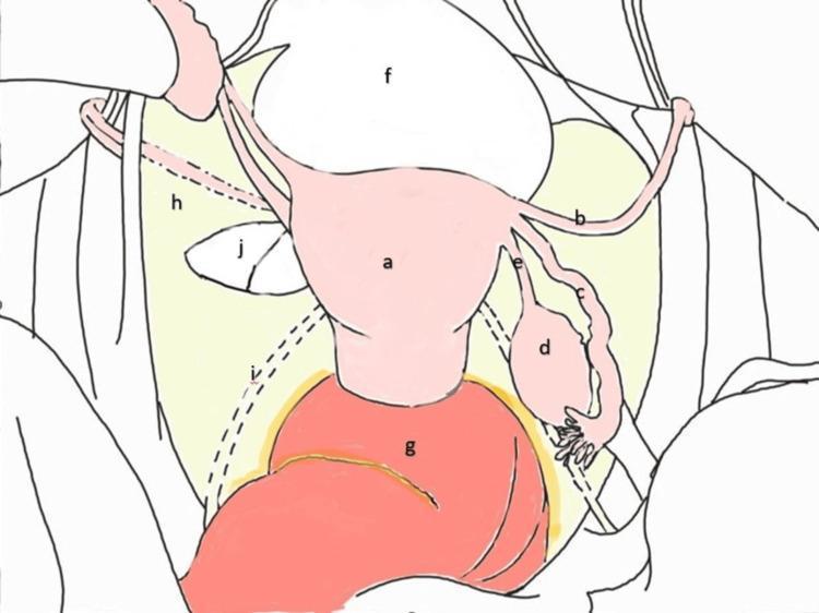
This medical anatomical diagram illustrates a superior view of the female pelvis and internal reproductive organs. Centrally, the uterus (a) is positioned posterior to the urinary bladder (f) and anterior to the rectum (g). Extending laterally from the uterine horns are the round ligaments (b), fallopian tubes (c), and the ovaries (d), which are anchored by the ovarian ligaments (e). The broad ligament (h) is depicted as a wide peritoneal fold draping over these structures and extending to the pelvic side walls. A dashed line indicates the path of the ureteric fold (i) beneath the peritoneum. Notably, the diagram highlights a broad ligament defect (j) on the left side, located caudal to the round ligament. This visual representation serves as an educational guide for identifying Type 1 defects according to the Cilley classification, which are clinically significant as potential sites for internal hernias of the bowel. The diagram uses a combination of solid lines for superficial structures and dashed lines for retroperitoneal or obscured landmarks, providing a clear topographic map of pelvic surgical anatomy.
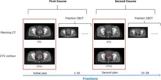
This medical schematic illustrates a two-course radiotherapy data acquisition protocol for cervical cancer, highlighting the relationship between planning CTs and inter-fractional imaging. The diagram is divided into 'First Course' and 'Second Course' sections along a chronological 'Fractions' timeline. For each course, axial CT images of the female pelvis are shown. The first row displays the 'Planning CT' (labeled CT1 and CT2) used for initial dose calculation. The second row displays corresponding 'CTV contour' images (labeled CTV1 and CTV2), featuring a red outline superimposed on the anatomy to delineate the Clinical Target Volume (CTV), which includes the primary tumor, uterus, and regional pelvic lymph nodes. To the right of each planning phase, smaller 'Fraction CBCT' (Cone Beam CT) images represent daily imaging sessions (fractions 1-20 for the first course and 21-28 for the second). The diagram demonstrates the clinical workflow for adaptive radiotherapy, where a second planning CT is acquired mid-treatment to account for anatomical changes and tumor shrinkage.
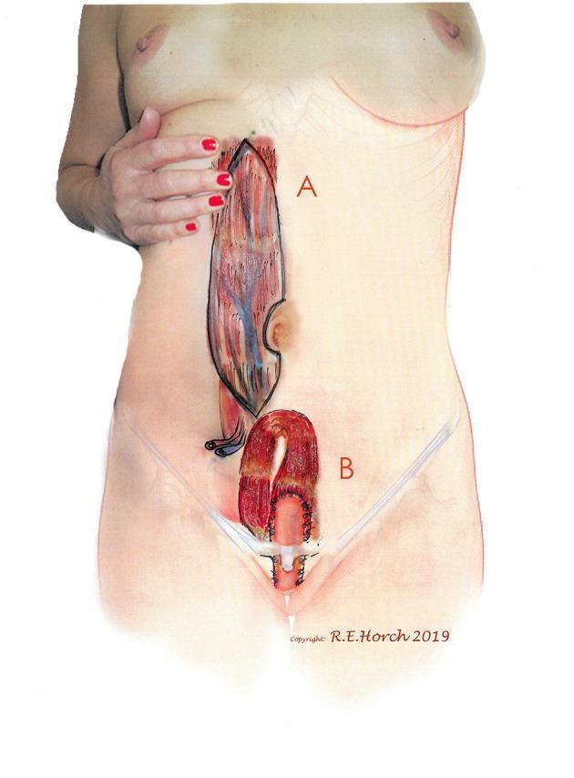
This medical illustration demonstrates the surgical planning for a vertical rectus abdominis myocutaneous (VRAM) flap. Image A shows a preoperative anatomical diagram of the flap donor site on a standing female torso. An elongated, vertical skin island is outlined on the right abdomen, revealing the underlying muscular anatomy, rectus abdominis fibers, and vascular supply, specifically the deep inferior epigastric vessels. The umbilical notch is clearly marked medially. Image B depicts the transposition of this VRAM flap into the pelvic region for reconstruction. The illustration shows the flap being tunneled into the pelvis to serve as a neovagina or to reconstruct the posterior vaginal wall. Surgical sutures and the tubular configuration of the flap are visible in the perineal region, highlighting the clinical application of pedicled muscle flaps in complex oncological or reconstructive pelvic surgery. This visual serves as an educational guide for understanding flap design, vascular pedicles, and reconstructive pathways in gynecological or plastic surgery.
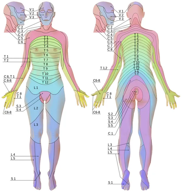
This is an anatomical illustration of human dermatomes showing anterior and posterior views in a female. Imaging modality: digital schematic diagram; technique: color-coded segmentation of cutaneous nerve territories mapped to spinal roots (C1–C8, T1–T12, L1–L5, S1–S5) with V1–V3 referenced around the head and neck. The front and back views reveal bilateral, overlapping dermatome bands that color gradient from light to dark to emphasize boundaries. Regions covered include the face (trigeminal V1–V3), neck, chest, abdomen, pelvis, upper and lower limbs. The torso shows thoracic dermatomes arranged in horizontal belts; the lumbar and sacral dermatomes extend into the hips, legs, and feet. Boundaries are schematic rather than exact anatomical demarcations, suitable for educational use. Pathophysiology implied includes sensory innervation by dorsal rami and ventral rami of spinal nerves; typical clinical relevance includes localization of neurologic deficits such as radiculopathy, herpes zoster reactivation along a dermatomal pattern, and planning of regional nerve blocks or sensory testing. This image supports medical education, anatomy review, clinical neurology practice, dermatology reference, and research on somatosensory mapping. Source attribution: Wikimedia Commons, Goran Tek-en, CC BY-SA 4.0; a freely available teaching resource. Users may print for classroom handouts or integrate into digital atlases and curricula for clinical training.
levator ani pelvic diaphragm muscles anatomy
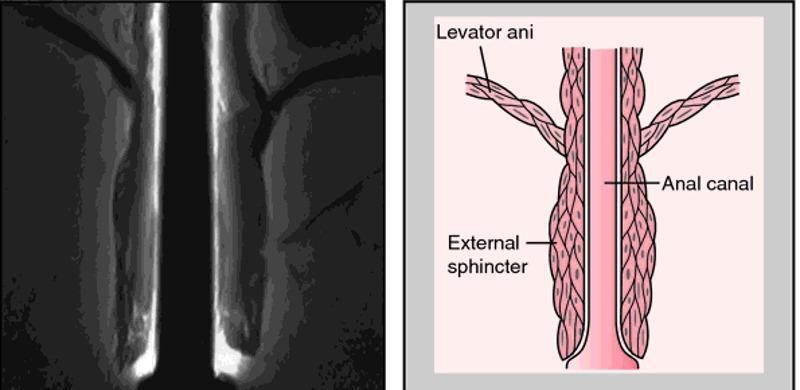
A composite image for medical education comprising a diagnostic radiological image (MRI) on the left and a corresponding anatomical diagram on the right, illustrating the pelvic floor and anal canal. The MRI shows a coronal view of the anorectal region, highlighting the muscular layers of the pelvic outlet. The anatomical diagram provides a schematic representation of the same structures for clarity. It labels the levator ani as a broad, striated muscle group superiorly that forms the pelvic diaphragm and descends to blend with the longitudinal muscle layer of the anal canal. The external sphincter is depicted as a distinct, thicker muscular cuff surrounding the distal portion of the anal canal, located inferior to the levator ani's main attachment points. This visual comparison is designed to teach the spatial relationships between the pelvic floor muscles and the terminal digestive tract, emphasizing the functional anatomy required for fecal continence. The educational focus is on identifying the levator ani and external sphincter during pelvic imaging and understanding their relative morphology.
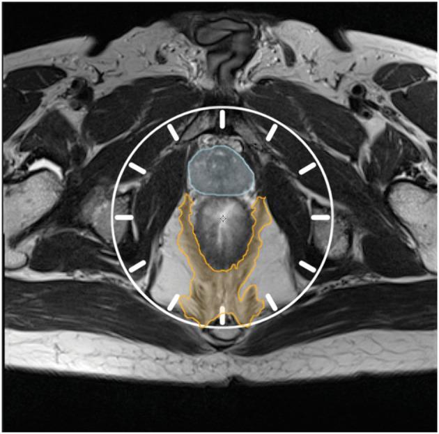
This diagnostic image is an axial T2-weighted MRI of the male pelvis, focusing on the rectal and perirectal anatomy. To facilitate standardized localization, a clock face overlay is centered on the mesorectum. The prostate gland is highlighted with blue hatching in the anterior compartment, corresponding to the 10 o'clock to 2 o'clock positions. Posteriorly, the pelvic diaphragm (levator ani and coccygeal muscles) is delineated with orange hatching, primarily occupying the posterior quadrants between the 3 o'clock and 9 o'clock positions. The image serves as an educational anatomical template for describing tumor location and surgical planes, particularly for extralevator abdominoperineal excision (ELAPE). Surrounding structures include the gluteal musculature, ischial tuberosities, and femoral heads, providing anatomical context for surgical oncology planning and postoperative assessment of the residual pelvic diaphragm.
Female Pelvis - Anatomy Notes
1. Bony Pelvis
Bones
- Two innominate bones - each formed by ilium, ischium, and pubis
- Sacrum and coccyx
Female vs. Male Pelvis Differences
| Feature | Female | Male |
|---|---|---|
| Shape | Larger, broader | Taller, narrower, more massive |
| Inlet shape | Oval | Heart-shaped |
| Subpubic angle | 90-100° (obtuse) | ~70° (acute) |
| Sacrum | Shorter, wider, less pronounced promontory; angulated at L3-L4 | Uniform curvature |
Pelvic Measurements (Obstetric Diameters)
- Pelvic inlet (conjugates): Measured front-to-back and transversely
- When standing upright, the ASIS and pubic symphysis lie in the same vertical plane - the pelvic inlet faces anteriorly, directing intra-abdominal pressure onto bone rather than muscles and fascia
2. Fascia and Peritoneum
| Stratum | Contents |
|---|---|
| Inner | Rectus fascia; Denonvilliers fascia (anterior rectal wall) |
| Intermediate | Surrounds uterus and supporting vessels; includes pubovesical, pubocervical fascia; parametrium (around uterus); paracolpium (around vagina) |
| Outer | Transversalis fascia, continuous with endopelvic and lateral pelvic fascia; iliac fascia covering iliacus and psoas |
- Uterosacral and cardinal ligaments - provide fixation
- Vesicovaginal septum - separates bladder from vagina
- Rectovaginal septum - separates vagina from rectum
3. Pelvic Floor (Pelvic Diaphragm)
Levator Ani Muscles
| Component | Origin | Insertion | Function |
|---|---|---|---|
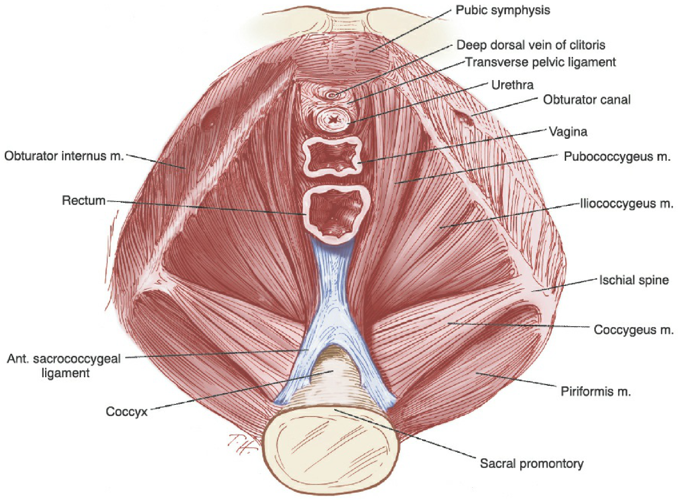
| Pubococcygeus (incl. pubovaginalis, pubourethralis) | Posterior pubis + ATLA | Anococcygeal raphe, coccyx, visceral organs | Sling around vagina/urethra |
| Puborectalis | Back of symphysis pubis | Forms U-loop behind rectum, joins opposite side | Anorectal sling; maintains anorectal angle |
| Iliococcygeus | Ischial spine + posterior obturator fossa | Last two sacral segments, coccyx, anococcygeal raphe | Pelvic shelf |
Coccygeus (Ischiococcygeus)
- Sits anterior to the sacrospinous ligament
- Arises from ischial spine; inserts on lower sacrum/coccyx
Levator Hiatus
4. Pelvic Sidewalls
- Obturator internus - covers most of the lateral pelvic sidewall; exits via lesser sciatic foramen to greater trochanter
- Piriformis - covers posterolateral walls; exits via greater sciatic foramen to greater trochanter
- Sacral plexus - lies medially on piriformis
5. Pelvic Ligaments
| Ligament | Description | Clinical Significance |
|---|---|---|
| Broad ligament | Double fold of peritoneum extending from uterine sides to pelvic walls; contains fallopian tubes, round ligament, ovarian vessels, ureter | Major surgical landmark |
| Round ligament | From uterine cornu through inguinal canal to labia majora; prevents uterine retroversion | Contains in the mesosalpinx |
| Ovarian ligament | Uterus to ovary | Tethers ovary medially |
| Infundibulopelvic (suspensory) ligament | Ovary to pelvic sidewall | Contains ovarian vessels - must be divided in oophorectomy |
| Cardinal ligament (Mackenrodt) | Cervix and upper vagina to pelvic sidewall | Primary support of cervix - divided in hysterectomy |
| Uterosacral ligament | Cervix/upper vagina posteriorly to sacrum (S2-S4) | Main apical support; used in sacrospinous fixation; carries autonomic nerves |
| Pubovesical ligaments | Bladder neck to pubic bone | Urethral support |
6. Pelvic Viscera
Uterus
- Pear-shaped muscular organ with three layers: perimetrium (serosa), myometrium (smooth muscle), endometrium (mucosa)
- Parts: fundus, body, isthmus, cervix
- Normally anteverted and anteflexed
- Uterine innervation from the inferior hypogastric plexus
Fallopian Tubes
- 10-12 cm long; draped in the broad ligament
- Four parts: interstitial (uterine), isthmus, ampulla (site of fertilization), infundibulum (with fimbriae)
- Enter uterus at the cornuа (horns)
Ovaries
- Attached to posterior broad ligament in the ovarian fossa
- Bounded by: obliterated umbilical artery, ureter, internal iliac artery
- Suspended by the infundibulopelvic ligament (containing ovarian vessels)
- Attached to uterus by the ovarian ligament
Vagina
- Fascial support: pubocervical fascia anteriorly, rectovaginal fascia posteriorly
- The paracolpium connects the vaginal walls to the pelvic sidewalls
Bladder
- Anterior to uterus, separated by vesicovaginal space
- Supported by pubovesical ligaments
Ureter (surgical danger!)
- Crosses below the uterine artery close to the cervix ("water under the bridge")
- Vulnerable to injury during hysterectomy when dividing the uterine pedicle
- Also at risk near the cardinal and uterosacral ligaments
7. Blood Supply
Arterial Supply
| Artery | Origin | Supplies |
|---|---|---|
| Uterine artery | Anterior division of internal iliac | Uterus, cervix, vagina; anastomoses with ovarian artery; crosses OVER ureter near cervix |
| Ovarian artery | Directly from aorta (L1-L2) | Ovary and fallopian tube; runs in infundibulopelvic ligament |
| Vaginal artery | Anterior division of internal iliac | Vagina |
| Internal pudendal artery | Internal iliac | Perineum, external genitalia, clitoris |
| Superior vesical artery | Internal iliac (obliterated umbilical) | Bladder, upper ureter |
| Middle/inferior vesical | Internal iliac | Bladder base, vagina |
| Middle rectal artery | Internal iliac | Rectum |
Venous Drainage
- Extensive pelvic venous plexuses associated with bladder, rectum, vagina, and uterus
- All drain into the internal iliac veins
- Clitoral veins drain into the retropubic plexus
- Superior rectal vein drains into inferior mesenteric vein (portal system - porto-systemic anastomosis site)
8. Lymphatic Drainage
| Node Group | Drains |
|---|---|
| Internal iliac nodes (presacral, obturator, internal pudendal) | Most pelvic viscera |
| External iliac nodes (external, middle, internal chains) | Anterior abdominal wall, clitoris (external chain); bladder and vagina (middle); lower abdominal wall, inguinal nodes, bladder neck, urethra (internal) |
| Common iliac nodes | Receive from both internal + external iliac chains; drain to lateral aortic nodes |
| Para-aortic (lateral aortic) nodes | Ovary, fallopian tube, uterine fundus |
| Superficial inguinal nodes | Round ligament drainage of uterus |
- Cervix → external iliac, internal iliac, and sacral nodes
- Uterine body → external iliac nodes
- Fundus → para-aortic and lateral aortic nodes
- Ovary/tubes → para-aortic (follows ovarian vessels)
- Labia and vulva → superficial inguinal nodes
9. Innervation
Somatic Innervation
- Exits via greater sciatic foramen
- Crosses ischial spine and SSL, enters lesser sciatic foramen
- Runs in Alcock's canal (pudendal canal) in the lateral wall of the ischiorectal fossa
- Branches: dorsal nerve of clitoris, perineal nerve (labial branches + muscular branches), inferior rectal nerve
- Provides sensation to perineum, external genitalia; motor to external sphincters and perineal muscles
Autonomic Innervation
| Plexus | Fibers | Supplies |
|---|---|---|
| Superior hypogastric plexus | Sympathetic (L1-L2) | Pre-sacral; descends as hypogastric nerves |
| Hypogastric nerves | Sympathetic | Connect to inferior hypogastric plexus |
| Pelvic splanchnic nerves (nervi erigentes) | Parasympathetic (S2-S4) | Join inferior hypogastric plexus |
| Inferior hypogastric plexus | Both sympathetic + parasympathetic | Bladder, uterus, vagina, rectum |
- Sympathetic (T10-L2): "fight or flight" - contracts bladder neck, inhibits detrusor, contracts internal anal sphincter, promotes ejaculation equivalent (orgasm)
- Parasympathetic (S2-S4): "rest and digest" - stimulates detrusor, relaxes bladder neck, promotes voiding, mediates clitoral erection
10. Perineum and External Genitalia
Urogenital Triangle (anterior)
- Deep transverse perineal muscles
- External urethral sphincter (striated, voluntary)
- Membranous urethra
- Innervated by perineal branch of pudendal nerve
- Ischiocavernosus muscle - covers crura of clitoris; promotes erection
- Bulbospongiosus muscle - surrounds introitus; covers vestibular bulbs; constricts vaginal orifice; innervated by pudendal nerve
- Superficial transverse perineal muscles - overlie posterior vagina; attach to perineal body
- Vestibular bulbs - erectile tissue; homologue of corpus spongiosum
- Greater vestibular glands (Bartholin's glands) - at posterior end of vestibular bulbs; site of Bartholin cyst/abscess
Anal Triangle (posterior)
- External anal sphincter (EAS): Striated voluntary muscle; three parts (subcutaneous, superficial, deep); innervated by inferior rectal nerve + pudendal nerve
- Internal anal sphincter (IAS): Smooth involuntary muscle; autonomic innervation from iliohypogastric plexus branches
- Ischiorectal (ischioanal) fossa: Contains fat; bounded by EAS, ischial tuberosity, urogenital diaphragm, gluteus maximus; Alcock's canal runs in its lateral wall
Perineal Body
11. Key Surgical Danger Points (Clinical Highlights)
| Structure at Risk | Procedure | Mechanism |
|---|---|---|
| Ureter | Hysterectomy | Crosses under uterine artery at the parametrium near cervix; also near uterosacral ligament |
| Pudendal nerve/vessels | Sacrospinous ligament fixation | Nerve passes just posterior to the SSL |
| Sacral plexus | Sacrospinous fixation | Plexus is medial on piriformis, exits same foramen |
| Obturator nerve/vessels | Para-aortic lymph node dissection | Runs on the lateral pelvic wall |
| Sciatic nerve | Prolonged lithotomy position | Stretch causes foot drop (L4-S1 via deep peroneal nerve) |
| Inferior epigastric vessels | Laparoscopic trocar insertion | Lie lateral to the rectus sheath |